Looks like no one added any tags here yet for you.
Articulation
area where bones are joined to each other
Process
General term for any prominence on body surface; usually attachment sites for muscles, ligaments, tendons, or articulation site
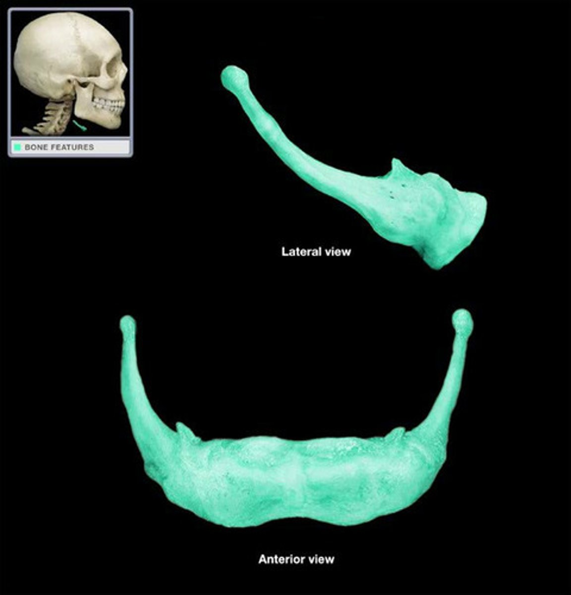
Mandible
only moveable bone of the skull at the temporomandibular joint
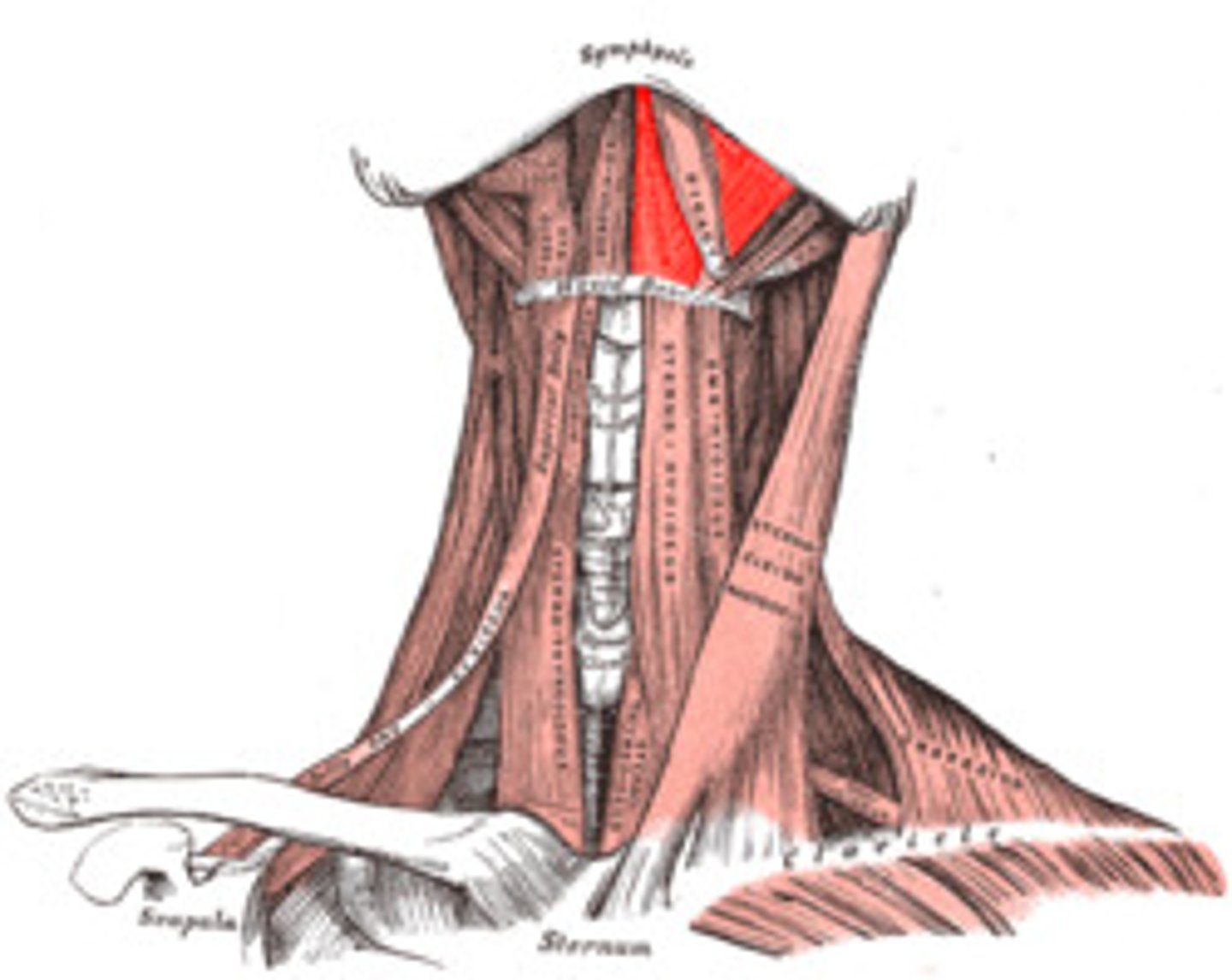
Cervical muscles
Muscles of facial expression
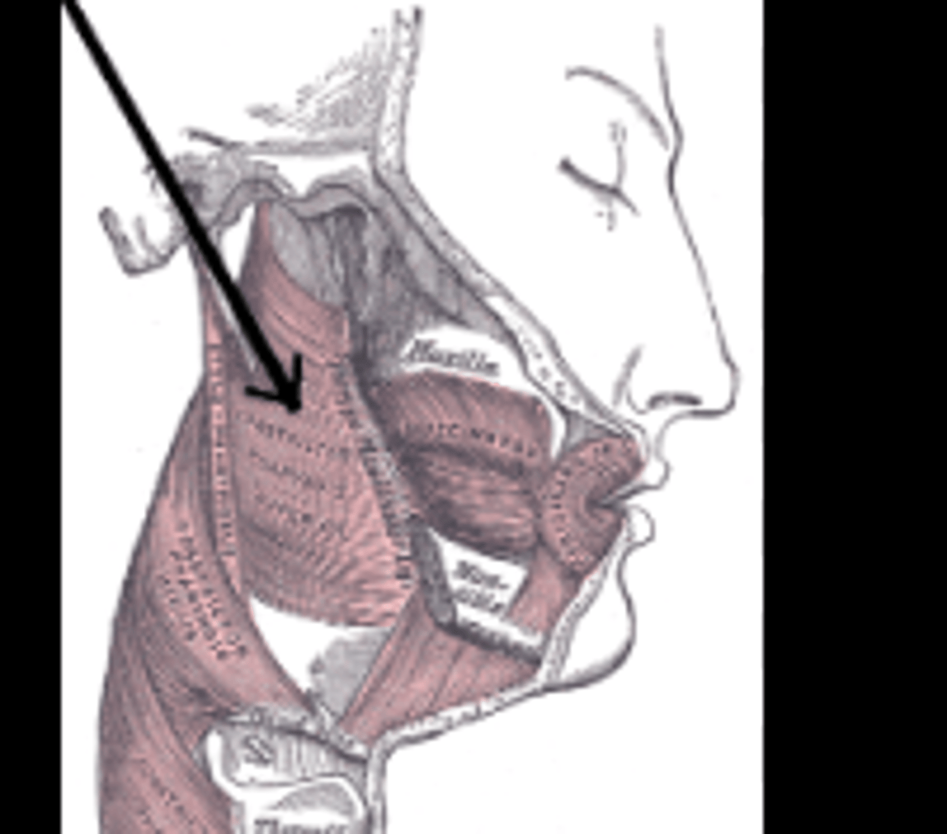
Muscles of mastication
Hyoid muscles
Muscles of the tongue
Muscles of the soft palate
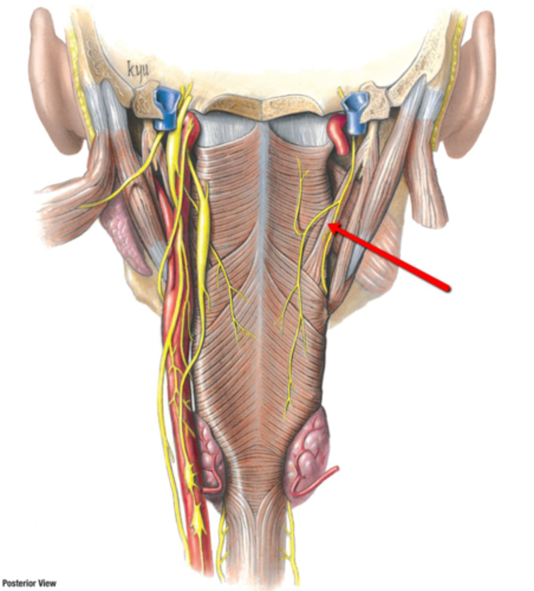
Muscles of the pharynx
Seven Main Muscle Groups of the Head and Neck:
Origin
Where the muscle originates
Insertion
Where the muscle attaches to the more moveable structure
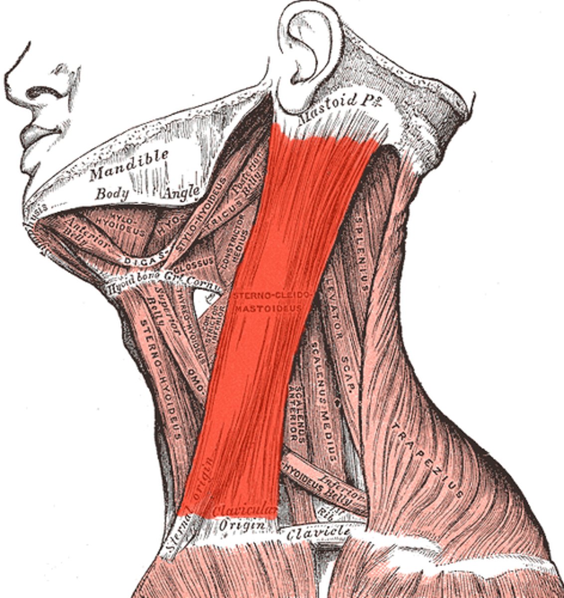
Sternocleidomastoid trapezius
Trapezius
what are the cervical muscles?
Sternocleidomastoid trapezius
Responsible for
-Tilting and rotating the head and neck
-Flexing the neck
-Stabilizing the neck
Trapezius
Responsible for:
-Lifting and rotating the shoulders
-Dorsal Flexion of the head
-Twisting Head
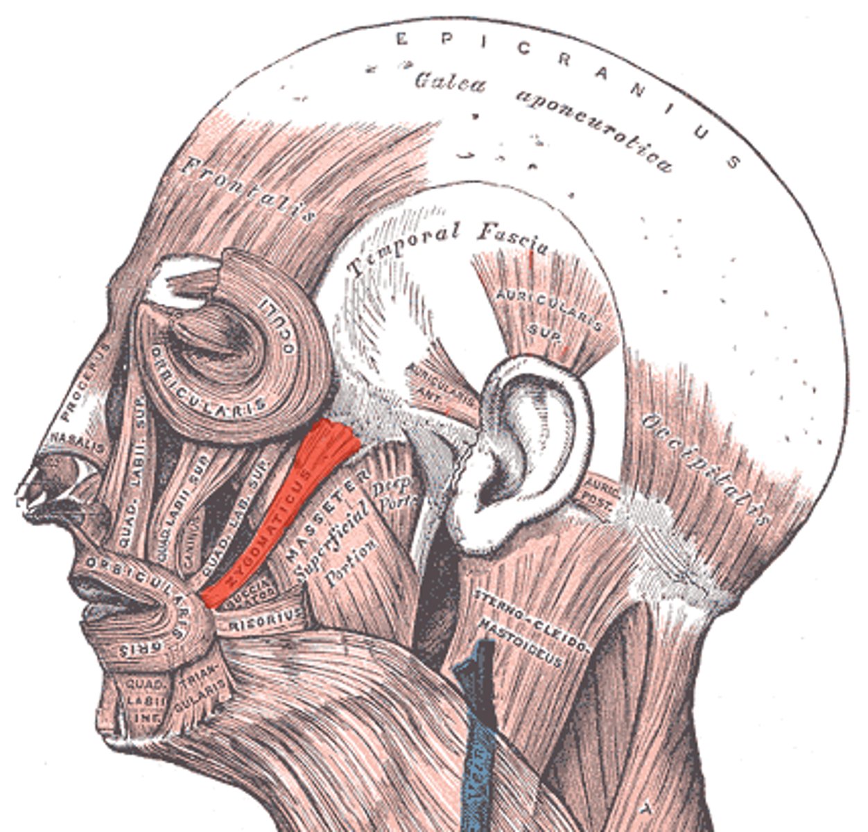
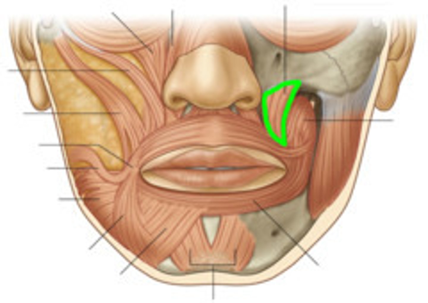
Orbicularis Oris
Buccinator
Risorius
Zygomaticus
Levator anguli oris
Depressor Anguli Oris
MUSCLES OF FACIAL EXPRESSION include
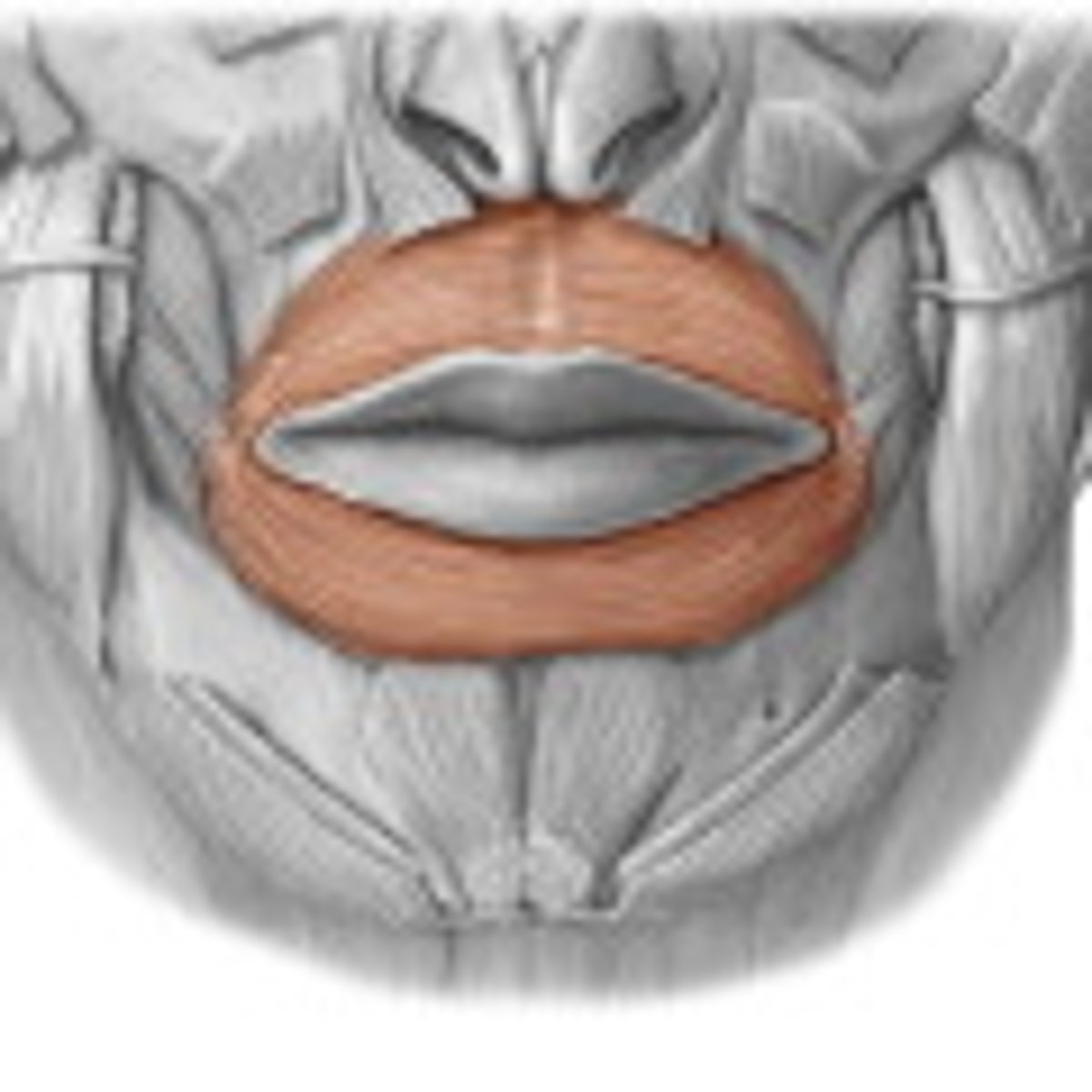
Orbicularis Oris
Muscle of facial expression responsible for Closing or pursing the lips
Buccinator
Muscle of facial expression responsible for:
Flattens cheek
Assists in chewing
Assists Muscles of mastication
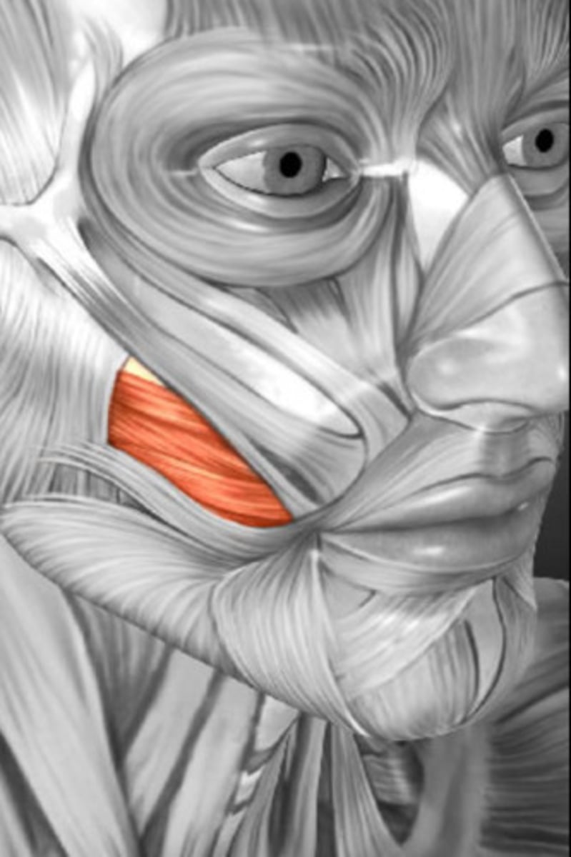
Risorius
Muscle of facial expression responsible for allowing a person to smile widely
Zygomaticus
Muscle of facial expression responsible for:
Smiling
Raising Upper Lip
levator anguli oris
muscle of facial expression responsible for smiling
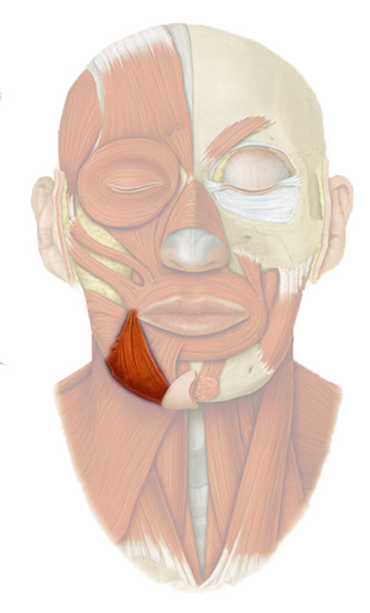
Depressor anguli oris
muscle of facial expression responsible for frowning
facial nerve (CN VII)
All facial muscles are innervated by the
facial artery
All facial muscles receive blood supply by the
bone
In general, facial muscles have origins with
skin tissue
In general, facial muscles have origins with bone, and insertions in
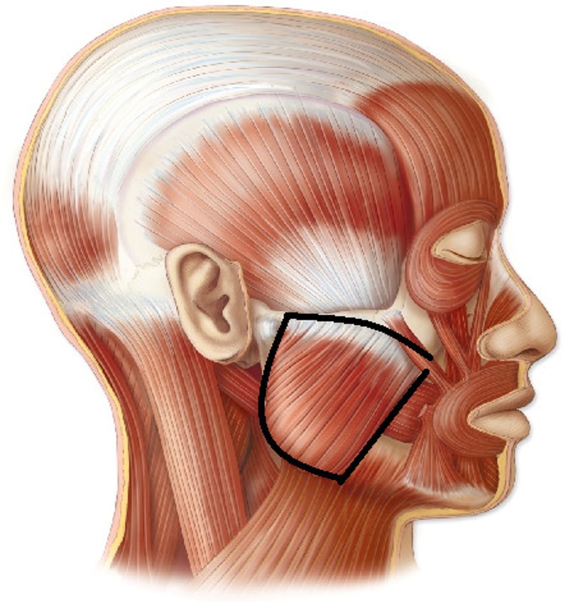
masseter
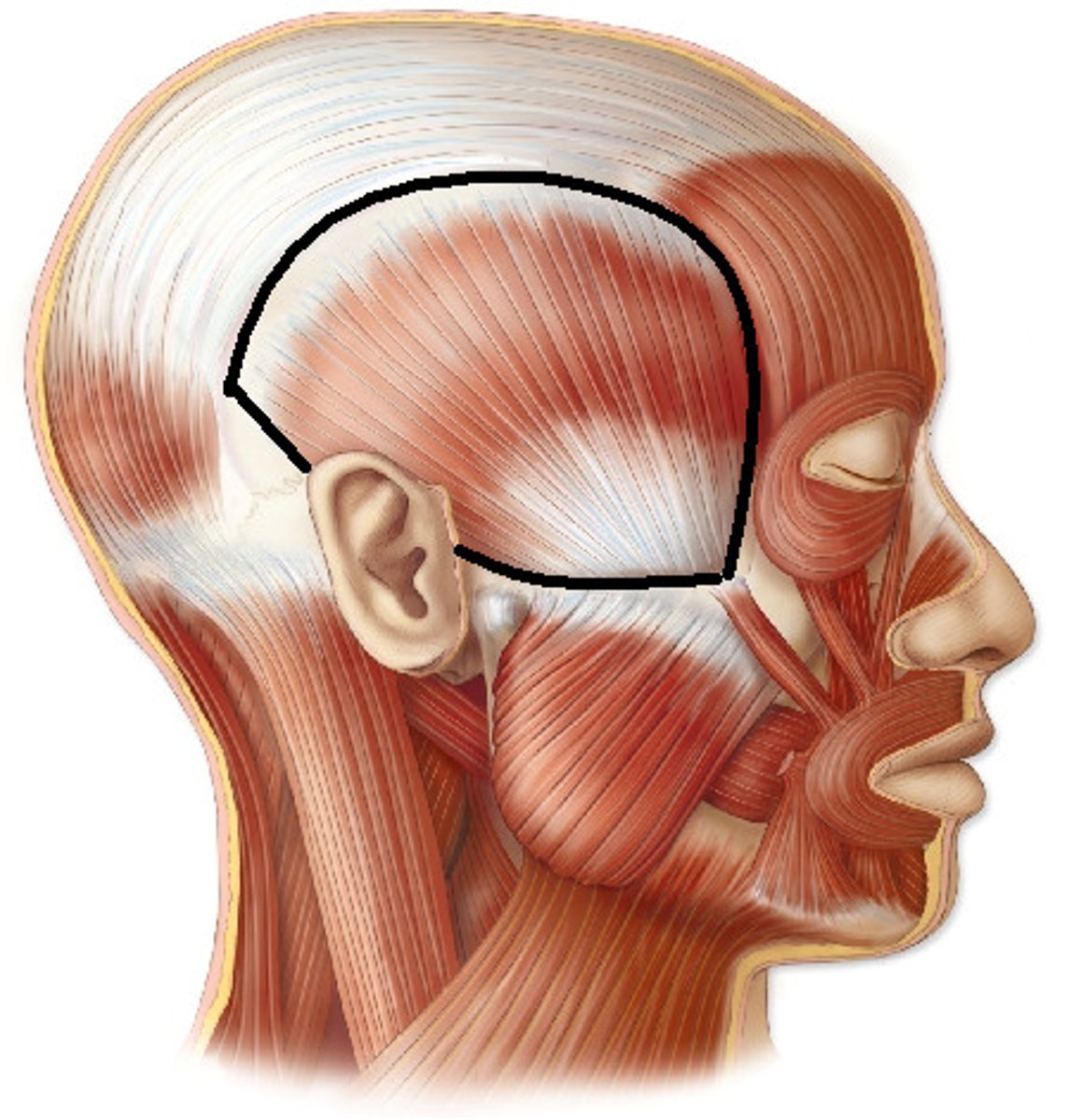
temporalis
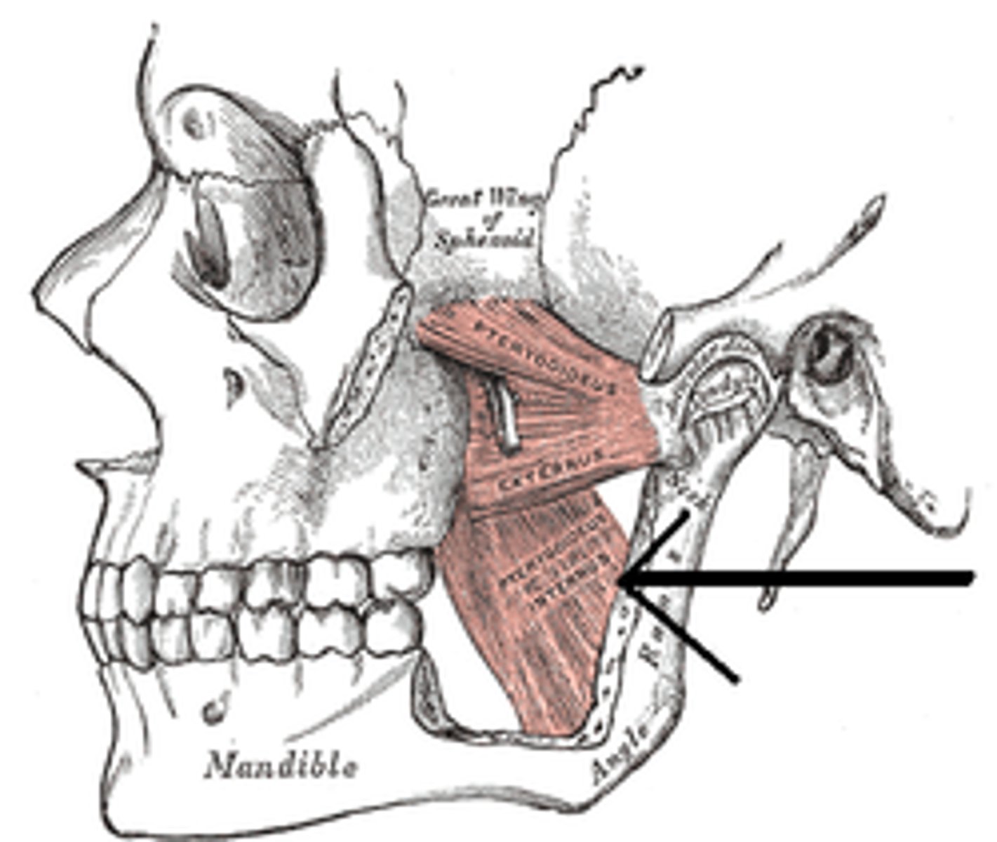
Medial pterygoid
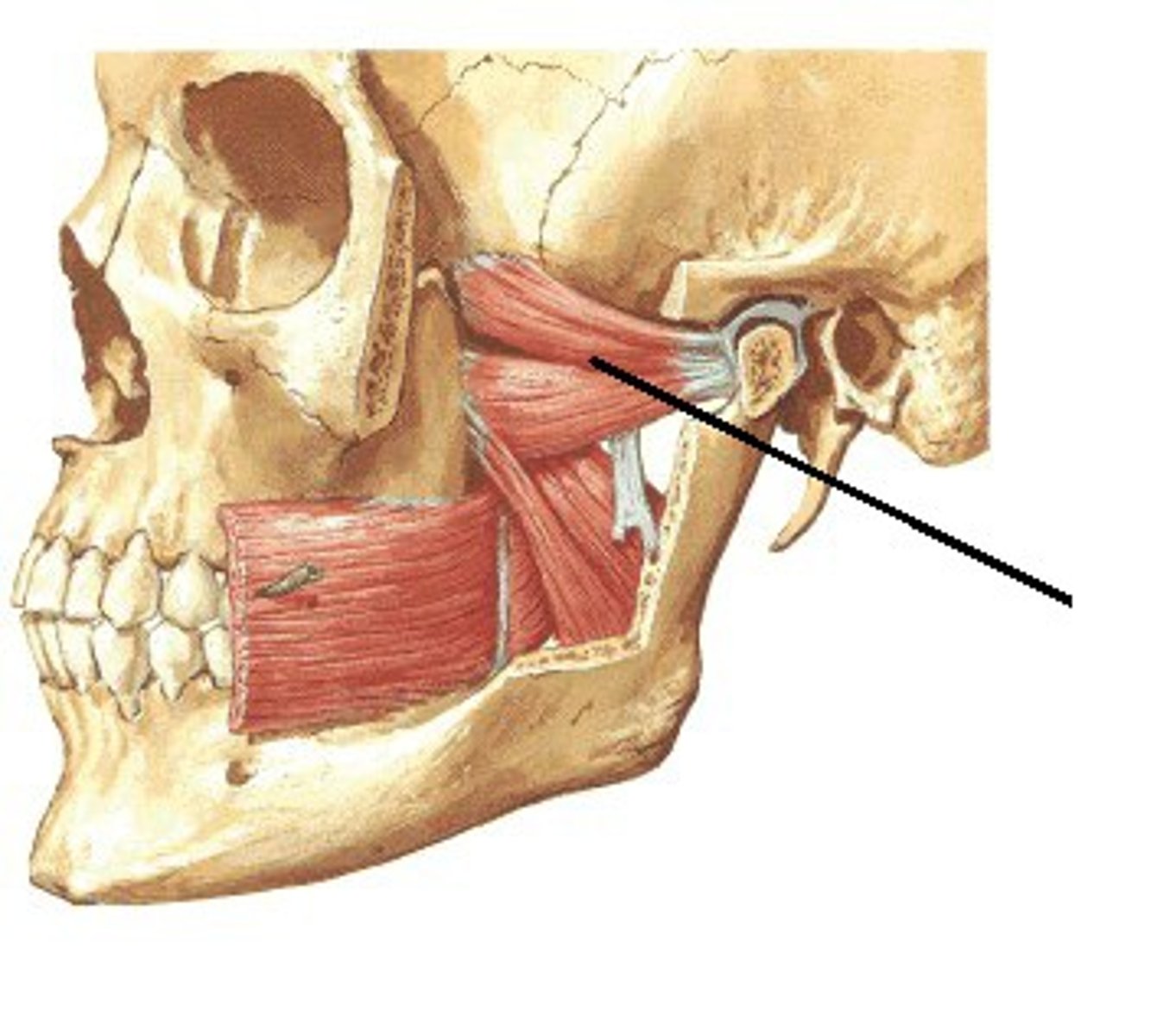
lateral pterygoid
what are the muscles of mastication
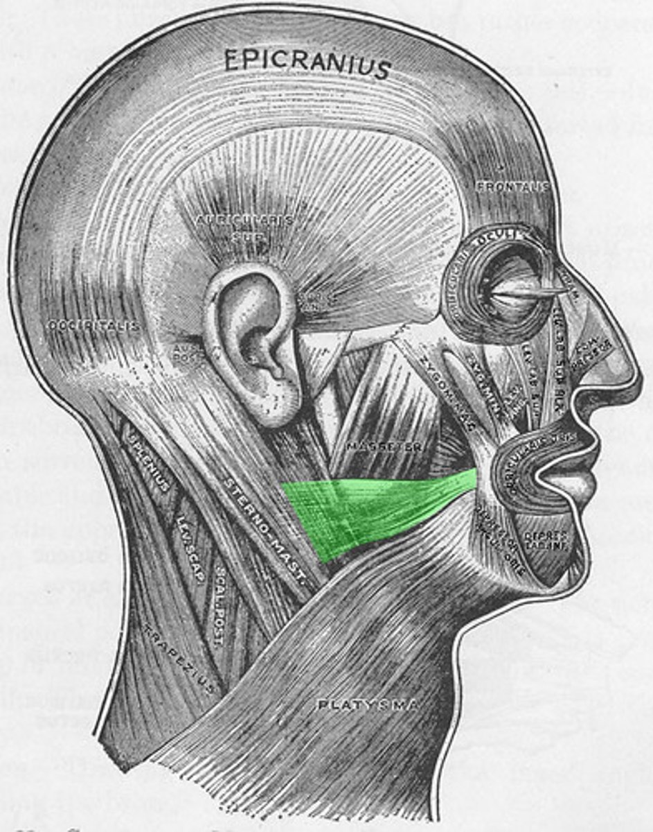
Masseter
Muscle of Mastication involved in Elevation of the mandible during jaw closing
Temporalis
Muscle of Mastication involved in
Elevation of the mandible during jaw closing.
Retraction of the mandible
medial pterygoid
Muscle of Mastication involved in Elevation of the mandible
Lateral pterygoid
muscle of mastication responsible for:
Slight depression of the mandible during opening.
Protrusion of the mandible
Lateral Deviation of the mandible (shift lower jaw)
masseter
what muscle of mastication is most likely to become enlarged due to bruxism?
lateral pterygoid
what muscle of mastication is most likely to be affected by trauma to TMJ?
mandibular division of trigeminal nerve (CN V3)
All muscles of mastication are innervated by the
maxillary artery
All muscles of mastication receive blood supply by the
mandible
All muscles of mastication INSERT into the
mastication
swallowing
muscles of hyoid and pharynx assist in actions of
Suprahyoid
Infrahyoid
hyoid and pharynx muscles are grouped into what categories?
hyoid bone
ALL HYOID BONES INSERT ON THE
none
what bones does the hyoid bone articulate with?
Mylohyoid
elevates hyoid bone and floor of mouth
supra hyoid muscles
Group of hyoid muscles responsible for depression of the mandible
Mylohyoid
Geniohyoid
Stylohyoid
Digastric
swallowing
what action are supra hyoid muscles responsible for
V3
VII
the supra hyoid muscles are innervated by what nerves?
Infrahyoid muscles
group of hyoid muscles responsible for stabilizing the hyoid bone:
Sternothyroid (C1-C3)
Sternohyoid (C1-C3)
Omohyoid (C1-C3)
Thyrohyoid (C1-C3)
swallowing
speech
the Infrahyoid muscles are responsible for what action
Speaking
Swallowing
Middle Ear Function
Muscles of pharynx are involved in:
Stylophayngeus
Pharyngeal Constrictor
Muscles of the Soft Palate
Muscles of the pharynx include:
Stylophayngeus
phayrnx muscle responsible for Lifting and widening the pharynx
Pharyngeal Constrictor
phayrnx muscle responsible for Raising the pharynx and larynx pushing food into the esophagus during swallowing
CN IX (glossopharyngeal nerve)
what is the Stylophayngeus innervated by?
pharyngeal plexus
what is the Pharyngeal Constrictor innervated by?
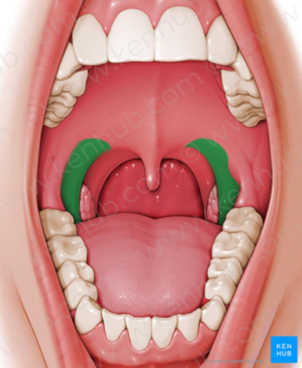
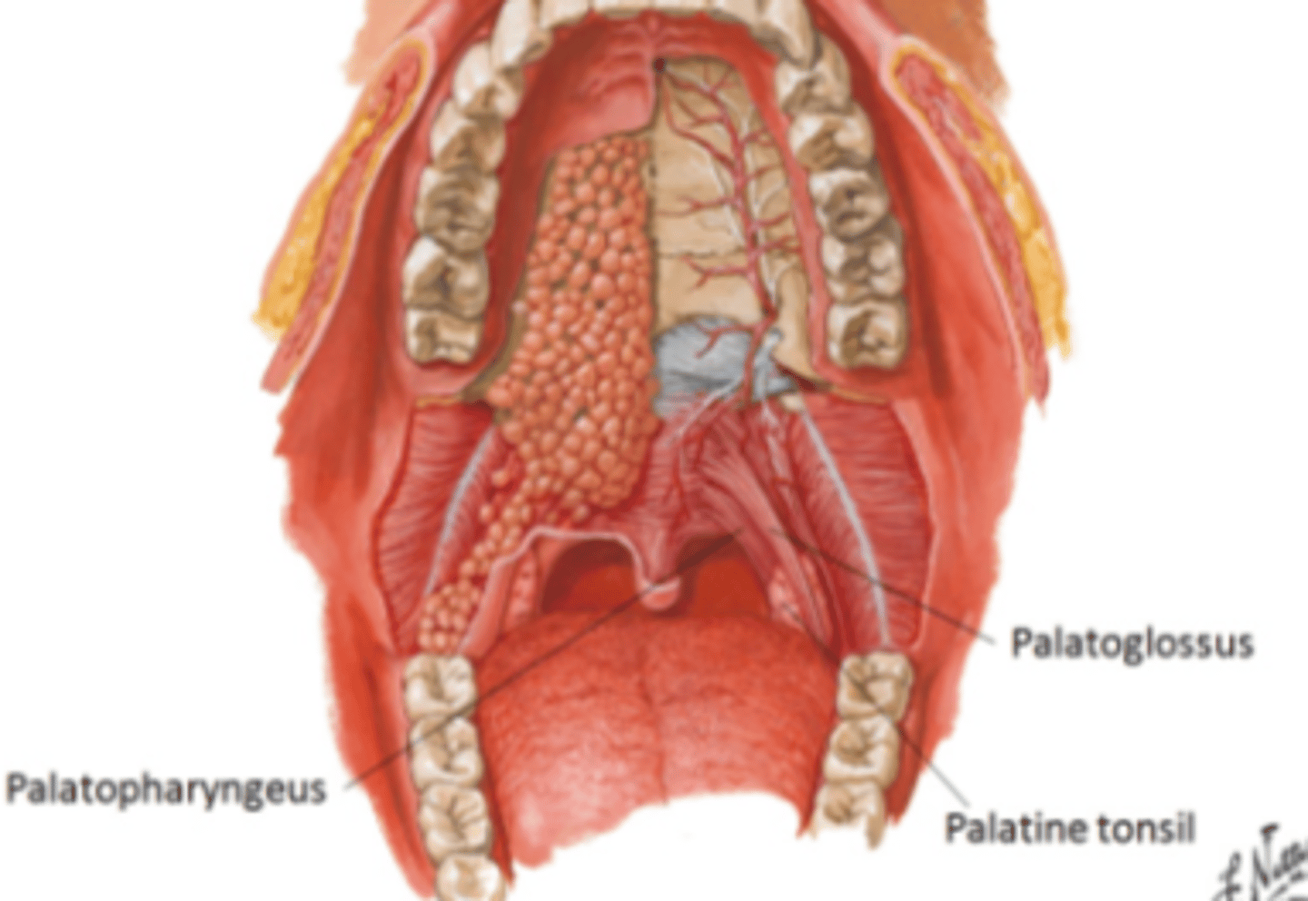
Palatoglossus muscle
palatophayngeus
levator veli palatini
tensor veli palatin
uvula muscle
pterygomandibular fold
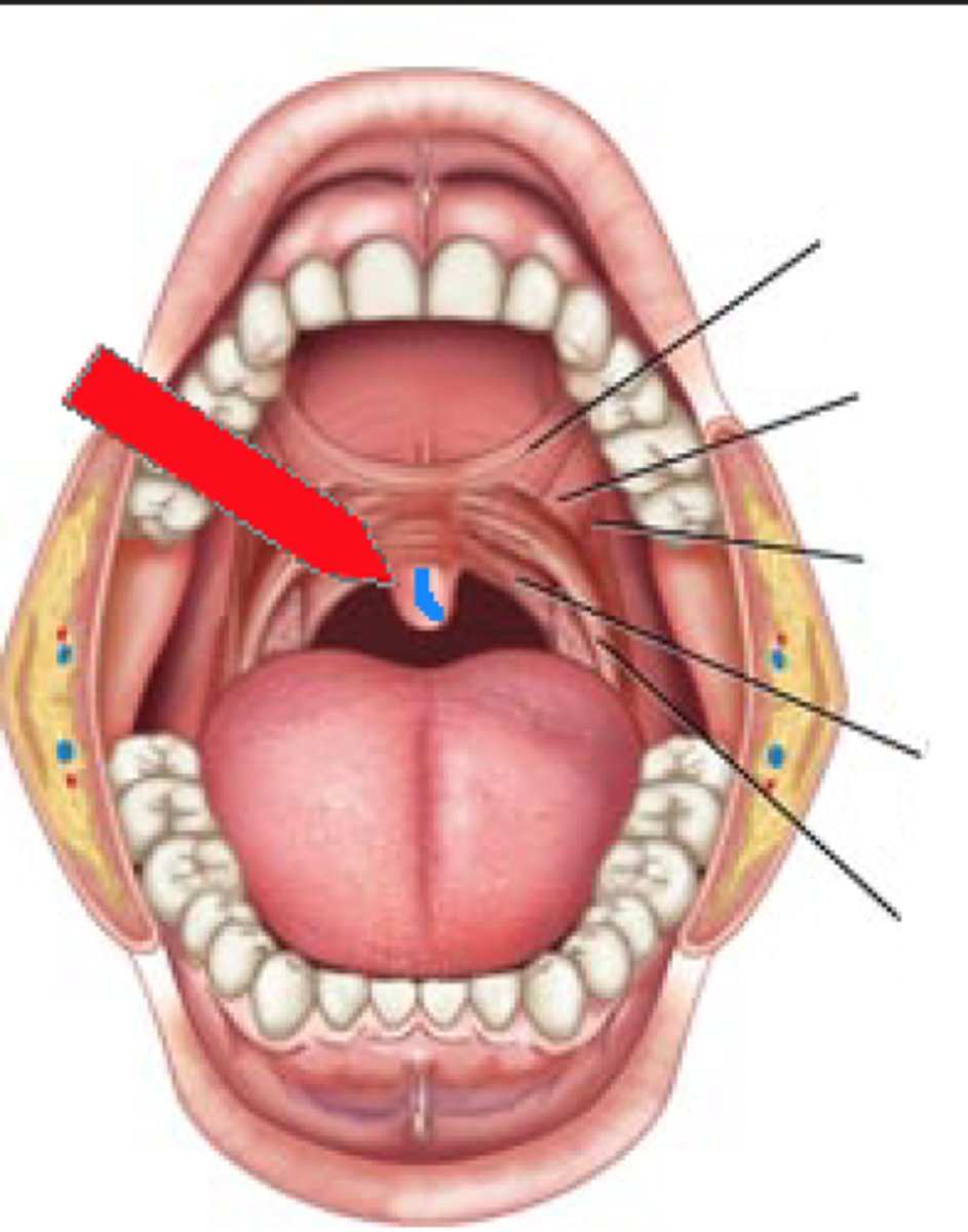
what are the muscles of the soft palate?
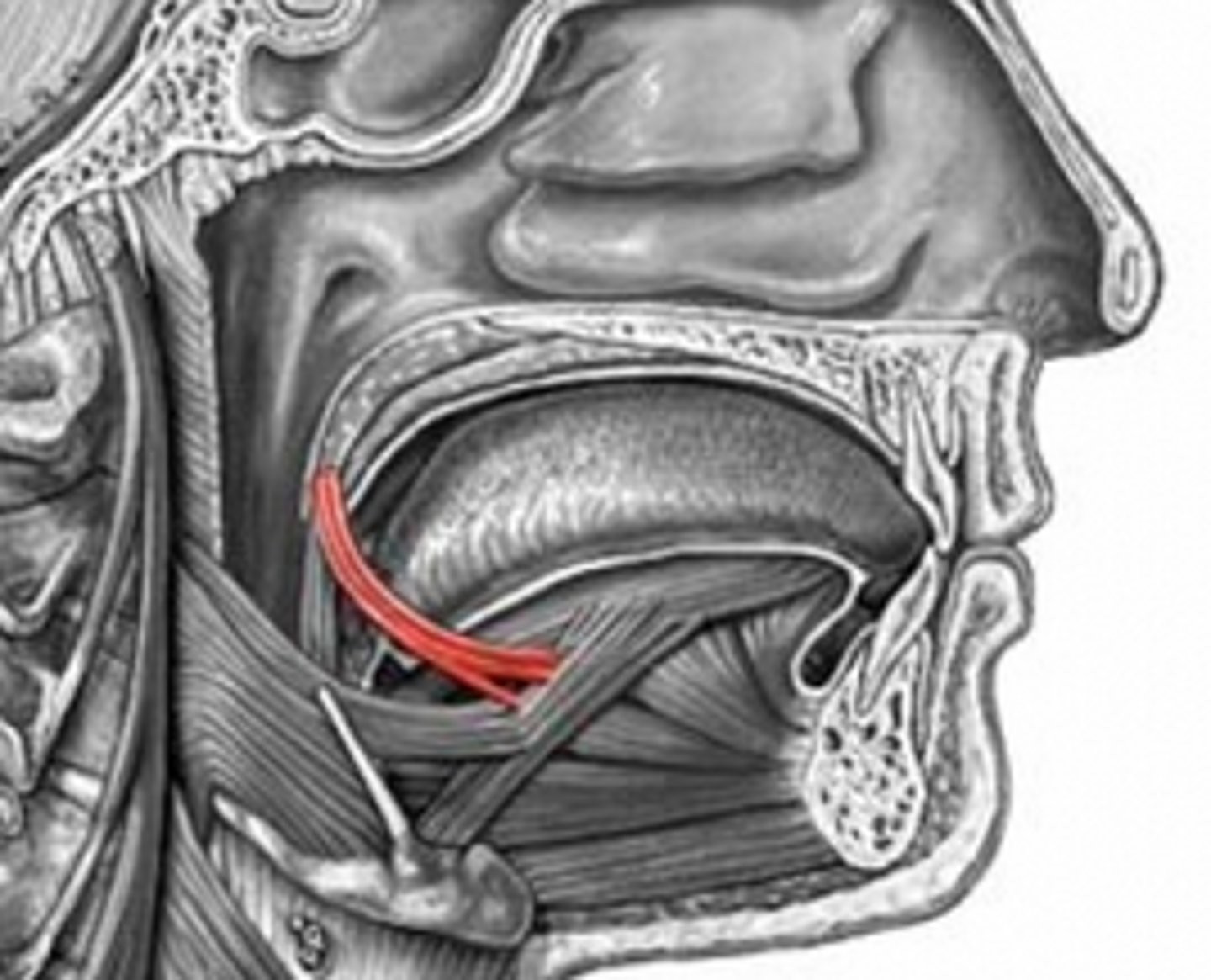
Palatoglossus muscle
Soft palate muscle responsible for elevating the posterior portion of the tongue
Palatopharyngeus
soft palate muscle responsible for tensing the soft palate to pull the pharyngeal walls superiorly, anteriorly, and medially during swallowing.
Levator veli palatini
soft palate muscle responsible for elevating the velum to create closure between oral and nasal cavities.
Tensor Veli Palatin
soft palate muscle responsible for assisting the levator veil palatini to prevent entry of food into the nasopharynx when swallowing.
Uvula Muscle
soft palate muscle responsible for shortening the uvula.
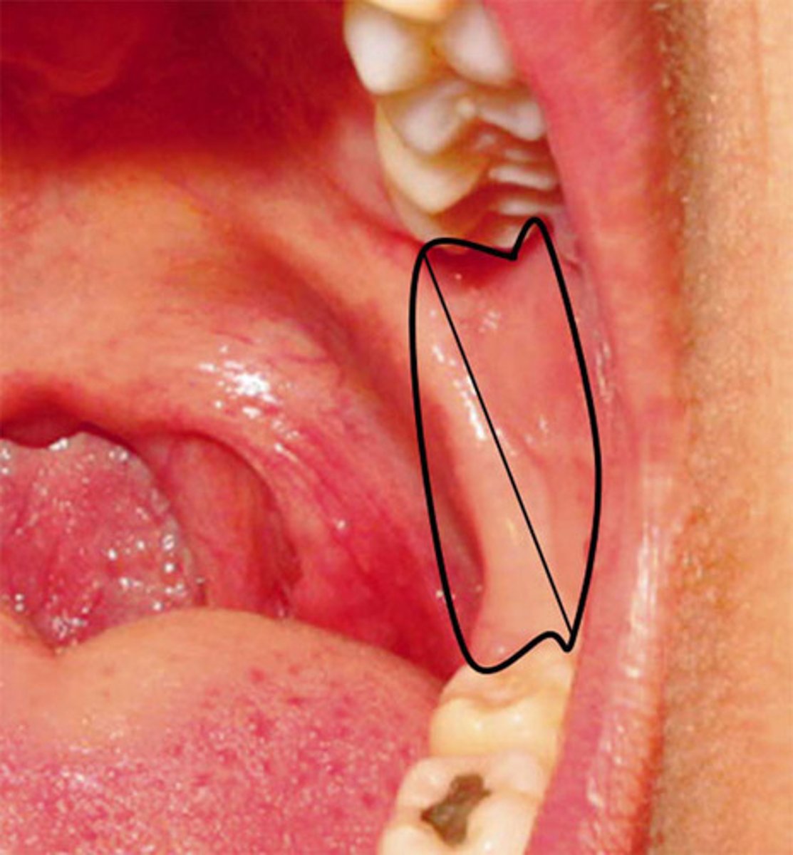
Pterygomandibular fold
Tissue that extends from the junction of the hard and soft palates down to the mandible and stretches upon opening (RAPHE)
Palatoglossus muscle
muscle of soft palate responsible for:
Elevates base of tongue
Depresses soft palate
INITIATES SWALLOWING
oral cavity from pharynx
the Palatoglossus muscle separates the
pharyngeal plexus
the Palatoglossus muscle is innervated by
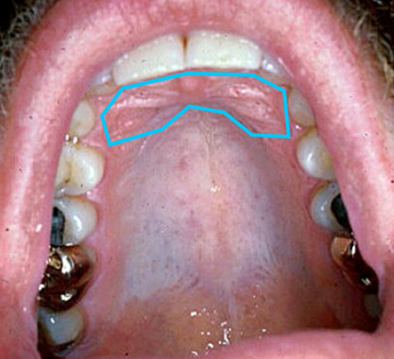
palatine rugae
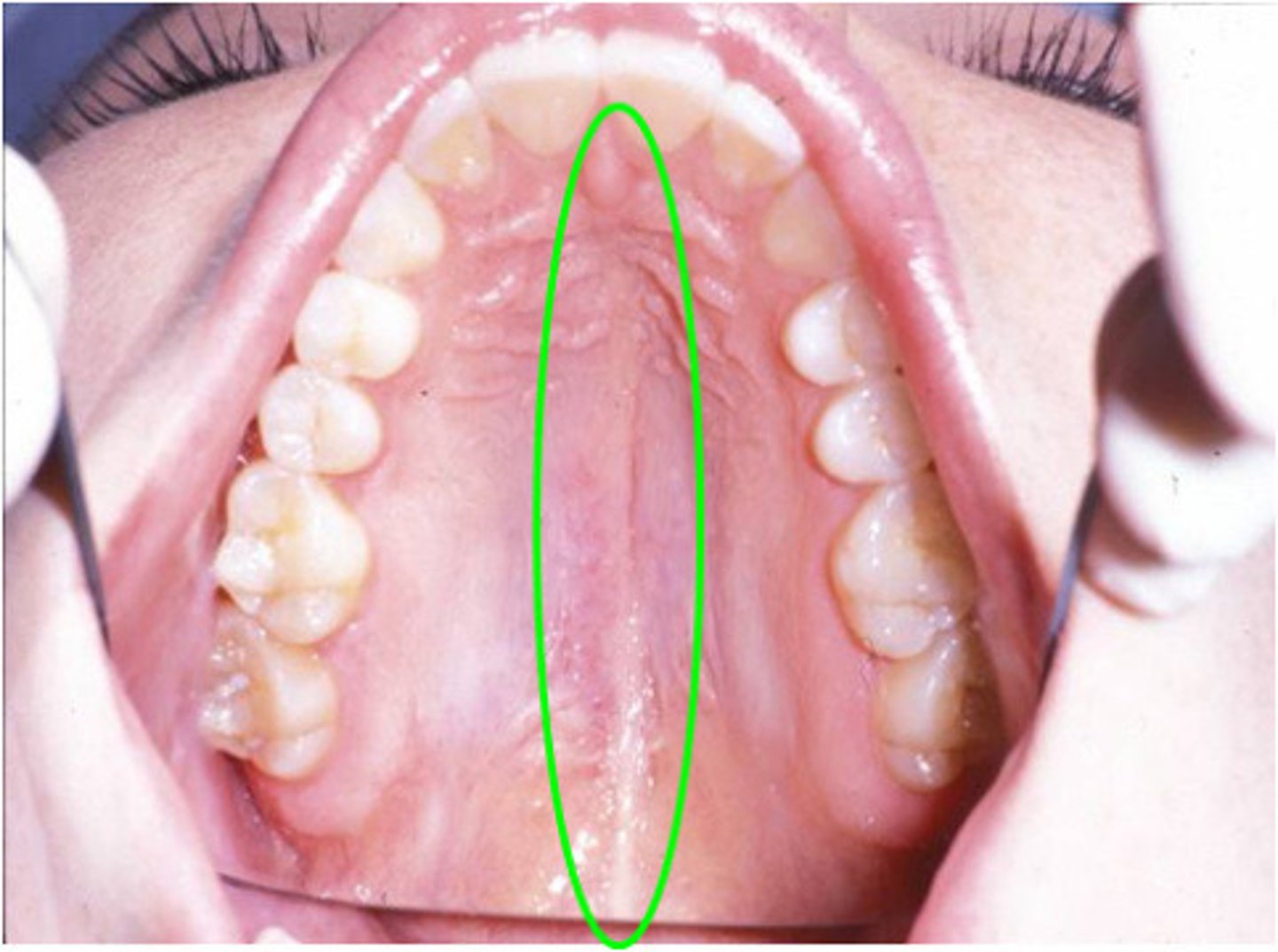
median palatine raphe
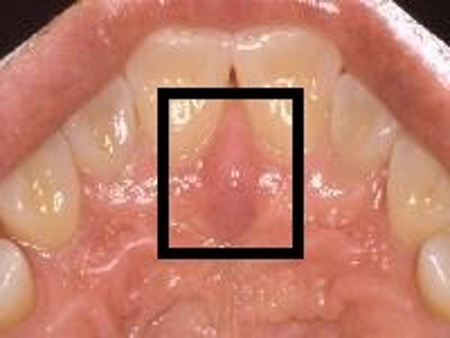
incisive papilla
palatal tori/torus
what are the structures of the hard palate?
Palatine Rugae
A series of transverse ridges on the anterior part of the palatal mucosa
Median Palatine Raphe
Raphe running across the palate from the palatine uvula to the incisive papilla
Incisive Papilla
An oval midline mucosal prominence of the anterior hard palate overlying the incisive fossa.
Palatal Tori/Torus
Benign bone tumors of the maxilla that elevate the midline of the palate
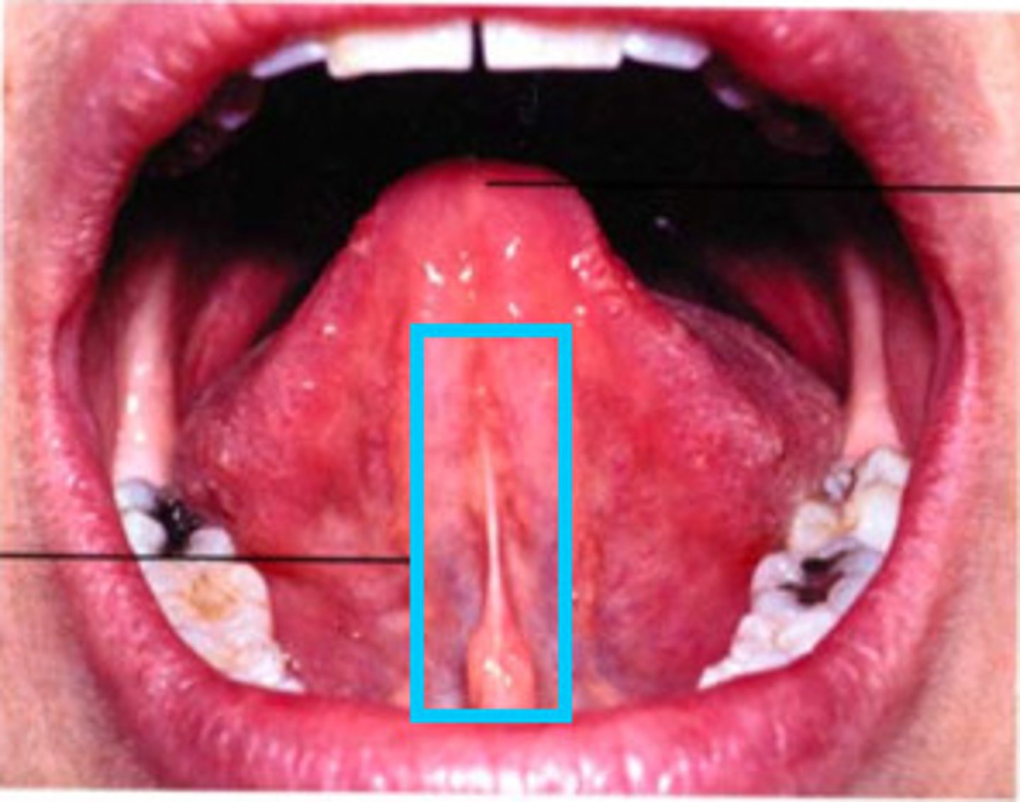
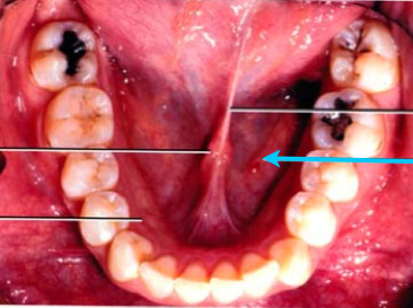
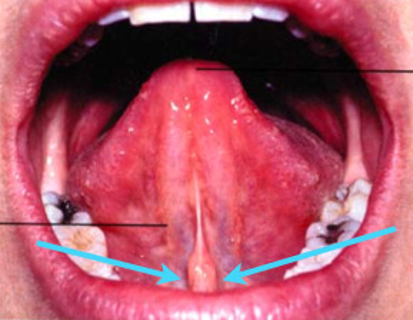
Lingual Frenum
structure on the floor of the mouth that is the midline fold of tissue from the ventral surface of the tongue to the floor of the mouth.
Sublingual Fold/Plica Sublingaulis
Structure on the floor of the mouth that appears as a v shaped ridge of tissue on each side of the frenum, that FUNCTION to empty the sublingual glands
Sublingual Caruncle
Structure on the floor of the mouth that is a small papilla on the lingual frenum, acting as duct openings for submandibular and sublingual salivary glands.
4
the muscles of the tongue include ___ sets of intrinsic muscles and three pairs of extrinsic tongue muscles.
3
the muscles of the tongue include four sets of intrinsic muscles and ____ pairs of extrinsic tongue muscles.
inside the tongue
The intrinsic muscles are entirely
lingual nerve (v3)
Chorda tympani
what nerves innervate the anterior 2/3 of the tongue
glossopharyngeal nerve (IX)
what nerves innervate the posterior 1/3 of the tongue
shaping the tongue
Intrinsic muscles of the tongue are responsible for _____ and consist of:
Superior longitudinal
Transverse
Verical
Inferior Longitudinal
Hypoglossal Nerve (XII) motor
innervation of the intrinsic muscles of the tongue is by the
lingual artery
blood supply of the intrinsic muscles of the tongue is by the
moving/controlling position of the tongue
extrinsic muscles of the tongue are responsible for ______ and consist of:
Genioglossus
Styloglossus
Hypoglossus
Genioglossus
extrinsic muscle of the tongue that is responsible for protruding the tongue.
Styloglossus
extrinsic muscle of the tongue that is responsible for retracting the tongue
Hypoglossus
extrinsic muscle of the tongue that is responsible for depressing the tongue
hypoglossal nerve (XII)
Motor innervation of the extrinsic muscles of the tongue include
Anterior ⅔ = lingual nerve (V3) and chorda tympani
Posterior ⅓ = glossopharyngeal nerve
Sensory innervation of the extrinsic muscles of the tongue include
lingual artery
blood supply of extrinsic muscles of the tongue is
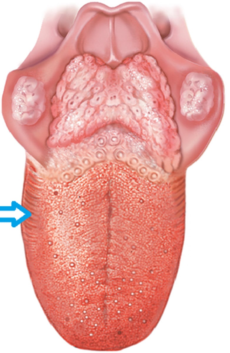
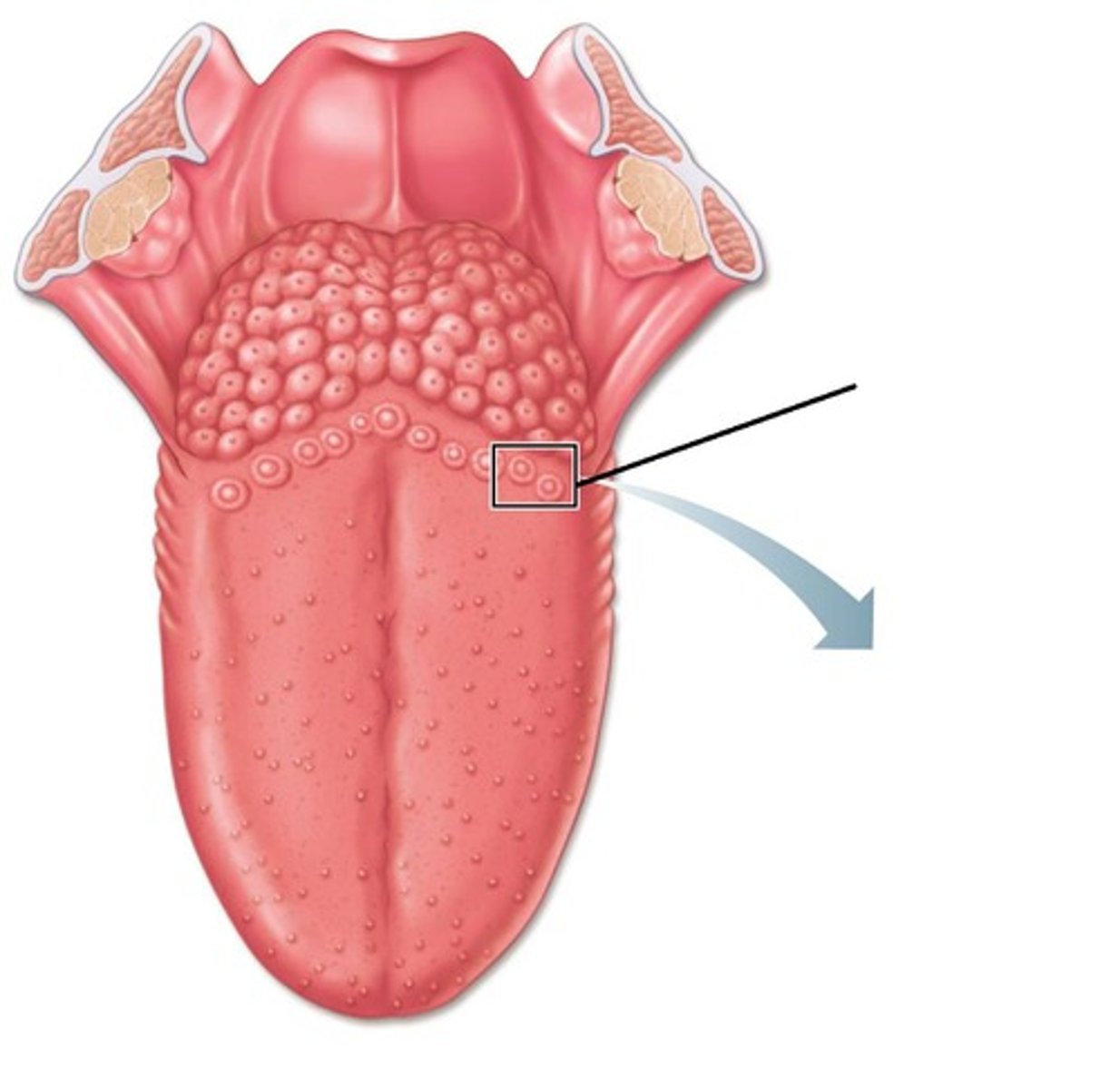
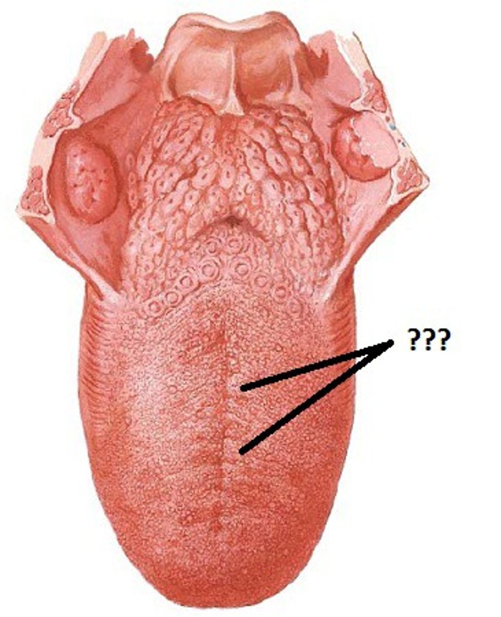
Foliate
Circumvallate
Fungiform
Filiform
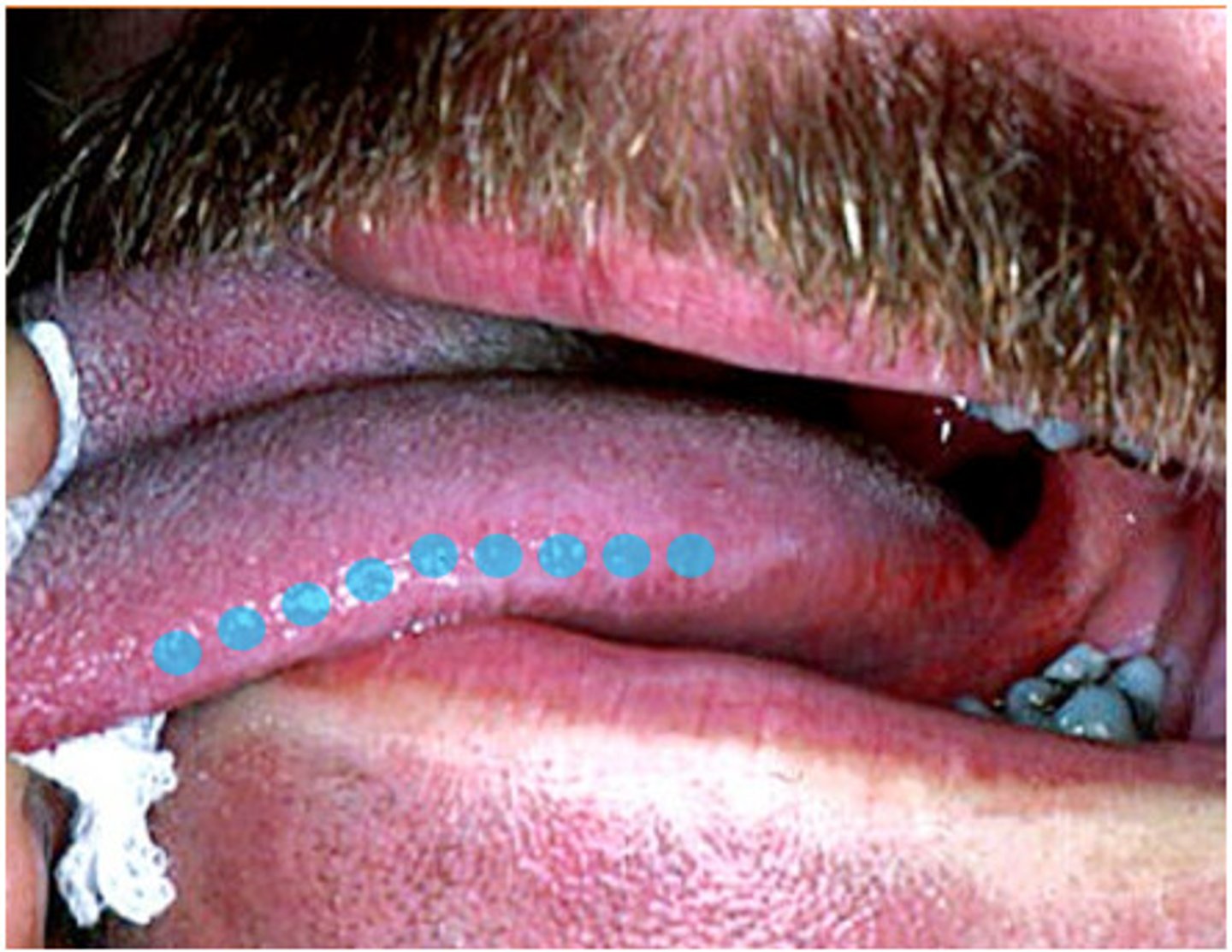
Foliate lingual papillae
what are the forms of tongue papillae?
Foliate
Tongue papillae that contain taste buds (taste buds are red in color)
Circumvallate
Large tongue papillae that contain taste (10-14 present), and are associated with Von Ebner's glands
Fungiform
Tongue papillae that are present in fewer concentrations and are mushroom shaped, that contain taste buds (red/pink in color)
Filiform
Tongue papillae that are most numerous, but DO NOT contain taste buds. - give tongue a velvety texture associated with geographic or hairy tongue.
Foliate Lingual Papillae
Tongue papillae that are located on the lateral surface of the tongue, some contain taste buds that are red in color.
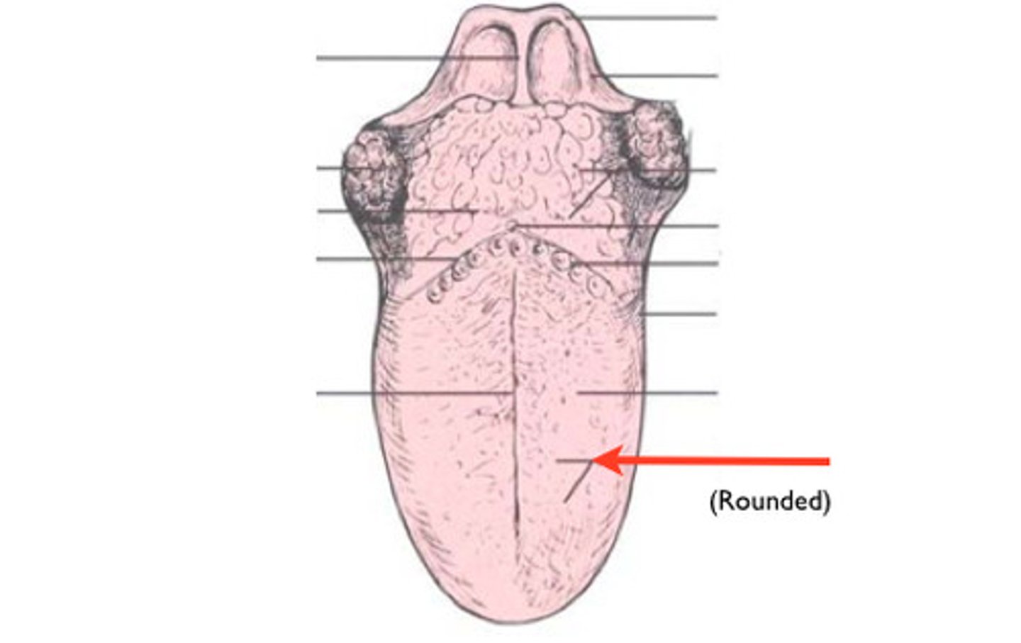
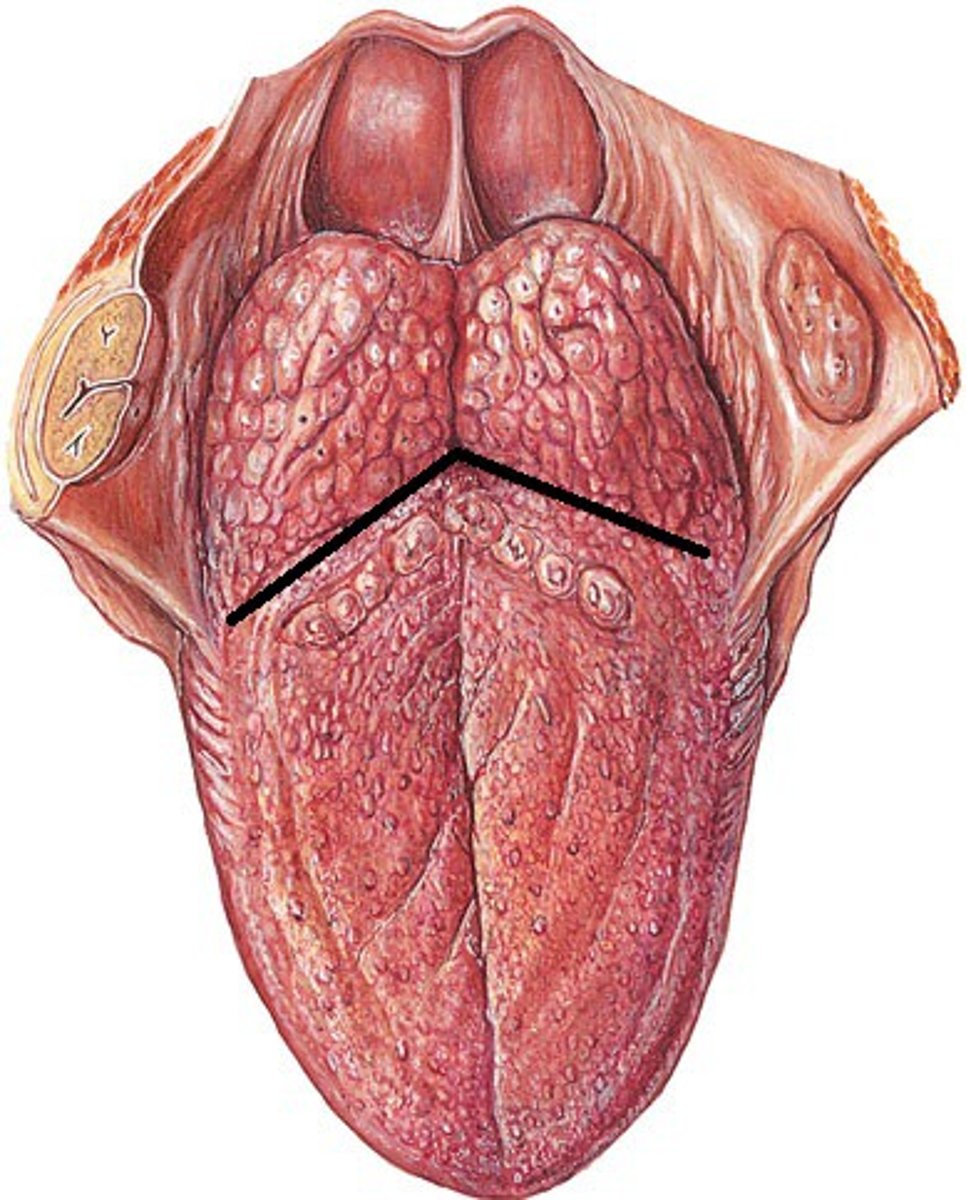
Sulcus terminalis
Separation of ⅓ posterior and ⅔ anterior portions of the tongue with the foramen cecum at the point
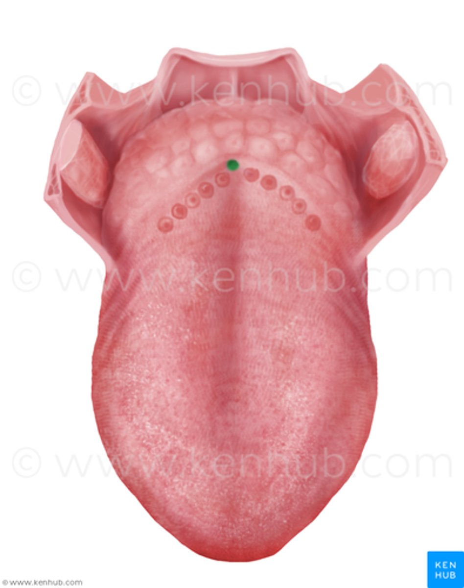
Foramen Cecum
Remnant of median thyroid diverticulum in early embryonic development/origin of the thyroid gland.
temporomandibular joint (TMJ)
the joint between the temporal bone and the mandible that is known as the hinge and sliding joint
Temporal Bone
part of TMJ that includes the articular eminence, articular fossa, and postglenoid process.
Manndible
Part of TMJ that includes the condyle of the mandible, the coronoid process, and the mandibular notch
Joint Capsule
Part of the TMJ that completely encloses the TMJ by wrapping around the margin of the articular eminence, articular fossa, and around the circumference of the condyle.
Articular Disc
Part of the TMJ that divides the TMJ into two compartments (synovial cavities), CREATED BY DENSE CT that can thin over time or dislocated.
Synovial Cavities
Two compartments of the TMJ created by the articular disc where synovial fluid is produced to lubricate the joint
TMJ ligament
Sphenomandibular ligament
Stylomandibular ligament
Ligaments of TMJ include
speech
mastication
movement of TMJ allows for movement of the mandible for
Gliding
Rotation
what are the two basic movements of the TMJ?
pain, headache, tinnitus, impaired hearing, pain around the tongue, pain in area of TMJ/muscles of mastication, limitation or deviation in movement of mandible, detectable sounds during movement of mandible
TMJ disorders are characterized by
Clenching
Grinding
Bruxism
Parafunctional Habits associated with TMJ Disorder include
Trismus
Limited ability to open the mouth due to contraction of the muscles of mastication (lockjaw)
Hypomobility
Limited ability to open the mouth