📸quiz 7 chapter 23
 Call Kai
 Learn
Learn Practice Test
 Spaced Repetition
 Match
Match Flashcards
 Knowt Play
1/24

Earn XP
Description and Tags
https://quizlet.com/930969583/quiz-7-chapter-23-flash-cards/?funnelUUID=3bf6df3f-b972-44e1-969e-6340daa7f743
Name | Mastery | Learn | Test | Matching | Spaced | Call with Kai | Chat |
|---|
No analytics yet
Send a link to your students to track their progress
25 Terms
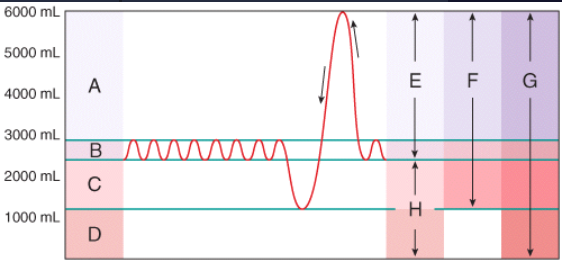
What area in the figure is the sum of the tidal volume and the inspiratory reserve volume and expiratory reserve volume?
H
B
G
E
F
F
—
A — Inspiratory reserve volume
B — Tidal volume
C — Expiratory reserve volume
D — Residual volume
E — Inspiratory capacity
F — Vital capacity
G — Total lung capacity
H — Functional residual capacity
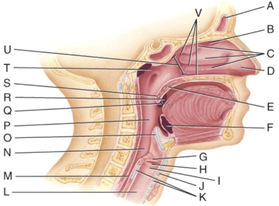
Where are the olfactory receptors found?
A
B
C
D
U
B
—
A – Frontal sinus
B – Olfactory epithelium
C – Nasal conchae
D – External naris
E – Soft palate
F – Lingual tonsil
G – Vestibular fold
H – Vocal fold
I – Larynx
J – Thyroid cartilage
K – Cricoid cartilage
L – Trachea
M – Esophagus
N – Laryngopharynx
O – Epiglottis
P – Oropharynx
Q – Fauces
R – Palatine tonsil
S – Uvula
T – Nasopharyx
U – Internal naris
V – Nasal meatuses)
Which structure prevents food or water from entering the trachea?
Arytenoid cartilage
Epiglottis
Nasopharynx
Thyroid cartilage
Paranasal sinus
Epiglottis
Which of the below tissues provides the functions of the inner layer of the conducting organs?
stratified squamous epithelium with keratin
ciliated pseudostratified columnar epithelium with goblet cells
ciliated cuboidal epithelium with goblet cells
transitional epithelium with cilia
columnar connective tissue with goblet cells
ciliated pseudostratified columnar epithelium with goblet cells
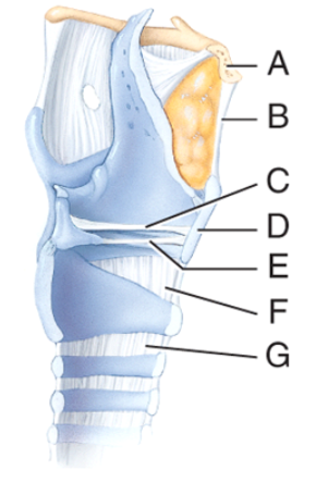
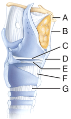
Which structure in the figure is the vocal fold?
F
D
C
E
G
E
—
A — Hyoid bone
B — Thyrohyoid membrane
C — Vestibular fold
D — Thyroid cartilage
E — Vocal fold
F — Cricothyroid ligament
G — Cricotracheal ligament
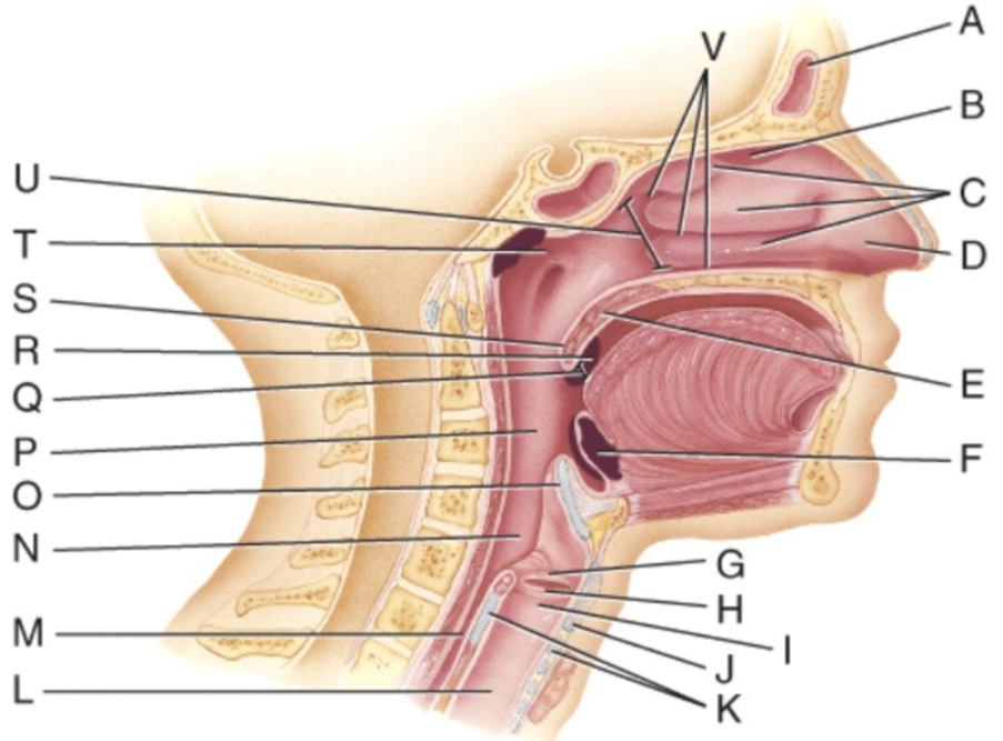
Where is the soft palate?
C
E
G
Q
S
E
—
A – Frontal sinus
B – Olfactory epithelium
C – Nasal conchae
D – External naris
E – Soft palate
F – Lingual tonsil
G – Vestibular fold
H – Vocal fold
I – Larynx
J – Thyroid cartilage
K – Cricoid cartilage
L – Trachea
M – Esophagus
N – Laryngopharynx
O – Epiglottis
P – Oropharynx
Q – Fauces
R – Palatine tonsil
S – Uvula
T – Nasopharyx
U – Internal naris
V – Nasal meatuses)
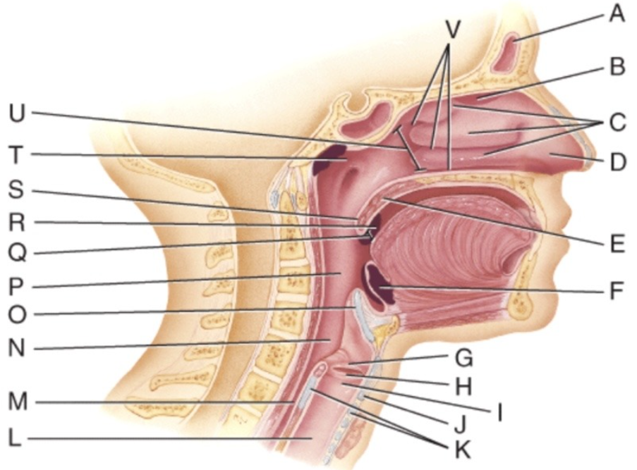
Where are the palatine tonsils?
E
F
R
U
None of these choices
R
—
A – Frontal sinus
B – Olfactory epithelium
C – Nasal conchae
D – External naris
E – Soft palate
F – Lingual tonsil
G – Vestibular fold
H – Vocal fold
I – Larynx
J – Thyroid cartilage
K – Cricoid cartilage
L – Trachea
M – Esophagus
N – Laryngopharynx
O – Epiglottis
P – Oropharynx
Q – Fauces
R – Palatine tonsil
S – Uvula
T – Nasopharyx
U – Internal naris
V – Nasal meatuses)
Which of the following is NOT a factor that determines the rate of pulmonary and systemic gas exchange?
Partial pressure difference of gases
surface are availability for gas exchange
Diffusion distance
Molecular weight and solubility of the gases
All of these are factors that determine the rate of pulmonary and systemic gas exchange.
All of these are factors that determine the rate of pulmonary and systemic gas exchange.
Which of the following is a long drawn and deep inhalation immediately followed by a shorter but forceful exhalation?
sobbing
yawning
sighing
hiccupping
Valsalva maneuver
sighing
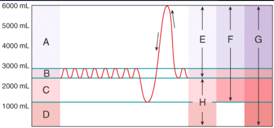
What area in the figure is the sum of the residual volume and the expiratory reserve volume?
H
A
G
E
F
H
—
A — Inspiratory reserve volume
B — Tidal volume
C — Expiratory reserve volume
D — Residual volume
E — Inspiratory capacity
F — Vital capacity
G — Total lung capacity
H — Functional residual capacity
Which of the following is a forced exhalation against the closed rima glottidis as may occur during periods of straining while defecating?
sobbing
coughing
crying
hiccupping
Valsalva maneuver
Valsalva maneuver
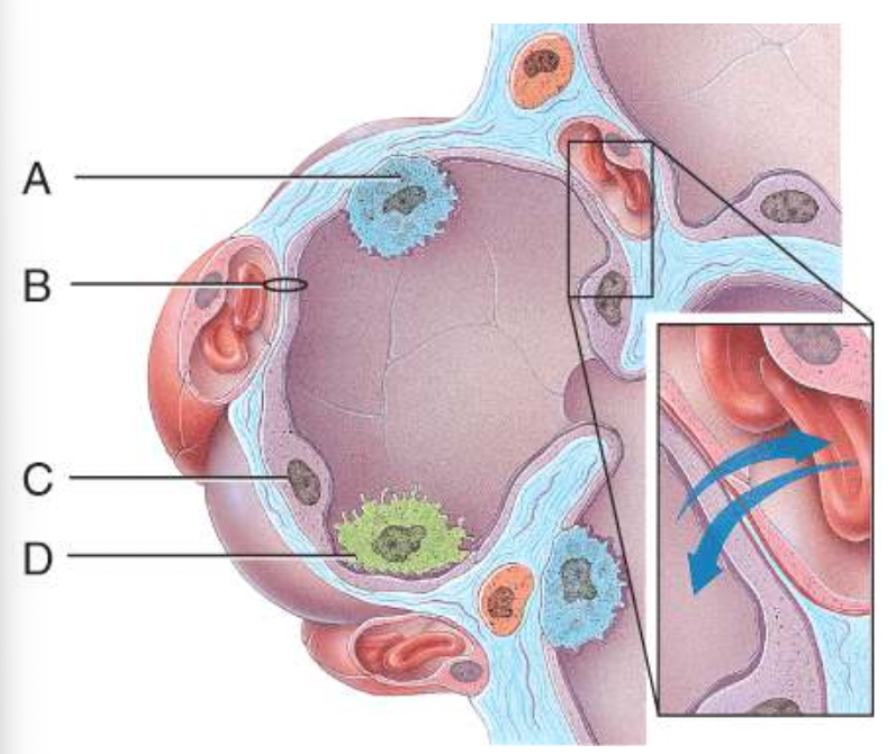
Which structure provides disease resistance within the lungs?
A
B
C
D
None of these choices
D
—
A (Type II alveolar cell)
B (Respiratory membrane)
C (Type I alveolar cell)
D (Alveolar macrophage)
Which of the following is the primary gas exchange site?
Trachea
Bronchiole
Nasal sinuses
Alveolus
Bronchus
Alveolus
Which of the following is a long drawn and deep inhalation followed by a complete closure of the rima glottidis, which results in a strong exhalation, pushing the rima glottidis open and sending a blast of air through the upper respiratory passages?
crying
sneezing
coughing
sighing
Valsalva maneuver
coughing
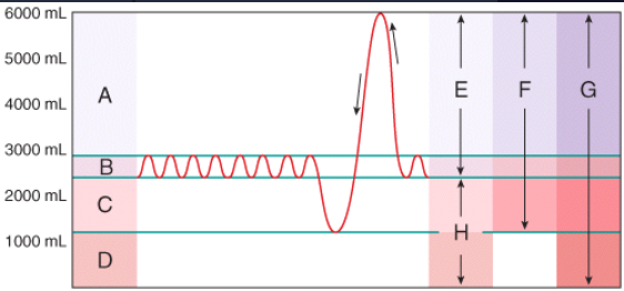
What area in the figure represents the volume of air remaining in the lungs after a deep exhalation?
C
H
D
B
G
D
—
A — Inspiratory reserve volume
B — Tidal volume
C — Expiratory reserve volume
D — Residual volume
E — Inspiratory capacity
F — Vital capacity
G — Total lung capacity
H — Functional residual capacity
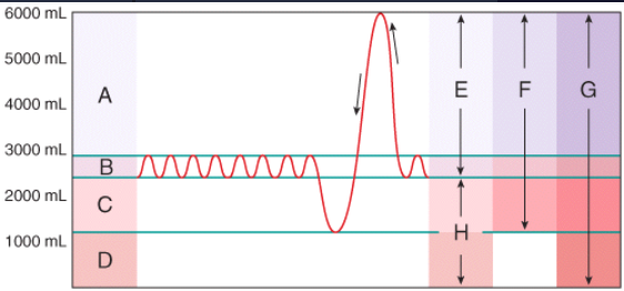
What area in the figure represents a very deep exhalation, much greater than the tidal volume?
C
H
D
B
G
C
—
A — Inspiratory reserve volume
B — Tidal volume
C — Expiratory reserve volume
D — Residual volume
E — Inspiratory capacity
F — Vital capacity
G — Total lung capacity
H — Functional residual capacity
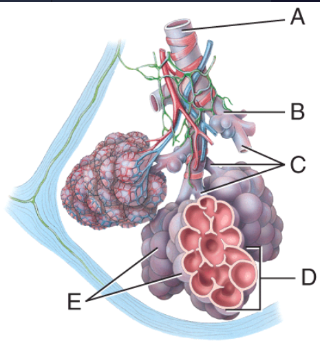
What is line C pointing to?
Terminal bronchiole
Respiratory bronchiole
Alveolar ducts
Alveolar sac
Alveoli
Alveolar ducts
—
A – Terminal bronchiole
B – Respiratory bronchiole
C – Alveolar ducts
D – Alveolar sac
E – Alveoli
Which of the below tissues maintains open airways in the lower respiratory system?
stratified squamous epithelium with keratin
ciliated pseudostratified columnar
epithelium with goblet cells
hyaline cartilage
mucus membrane
bone
hyaline cartilage
Which of the following is NOT a factor that the rate of pulmonary and systemic gas exchange depends on?
Partial pressure difference of the gases
Surface area for gas exchange
Diffusion distance
Molecular weight and solubility of the gases
Force of contraction of diaphragm
Force of contraction of diaphragm
The point where the trachea divides into right and left primary bronchi is a ridge called?
Carina
Secondary bronchioles
Parietal pleura
Visceral pleura
Diaphragm
Carina
The gas law that describes the pressure changes that occur during pulmonary ventilation is?
Boyle's law
Charles' law
Henry's law
The inhalation law
The law of partial pressure
Boyle's law
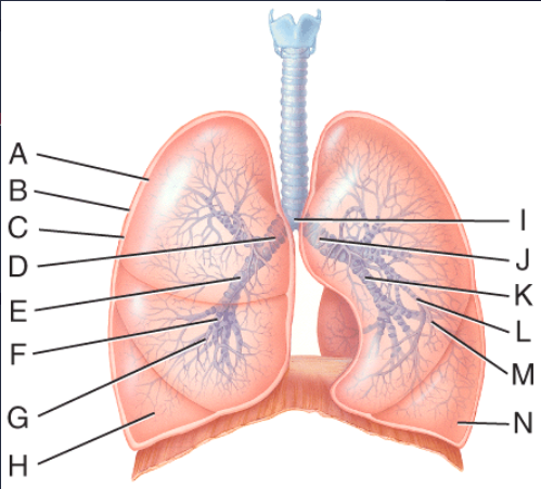
What lines are pointing to tertiary bronchi?
E and K
D and J
F and L
H and M
A and B
F and L
—
A — Visceral pleura
B — Parietal pleura
C — Pleural cavity
D — Right main bronchus
E — Right lobar bronchus
F — Right segmental bronchus
G — Right bronchiole
H — Right terminal bronchiole
I — Carina
J — Left chain bronchus
K — Left lobar bronchus
L — Left segmental bronchus
M — Left bronchiole
N — Left terminal bronchiole
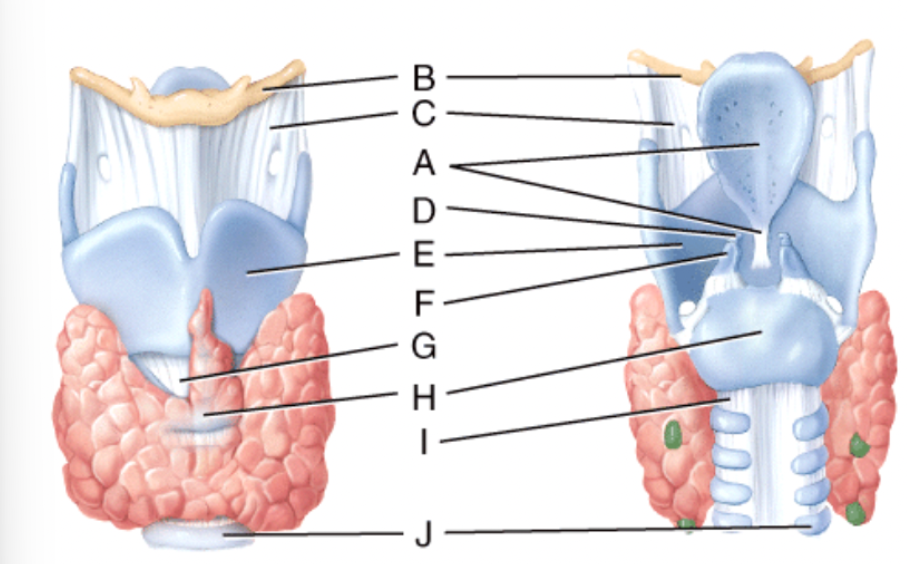
What is line D pointing to?
Thyrohyoid membrane
Corniculate cartilage
Cricothyroid ligament
Cricoid cartilage
Tracheal cartilage
Corniculate cartilage
—
B — Hyoid bone
C — Thyrohyoid membrane
A — Epiglottis
D — Corniculate Cartilage
E — Thyroid Cartilage
F — Arytenoid cartilage
G — Cricothyroid ligament
H — Cricoid Cartilage
I — Cricotracheal ligament
J — Tracheal cartilage
Which of the following indicates the direction of diffusion of gases at the alveoli of the lungs?
Oxygen into blood, Carbon dioxide into blood
Oxygen out of blood, Carbon dioxide into blood
Oxygen into blood, Carbon dioxide out of blood
Oxygen out of blood, Carbon dioxide out of blood
Oxygen into blood, Carbon dioxide out of blood
What area in the figure is the sum of the vital capacity and residual volume?
B
A
G
D
F
G
—
A — Inspiratory reserve volume
B — Tidal volume
C — Expiratory reserve volume
D — Residual volume
E — Inspiratory capacity
F — Vital capacity
G — Total lung capacity
H — Functional residual capacity