2.2 anatomy
1/64
There's no tags or description
Looks like no tags are added yet.
Name | Mastery | Learn | Test | Matching | Spaced |
|---|
No study sessions yet.
65 Terms
External ear
auricle, external acoustic meatus
elastic cartilage
Middle ear
Auditory ossicles, stapes, incus, mallus, tympanic cavity, tympanic membrane, oval window, round window, auditory tube
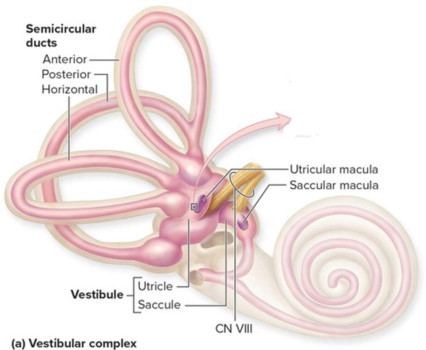
inner ear
semicircular canals, vestibulocochlear nerve (NC VIII), cochlea, oval window, round window, vestibule, auditory tube
What is equilibrium in the context of the vestibule system?
Awareness and monitoring of head position
Where does equilibrium occur in the body?
In the vestibular apparatus
What do semicircular ducts detect?
Angular acceleration
What do the utricle and saccule detect?
Static equilibrium and linear acceleration
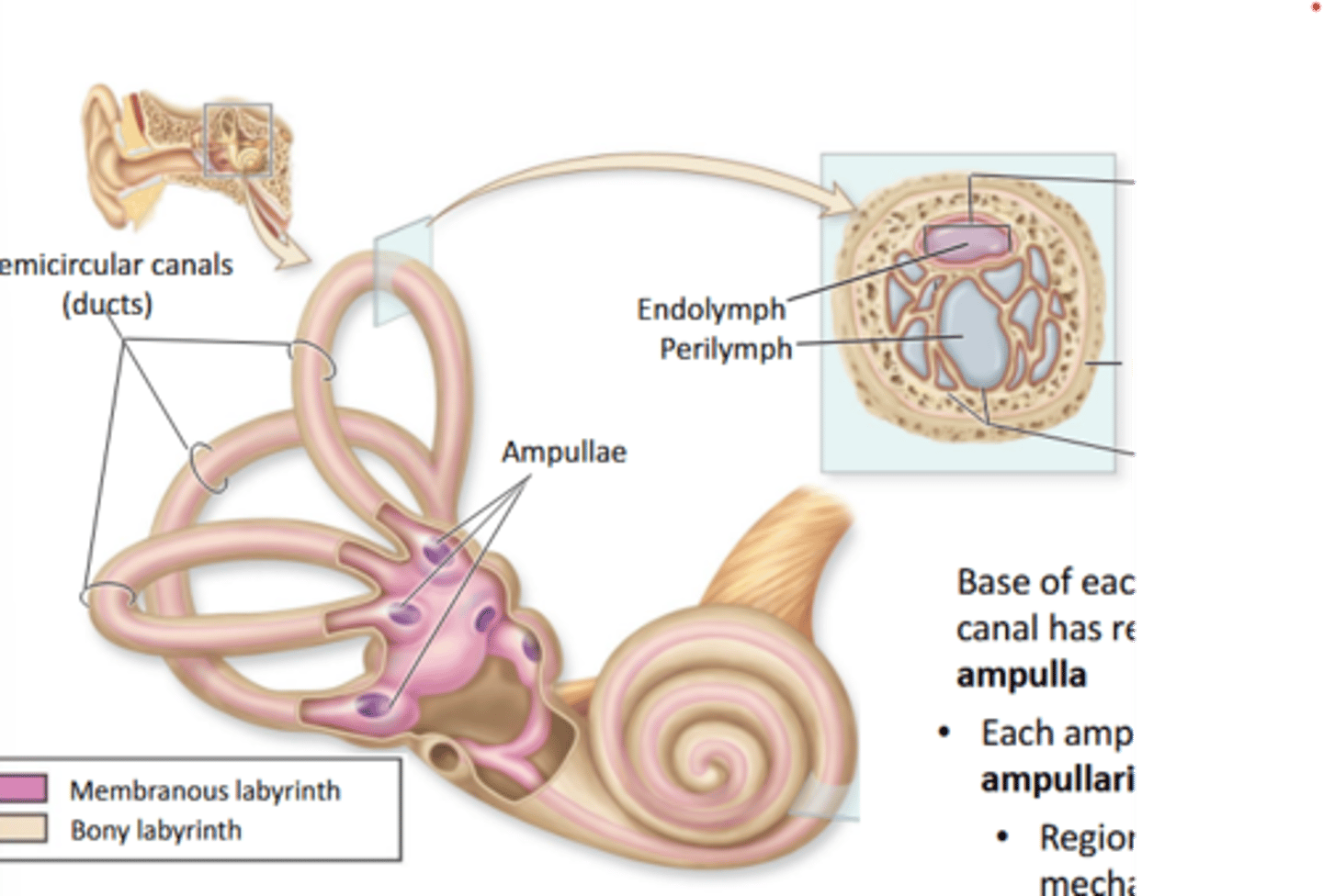
Ampulla
region at the Base of each semiciruclar canal
- each ampulla contains crista ampullaris
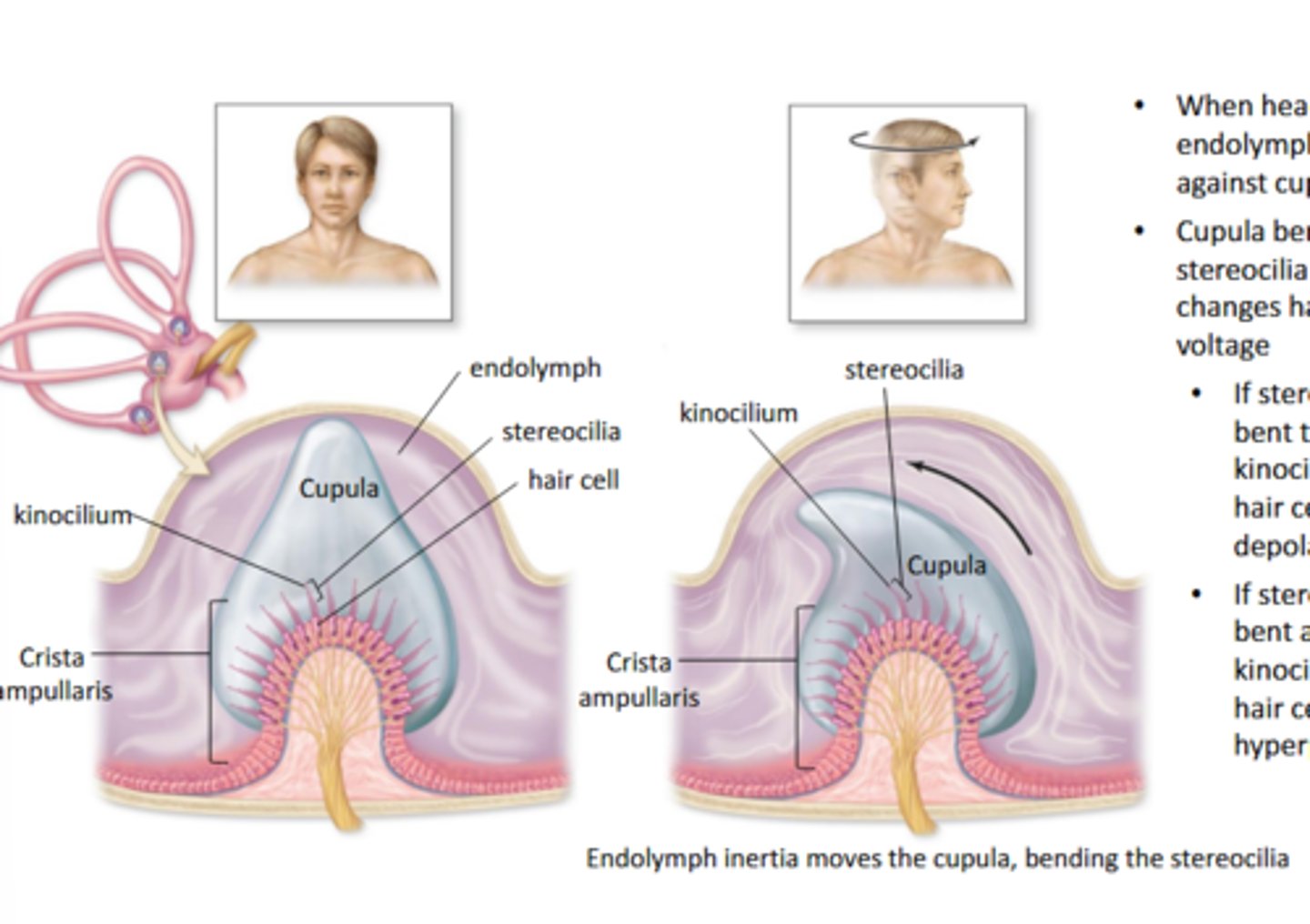
Crista ampullaris
Region of mechanoreceptors for dynamic equilibrium
Crista Ampullaris mechanisms
- when head roatates, enolymph pushes against cupula
- cupula bends stereocilia and changes hair cell voltage
- if stereocilia bent towards kinocilium, the hair cell depolarizes
- if the stereocilia bent away from kinocilum, the hair cell hyperpolarizes
vestibule
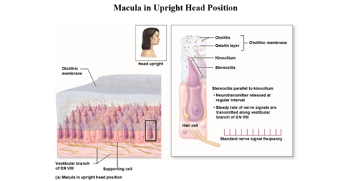
Macula- receptor for static equilibrium and linear acceleration located in utricle and saccule of vestibule
- composed of hair cells with a single kinocilium and stereocilia which are embedded in a gelatinous otolithic membrane (gel like matrix containing otoliths (calcium carbonate crystals))
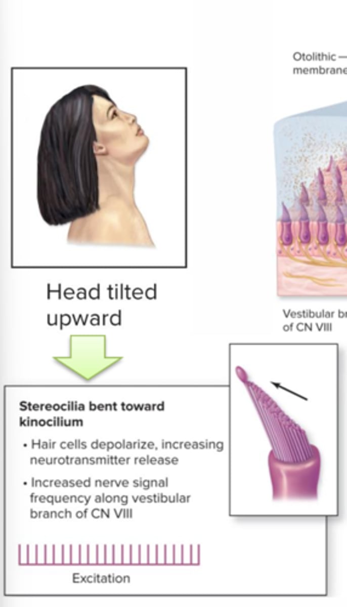
Macula: head tilted upward
Stereocilia bent toward kinocilium
-hair cells depolarize, increasing neurotransmitter release
-increased nerve singal frequency along vestibular branch of CN VII
-excitation
Macula: head upright
Stereocilia parallel to kinocilium
- neutransmitter released at regular interval
- steady rate of nerve signals are transmitted along vestibular branch of CN VII
- standard nerve signal frequency
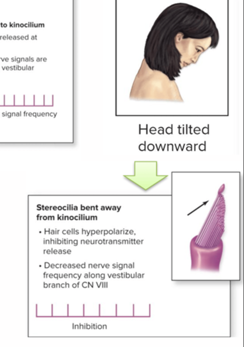
Macula: head tilted down
- sterreocilia bent away from kinocilium
- hair cells hyperpolarize, inhibitng neutrotransmitter release
-decreased nerve signal frequency along vestibular branch of CN VIII
- inhibition
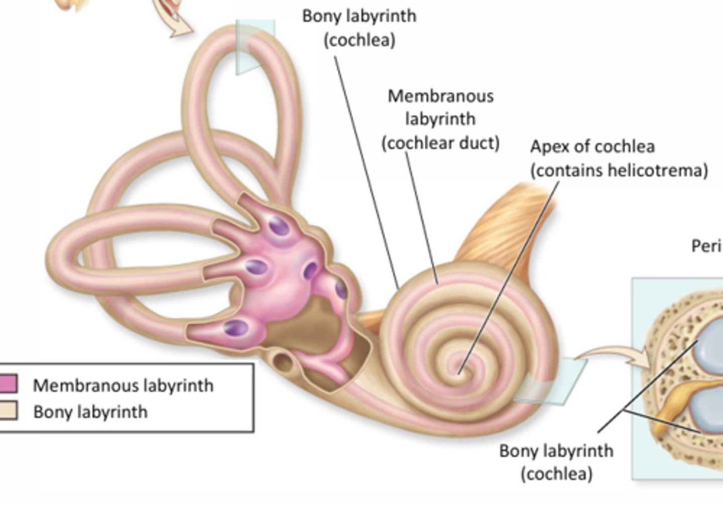
Cochlea
Bony labyrinth:(cochlea) outer layer of the snail shell looking thing
Membranous labyrinth(cochlear duct): second most outer layer
Apex of cochlea: Contains helicotrema --> Middle part of snail looking shell
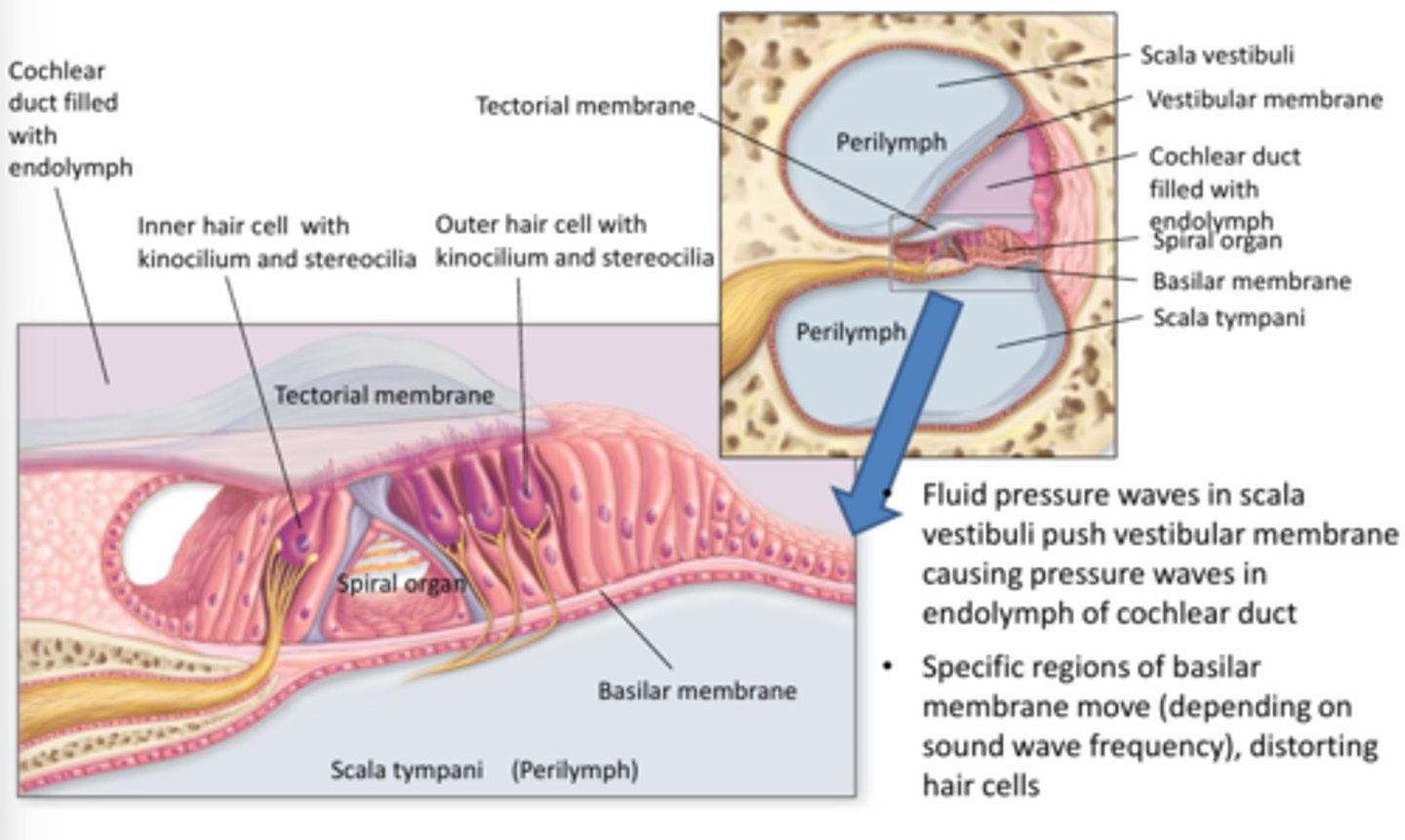
Cochlea
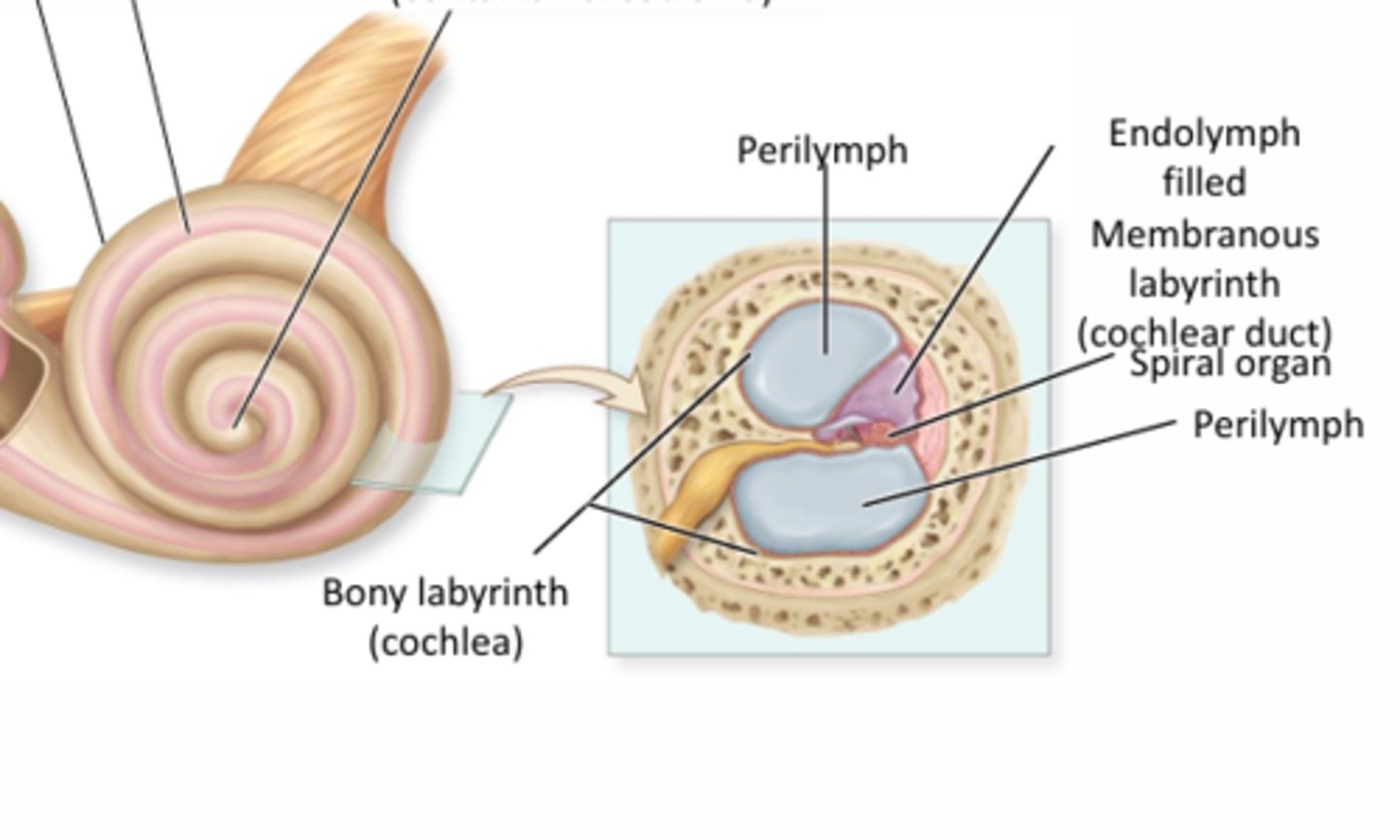
Spiral organ
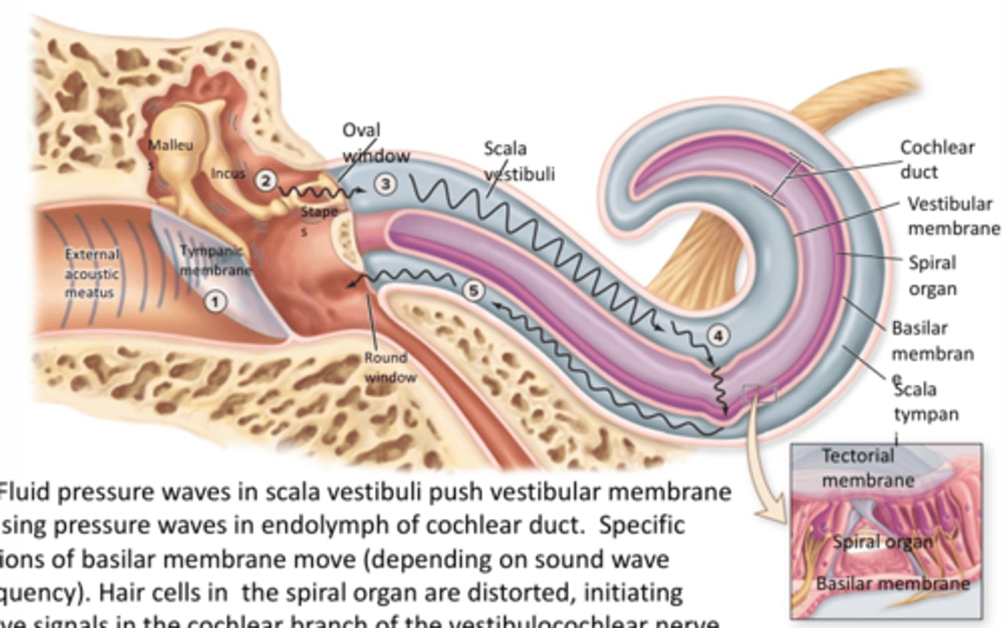
Fluid pressure waves in scala vestibuli push vestibular membrane causing pressure waves in endolymph of cochlear duct
- specific region of basilar membrane move (depending on sound wave frequency) distorting hair cells
cochlear hair cell stimulation step 1
-inner hair cells contain ion channels at their tips and tip link proteins connect them
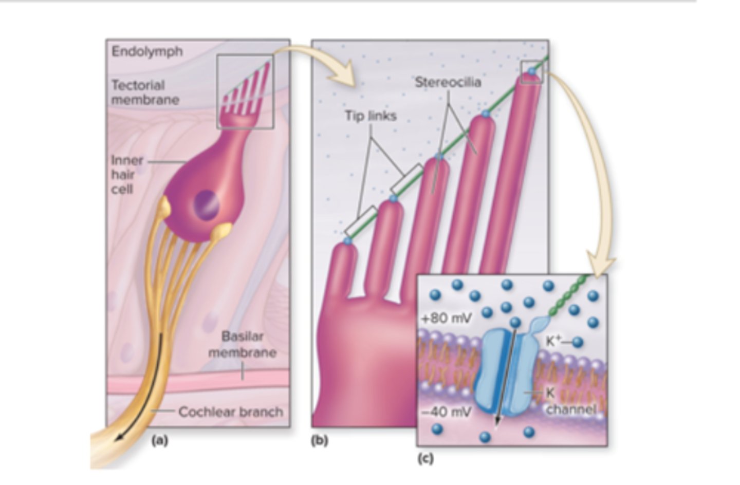
Cochlear hair cell stimulation step 2
- hair cells are bathed in K+ rich endolymph that is far more positive than the fluid inside the cell
Cochlear hair cell stimulation step 3
-when basilar membrane moves up, hair cells are pushed into tectorial membrane and their tips are tilted, causing the tip links to pull open ion channel causing K+ to diffuse into hair cell depolarization
Cochlear hair cell stimulation step 4
hair cells releases neurotransmitters from its base, exciting the sensory neuron and initiating an action potential
Cochlear hair cell stimulation step 5
- when basilar membrane moves down, the process quickly reverses
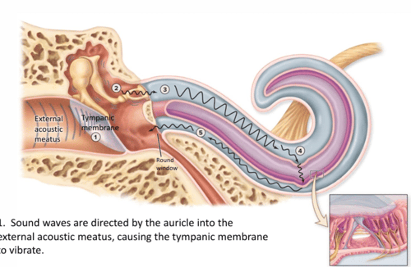
step 1
1. Sound waves are directed by the auricle into the external acoustic meatus, causing the tympanic membrane to vibrate
2.
tympanic membrane vibration moves auditory ossicles (malleus, incus, and stapes); amplifying the sound waves and transmit the waves to oval window

3.
the stapes at the oval window generates pressure waves in the perilymph within the scala vestibule
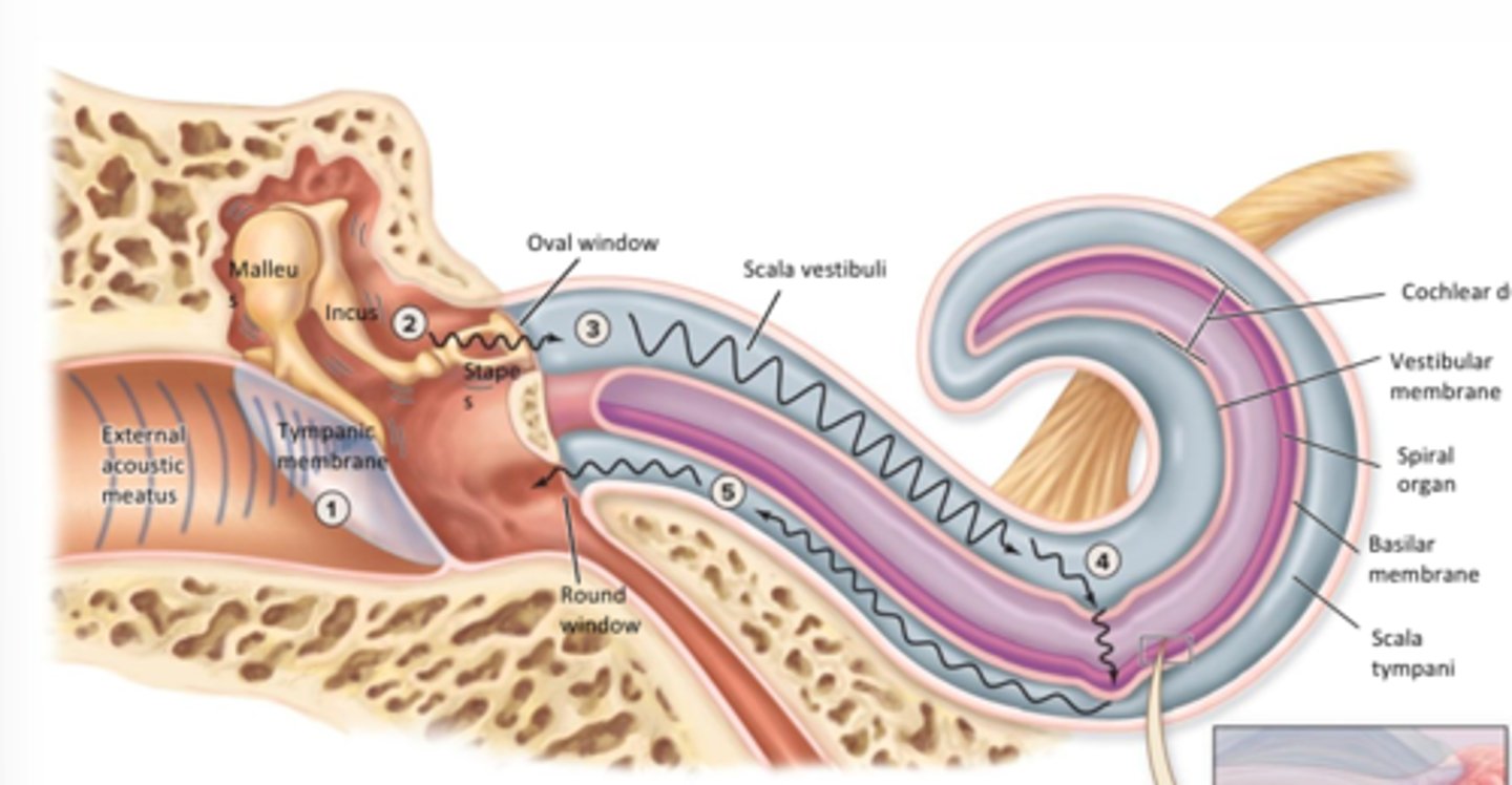
4.
Fluid pressure waves in scala vestibuli push vestibular membrane causing pressure waves in endolympth of cochlear duct. specific regions of basilar membrane move (depending on sound wave frequency). hair cells in the spiral organ are distorted, initiating nerve signals in the cochlear branch of the vestibulocochlear nerve (CN VIII)
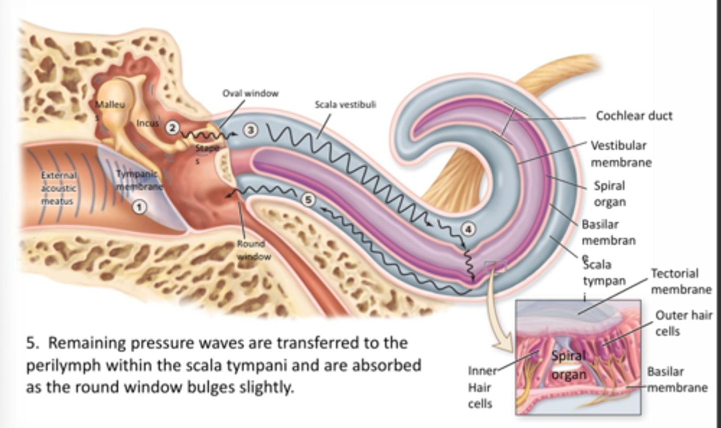
5.
Remaining pressure waves are transferred to the perilymph within the scala tympani and are absorbed as the round window bulges slightly
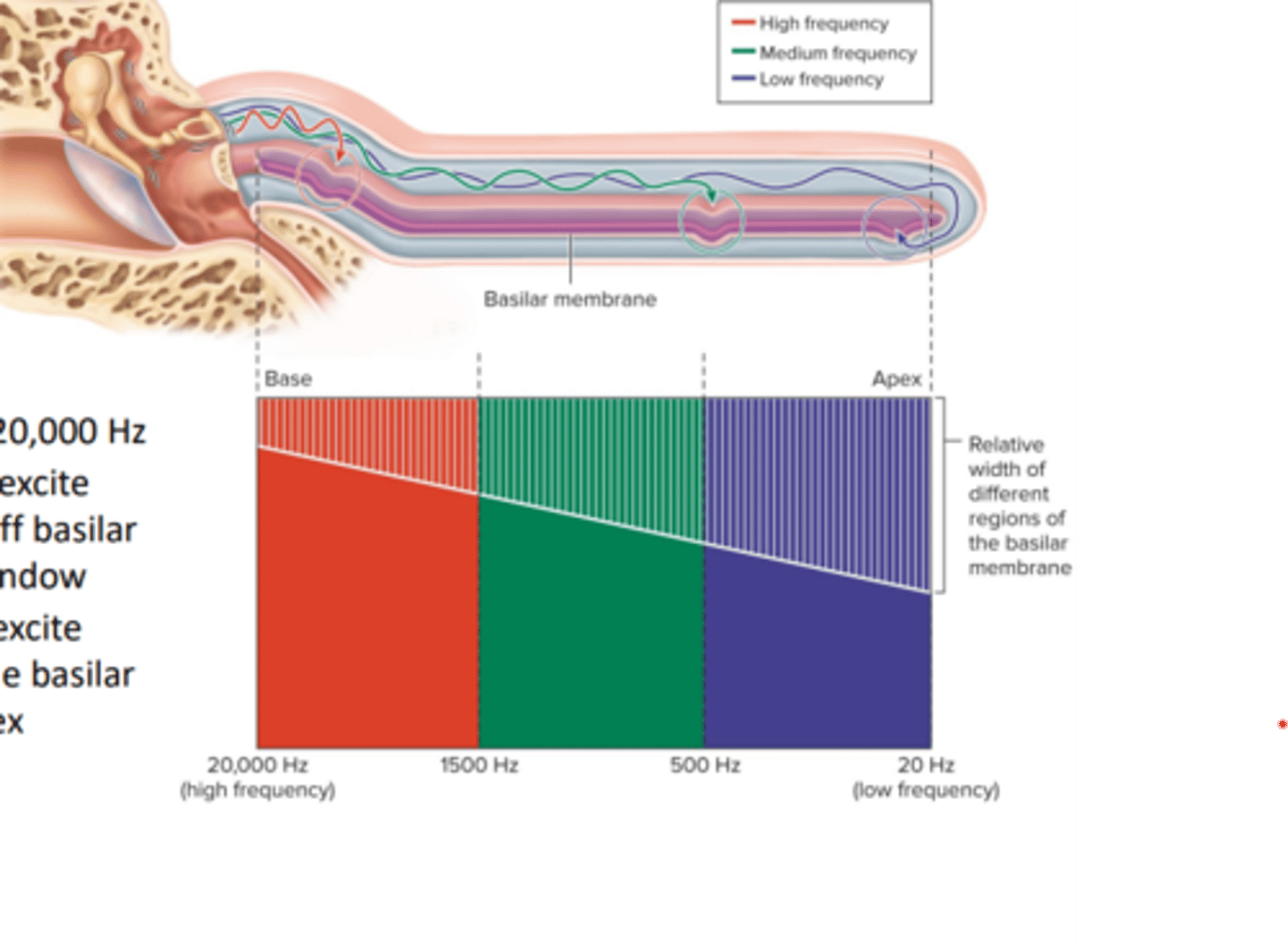
pitch
depends on the frequency (meausred in Hertz) of the vibrating object
- humans can hear 20-20,000 Hz
- high frequency sounds excite cells in narrower, stiff basilar membrane near oval window
- low frequency sounds excited cells in the wider, flexible basilar membrane near the apex
Eye anatomy
-Fibrous tunic
-sclera
-cornea
- vascular tunic
-iris
-ciliary body
-choroid
-retina
Fibrous tunic
-sclera
-cornea
sclera
tough outer vascularized "white" of the eye
Cornea
Clear convex avascular layer that refracts light
Vascular tunic
iris
ciliary body
choroid
Iris
"colored" muscular portion of eye that forms pupil
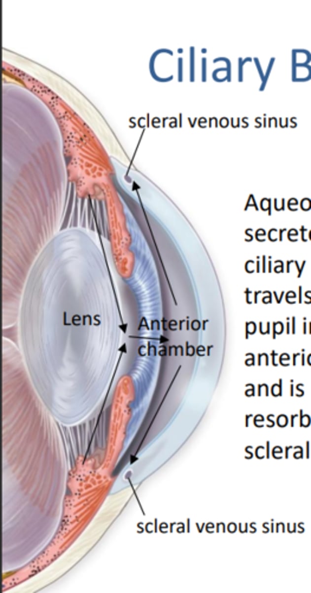
Ciliary body
Muscles that support and change the shape of the lens (attached to suspensory ligaments) and processes tha secrete aqueos humor of the eye
Choroid
blood vessels supply retina and melanin absorbs light preventing it form scattering
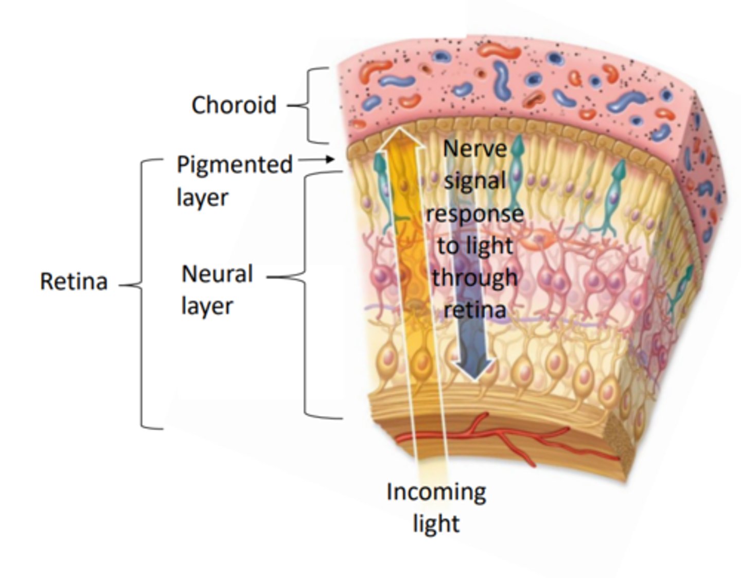
Retina
Pignmented layer
nueral layer
Pigmented layer
Absorbs extraneous light and provides Vitamin A for photoreceptors
Neural layer
where phototransduction occurs (light energy converted into an electrical signal)
Vision summary 1.
Light rays enter the eye as they pass through the cornea, aqueous humor (anterior chamber) and go through the pupil
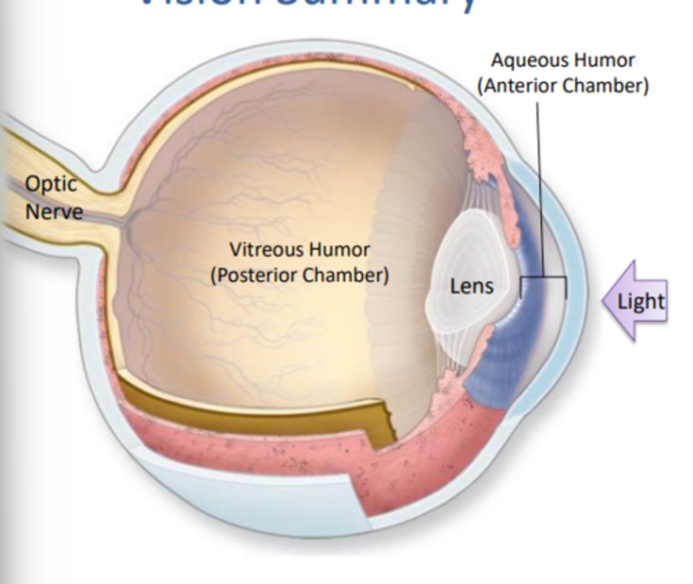
Vision summary 2.
the lens focuses the light on the retina through the vitreous humor (posterior chamber)
Vision Summary 3.
Photoreceptors respond to the light by creating an electrical signal which gets carried through the optic nerve to the brain
Iris
Pigmented, vascularized and contains muscles which control the pupil (opening) diameter therefore controlling the amount of light entering the eye (pupillary reflex)
- iris separates the anterior from the posterior chambers
pupil constriction: Bright light, sphincter pupillae contracts (parasympathetic innervation)
Pupillary dilation: low light, dilator pupillae (sympathetic innervation)
FOUND IN VASCULAR TUNIC
Ciliary body
FOUND IN VASCULAR TUNIC
- aqueous humor is secreted by the ciliary processes, travels through the pupil into the anterior chamber and is eventually resorbed into the scleral venous sinus
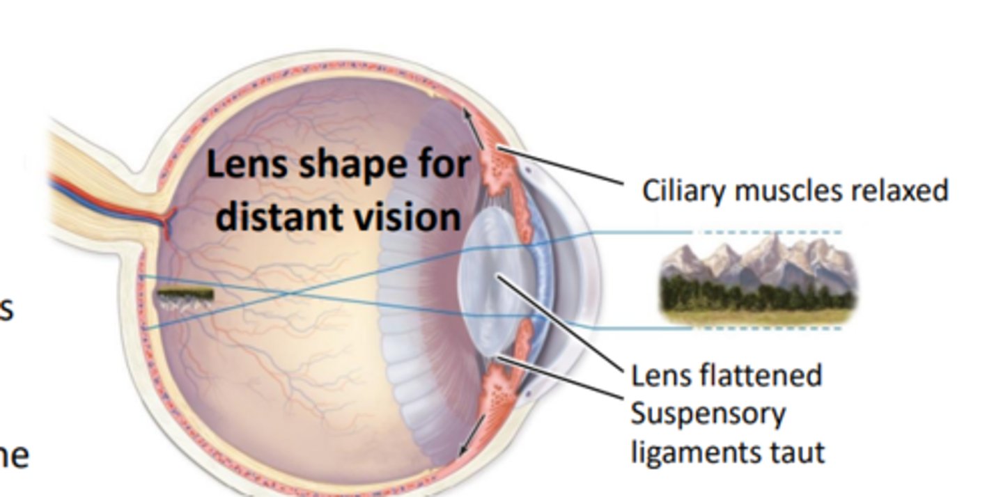
Lens shape for distant vision:
ciliary muscles relaxed, lens flattened, suspensory ligaments taut
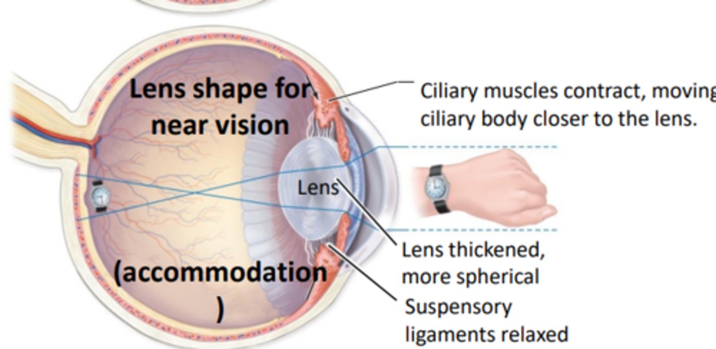
lens shape for near vision (accommodation)
Ciliary muscles contract, moving ciliary body closer to the lens
- lens thickened, more spherical, suspensory ligaments relaxed
Retina
Pigmented layer: attached to choroid (internal to it)
- provides vitamin A for photoreceptors
- absorbs stray light to prevent light scatter
Neural layer: houses photoreceptors and associated neurons
- receives light and converts it to nerve signals
Neural layer of retina
Photoreceptors cells
rods
cones
bipolar cells
ganglion cells
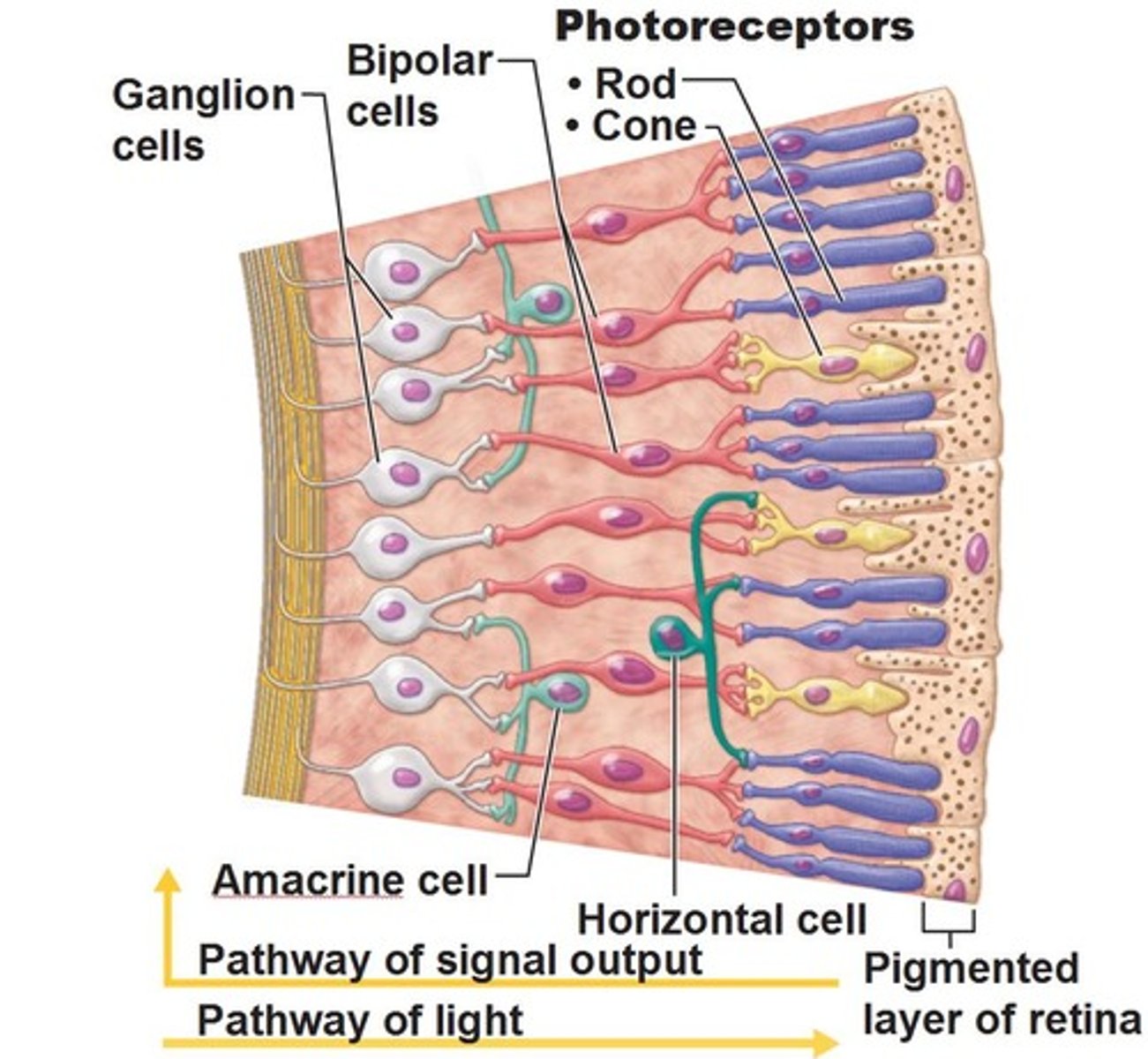
Photoreceptor cells
FOUND IN NEURAL LAYER OF RETINA
contains photopigments that react to light which triggers a change in the membrane potential
Rods
NUERAL LAYER OF RETINA
- more numerous than cones
- specialized for DIM light, night vision
- cannot distinguish color; poor at sharpness of vision
Cones
NEURAL LAYYER OF RETINA
- less numerous than cones
- responds to stimulation by bright light
- specialized for color recognition and sharpness of vision
subdiided into blue green and red cones
- absence or deficit in one type of cone cell
Bipolar cells:
NEURAL LAYER OF RETINA
- reciev input from photoreceptor cells and send infromation to ganglion cells
Ganglion cells
NEURAL LAYER OF RETINA
- responds to bipolar cells, send of action potential when stimulated, axon gather at optic disc and form optic nerve (Axon of ganglion cells become optic nerve)
Photoreceptors
The outer region of photoreceptors contain photopigment containing discs
-photopigments are embedded in the membrane and are composed of the opsin protein and the light absorbing retinal (formed from vitamin A)
-type of opsin protein determines the wavelength range of the light transduced (each photoreceptor contains one type)
- rods contain rhodopsin which is most sensitive to 500 nm wavelength
Cones contain photopsin in which there are three types (Blue, green, and red cones)
bleaching reaction and regeneration of rhodopsin step 1
Rhodopsin (opsin +cis retinal) absorbs light rays
bright light
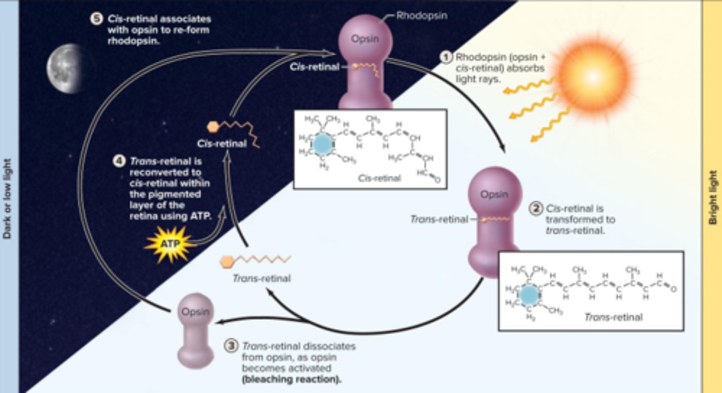
bleaching reaction and regeneration of rhodopsin step 2
cis-retinal is transformed to trans-retinal
bleaching reaction and regeneration of rhodopsin step 3
trans-retinal dissociates from opsin, as opsin becomes activated (bleaching reaction)
bleaching reaction and regeneration of rhodopsin step 4
Trans-retinal is reconverted to cis-retinal within the pigmented layer of the retina using ATP
bleaching reaction and regeneration of rhodopsin step 5
cis-retinal associates with opsin to re-form rhodopsin
dark or low light
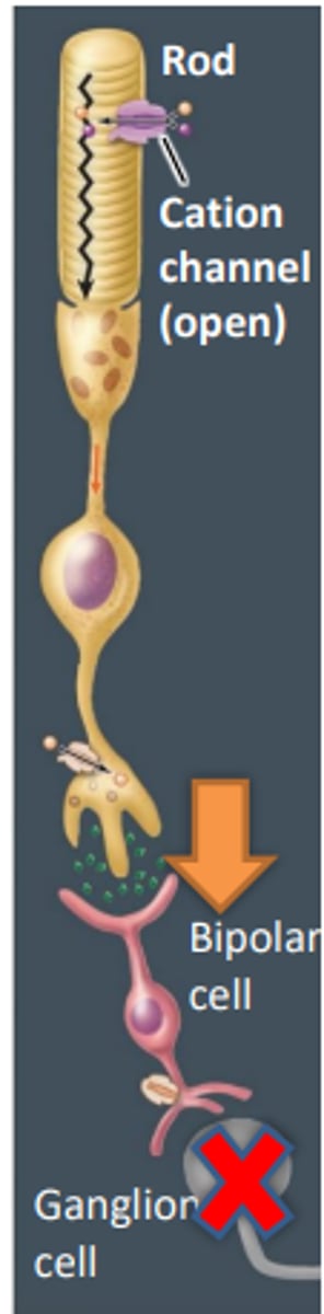
In the dark (photoreceptors)
Photoreceptors depolaried due to the opne cation channel causing it to continually release the neurotransmitter gluatmate
- gllutamate casues hyperpolarization of teh bipolar cells (inhibits signal)
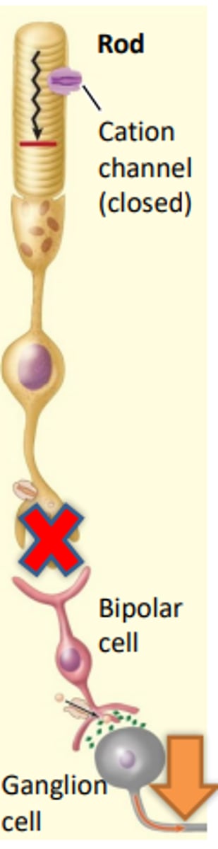
In the light (photoreceptors)
Stimulation by light causes the photoreceptor cells to hyperpolarize due to the closing of the cation channels
- lack of glutamate means that the bipolar cell is no longer inhibited and therefore becomes depolarized and releases glutamates stimulating the ganglion cells to fire sending a nerve signal to the brain
retinal regions
Optic disc
macula lutea
peripheral retina

Optic disc:
Contains no photoreceptors- blind spot
where ganglion axons exit towards the brain
Macula Lutea
Rounded, yellowish region lateral to optic disc
macular degeneration (leading cause of blindness) is the physical deterioration of the macula lutea
contains fovea centralis (central pit)
-highest proportion of cones (hardly any rods) area of sharpest vision
Peripheral retina
Contains primarily rods
functions most effectively in low light