Abdomen 1 Final Images
1/14
There's no tags or description
Looks like no tags are added yet.
Name | Mastery | Learn | Test | Matching | Spaced |
|---|
No study sessions yet.
15 Terms
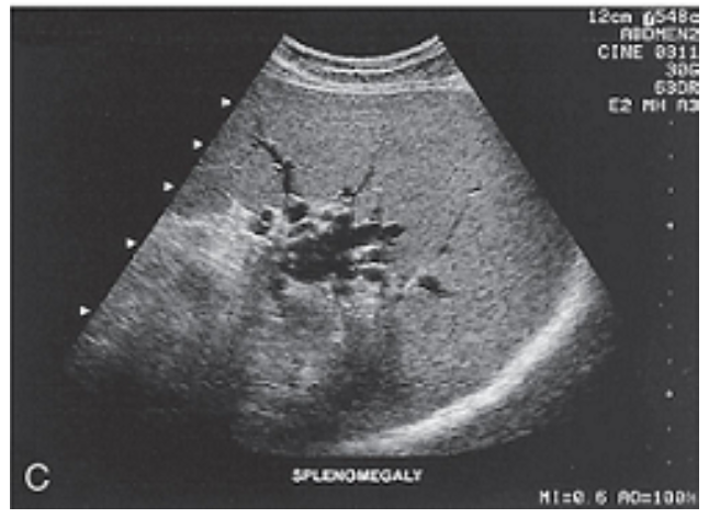
Splenomegaly: The dilated splenic hilum is secondary to portal hypertension, with hepatosplenomegaly.
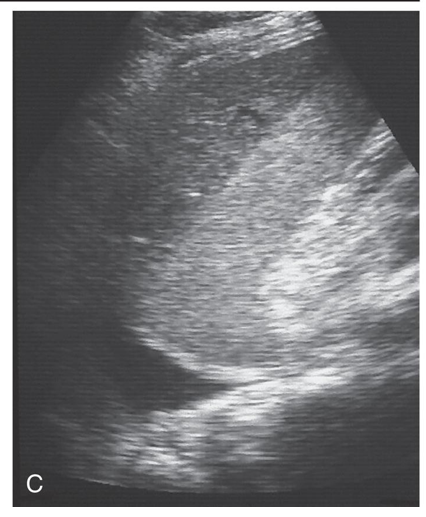
Splenic hematoma: Small hypoechoic separation medial to the splenic capsule represents a splenic hematoma.
Splenic hematoma: Separation of the splenic capsule from the spleen secondary to a large hematoma resulting from an automobile accident.
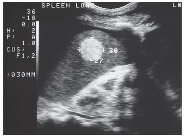
Hamartoma of the spleen: Small, solitary hyperechoic lesion within the spleen was seen in a young patient with ascites.
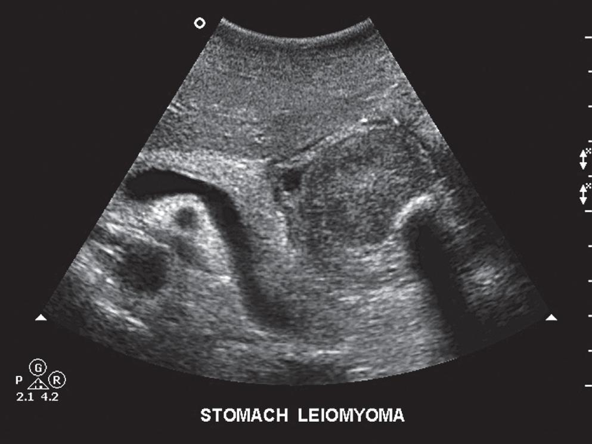
Leiomyoma
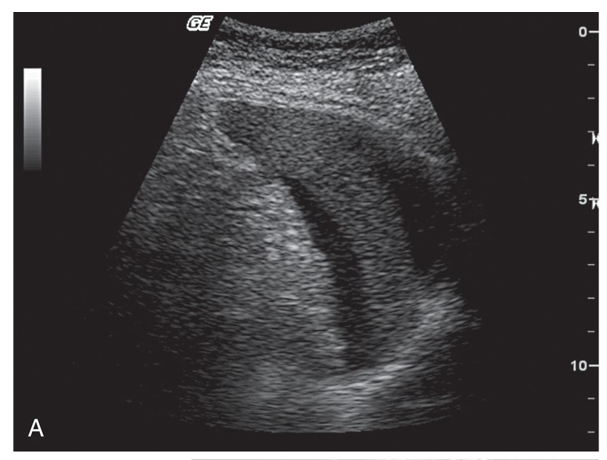
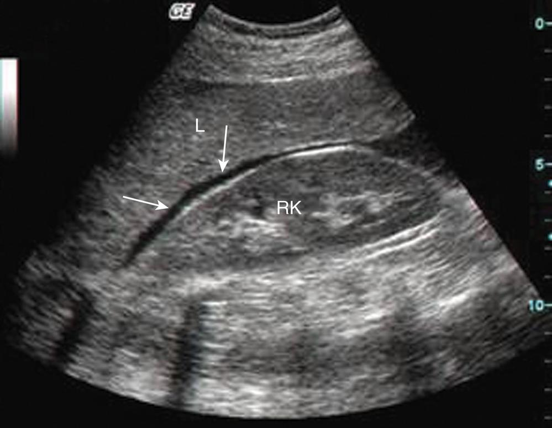
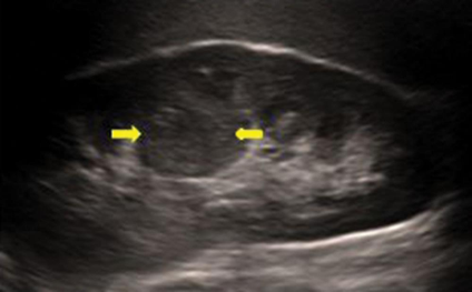
Fluid in Morison’s pouch is seen anterior to the right kidney in this sagittal view. Arrows, Fluid; L, liver; RK, right kidney. **Ascites**
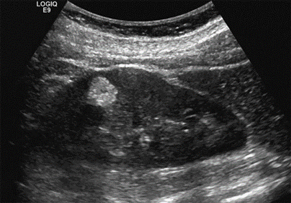
Angiomyolipoma appears as an echogenic focal mass in the renal parenchyma.
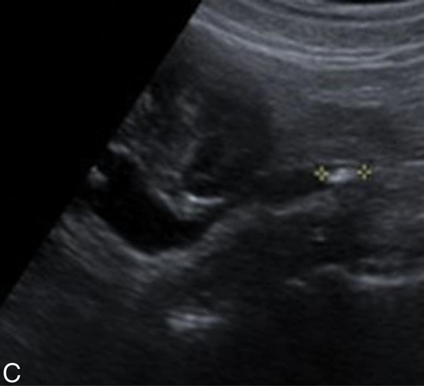
Urolithiasis: An 8-mm partially obstructed proximal left ureteral stone.
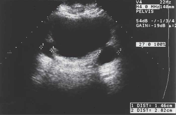
Bladder diverticula
Columns of Bertin
Dromedary Hump
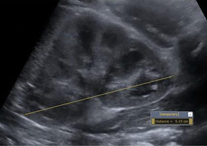
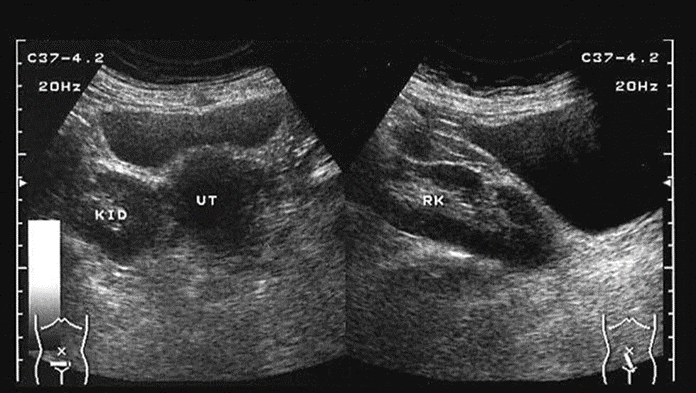
Pelvic kidney
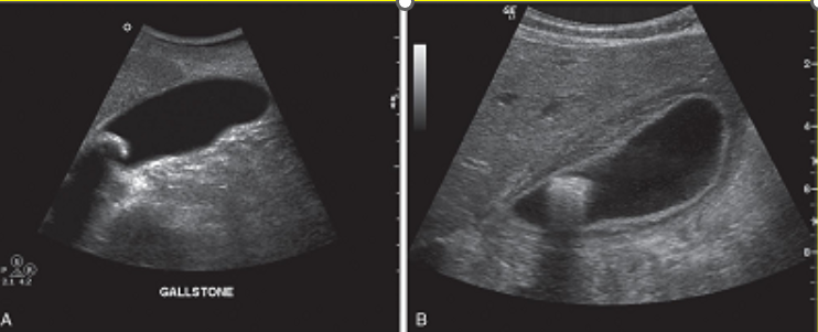
A single large gallstone near the neck of the gallbladder.
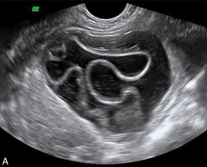
echinococcal cyst
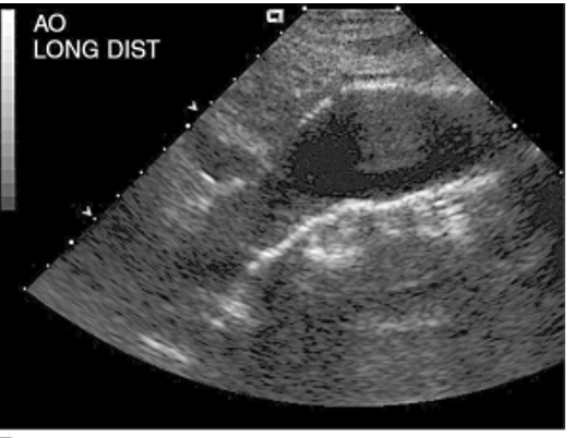
abdominal aortic aneurysm