Anatomy Exam 2 - Mizzou
1/262
There's no tags or description
Looks like no tags are added yet.
Name | Mastery | Learn | Test | Matching | Spaced |
|---|
No study sessions yet.
263 Terms
Functions of the Skeletal System (5)
Support: entire body
Protection: Viscera (organs)
Movement: Attachment for the muscles
Hemopoiesis: Blood cell production
Energy & Mineral Reserves: bones have calcium
Are bones organs?
Yes - bones consist of various types of tissue -- Including blood
Is the skeleton internal or external?
Internal
Where do bones meet?
At joints
Skeleton consist of
Bones, cartilages, joints, and ligaments
How many named bones are there?
206
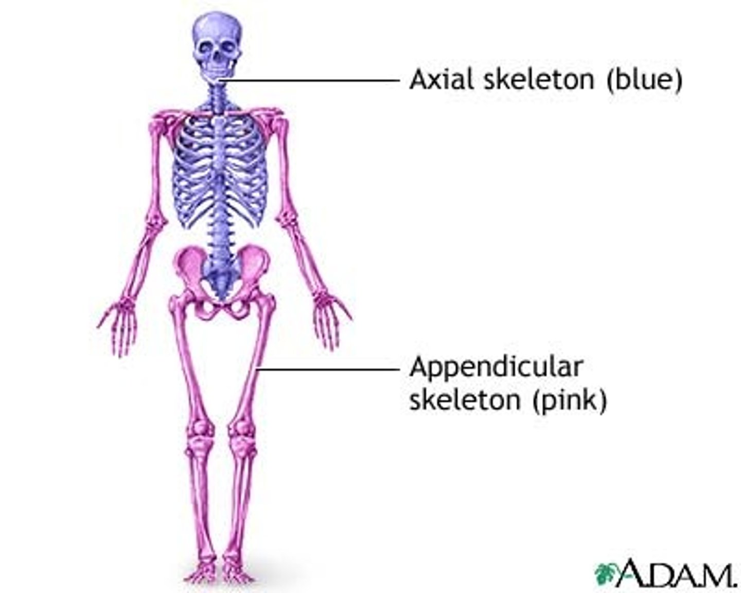
Skeleton subdivides into what?
Axial & appendicular
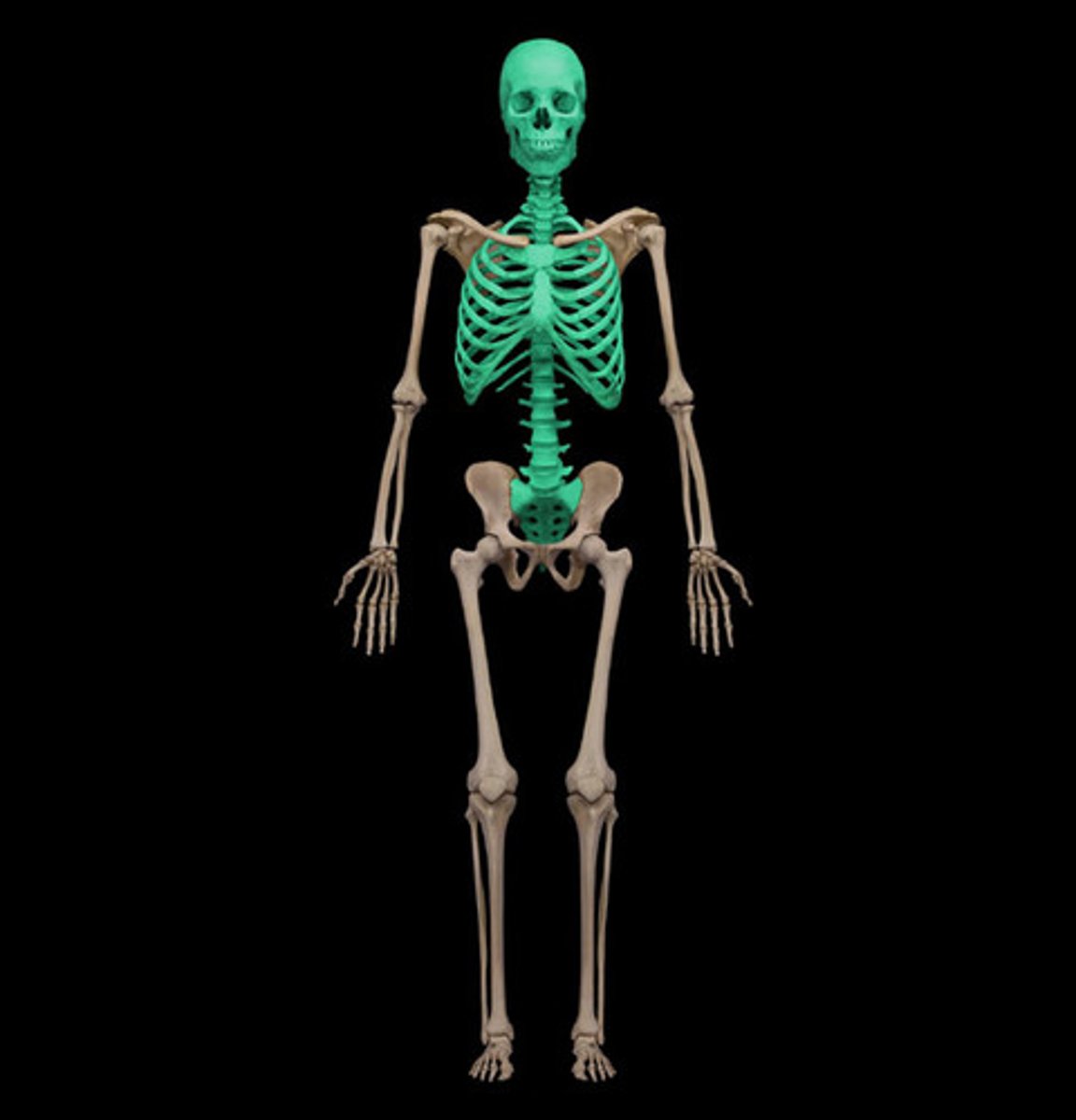
Axial Skeleton includes
Includes: Skull, vertebral, column, thoracic cage(sternum and ribs)
Appendicular Skeleton includes
Includes: pectoral girdle, upper limb, pelvic gridle, lower limbs
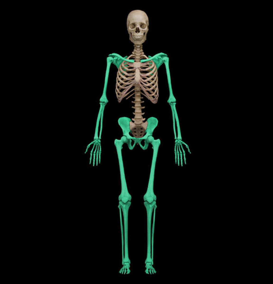
Axial & Appendicular Skeleton

Axial Skeleton amount of bones
80 named bones
Axial Skeleton Functions
Supports head, neck, and trunk
Axial Skeleton protects..
protects brain, spinal cord, thoracic organs
Bone Markings
characteristics on the surface of the axial and appendicular bones that indicate attachments, articulations or openings for nerves and blood vessels, explains Boundless.
Examples: Foramen, fossa, process, meatus, canal
Foramen (foramina) & example
a hole in a bone (typically for nerves or blood vessels)
Examples: foramen magnum, infraorbital foramen)
Fossa (fossae) & example
a depression in a bone
Examples: mandibular fossa, lacrimal fossa
Process & examples
projection from bone, narrow or wide, protrudes from surrounding bone
ex.: styloid or mastoid process
Meatus & examples
a hole or tube-like structure
(e.g. auditory meatus)
Canal & examples
a groove or tube-like structure
(e.g. optic canal)
Cartilage Tissue Structure
Avascular (no blood supply)
Cell Type: chondrocytes (in lacunae)
Cartilage Functions
Support soft tissues
Model for formation of bone
Gliding surface at articulations
Three types of cartilage
Hyaline, elastic, fibrocartilage
Hyaline Cartilage
most common kind of cartilage. Has tiny nearly invisible collagen fibers called fibrils
Fibrils
Tiny nearly invisible collagen fibers
Hyaline Cartilage functions
ends of long bones, costal cartilages, respiratory structures, fetal skeleton
Elastic Cartilage
Similar to hyaline but lots of elastic fibers. Very resilient and flexible, tolerates repeated bending
Elastic Cartilage is found where?
in pinna (outer ear) and epiglottis
Fibrocartilage
has little ground substance & matrix has thick, dense collagen fibers.
Resists strong compression
Fibrocartilage is found where?
in inter-vertebral disks, knee joint, public symphysis
Cartilage Locations
Bone Tissue
Much denser than cartilage, very little fluid. Resists compression and tension; very strong. Well vascularized, so it heals/remodels easy. Made of organic and inorganic materials
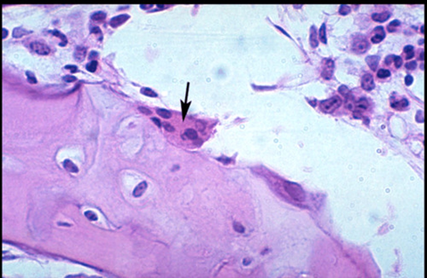
Osteoblasts & Osteoclasts
Bone is constantly being built up or broken down - growth, strengthening, remodeling, healing, maintenance.
Osteoblasts
Builds new bone
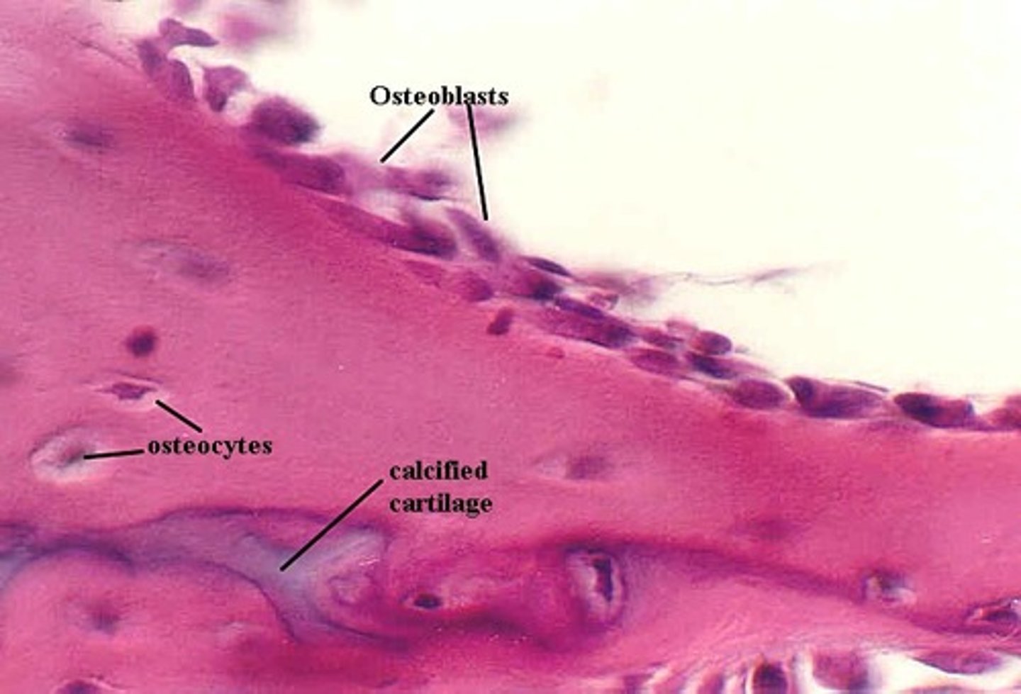
Osteoclasts
Break down (consume) bones and are mature bone cells
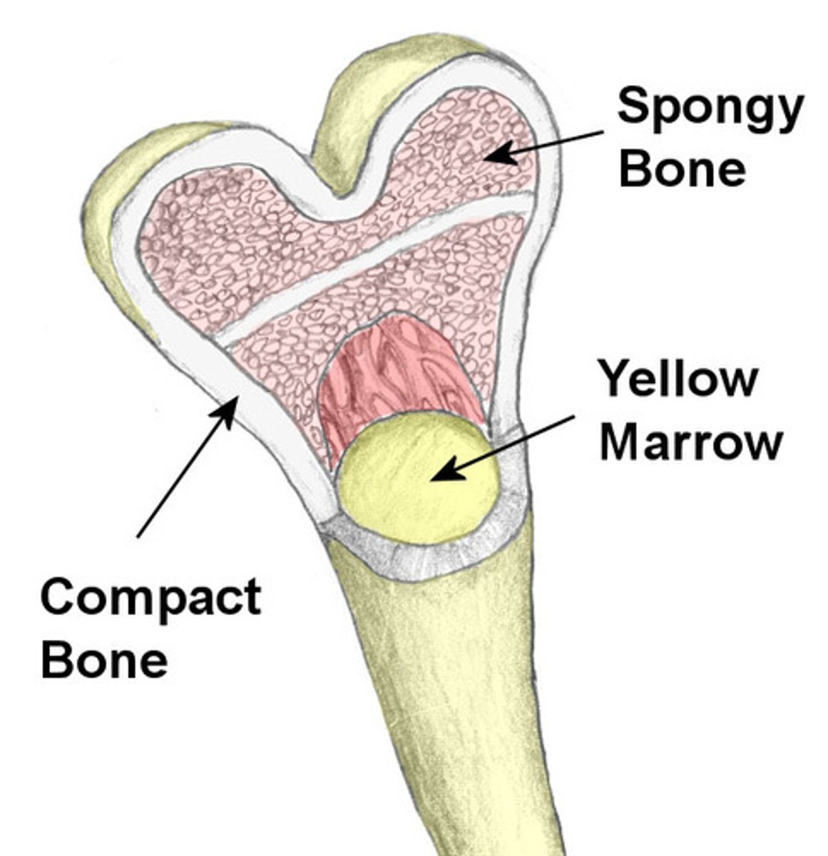
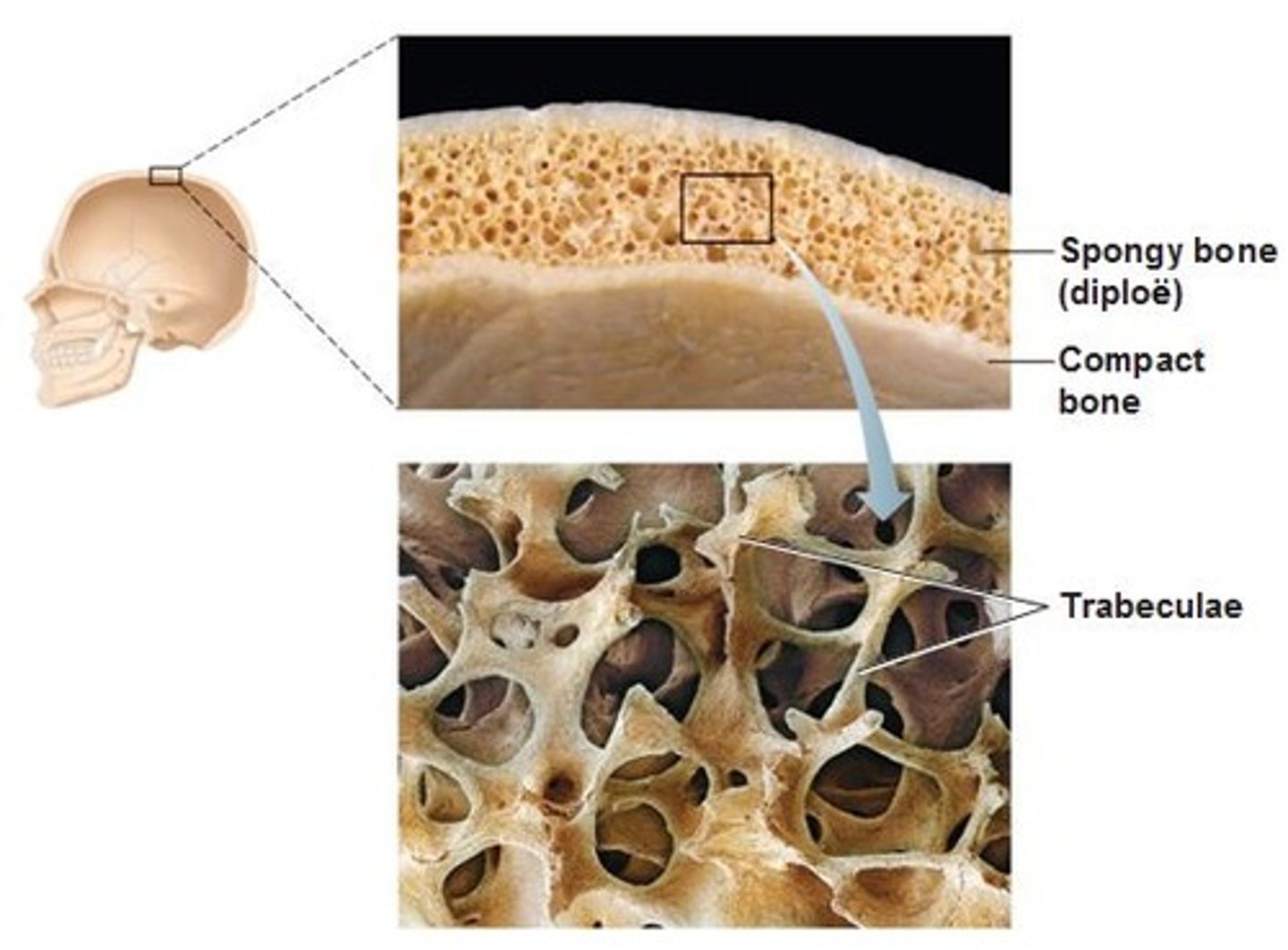
Spongy Bone
(Trabecular bone)
Inside bones
Better at shock absoption
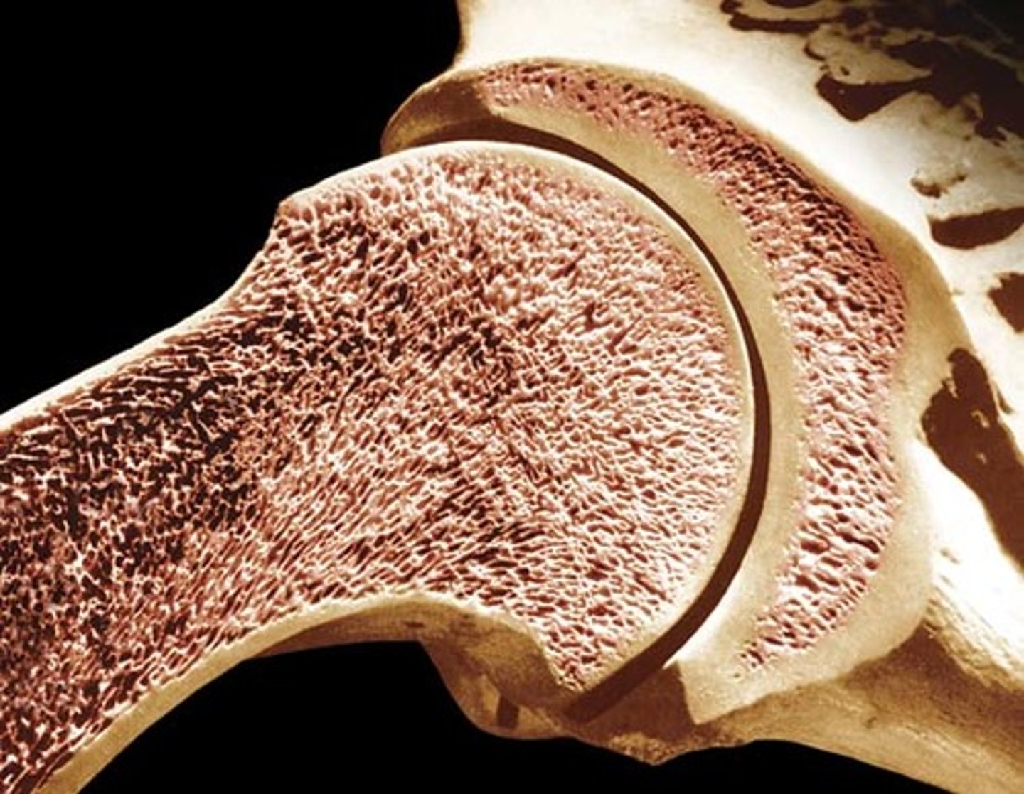
Compact Bone
(Cortical)
Smooth, dense, external portion of bones
Strong, rigid
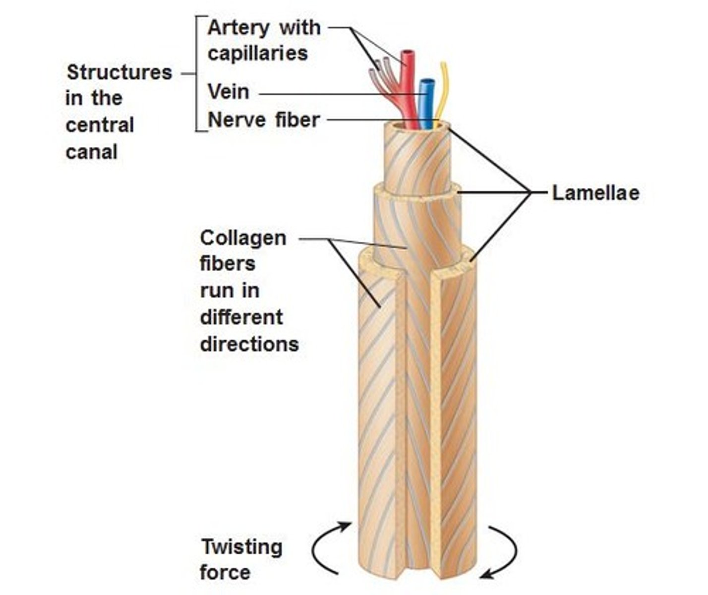
Compact Bone Structure
Osteon - structure unit
Osteon
-Made up of concentric tubes are called lamellae
-Oriented parallel to the long axis and main compression stresses
- Haversian (central) canal
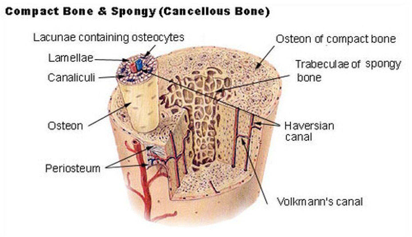
Lamellae
what makes up osteon
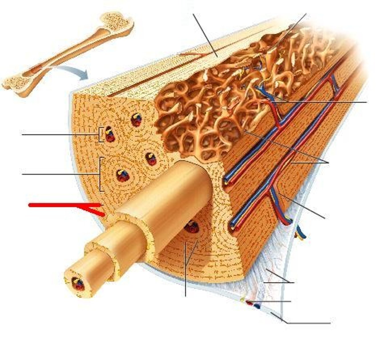
Haversian (central) canal
runs through core of each osteon & provides blood supply, nutrients, nerves
Bones shapes and sizes
Long bone - humerus
Short bone - talus
Flat bone - sternum
Irregular bone - vertebra
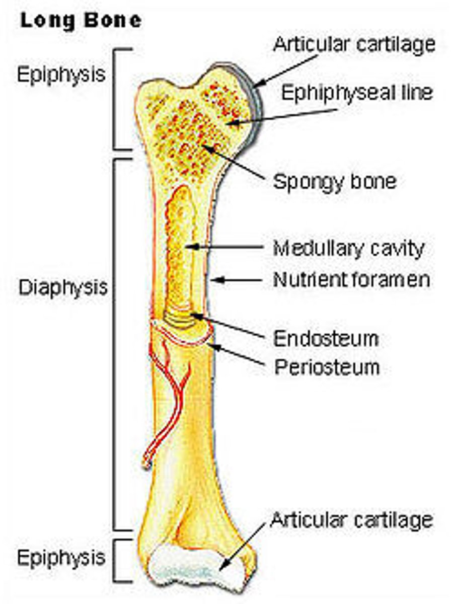
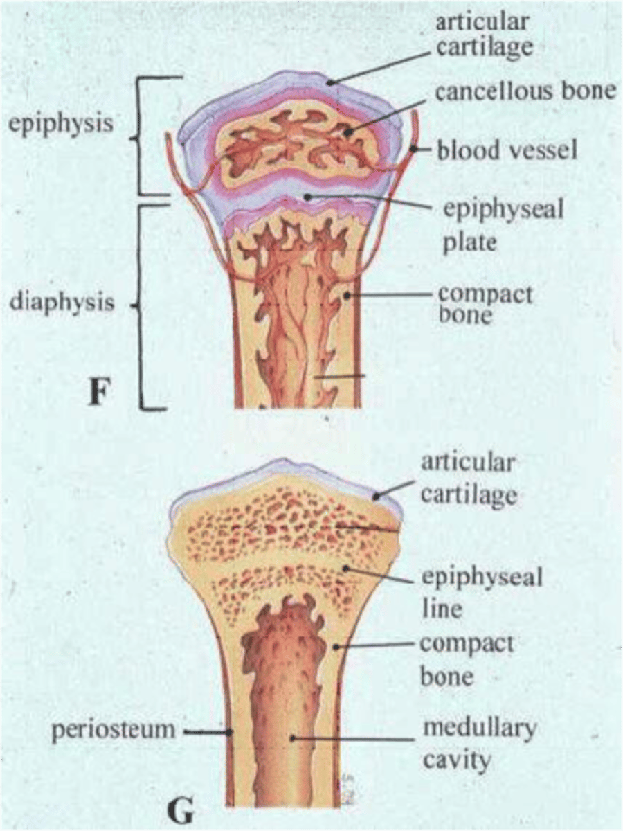
Structure of a Long Bone 1
Epiphysis - ends
Epiphyseal Line - growth plate
Diaphysis - shaft
Compact bone - superficial
Spongy bone - deep
STRUCTURE OF LONG BONE 1

Structure of a Long Bone 2
Periosteum - sheath on outside of bone
Endosteum - lines internal cavity
Medullary Cavity - Bone marrow
Nutrient Arteries - Feed bone
Articular cartilage is on ends

Flat, Irregular, and Short Bones
-Compact bone with periosteum on outside
-Spongy bone with endosteum inside
-Contain marrow but don't have a marrow cavity
Bone Development & Growth
-Osteogenesis/Ossification
-Being in the embryo & continues through life (more slowly in the adult)
-Before week 8, skeleton made of hayaline cartilage or mesenchyme
-Before week 8, bone tissue begins to replace most cartilage & mesenchyme
-Ossification may be endochondral or intramembranous
Osteogenesis/Ossification
Process of bone formation
-May be endochondral or intramembranous
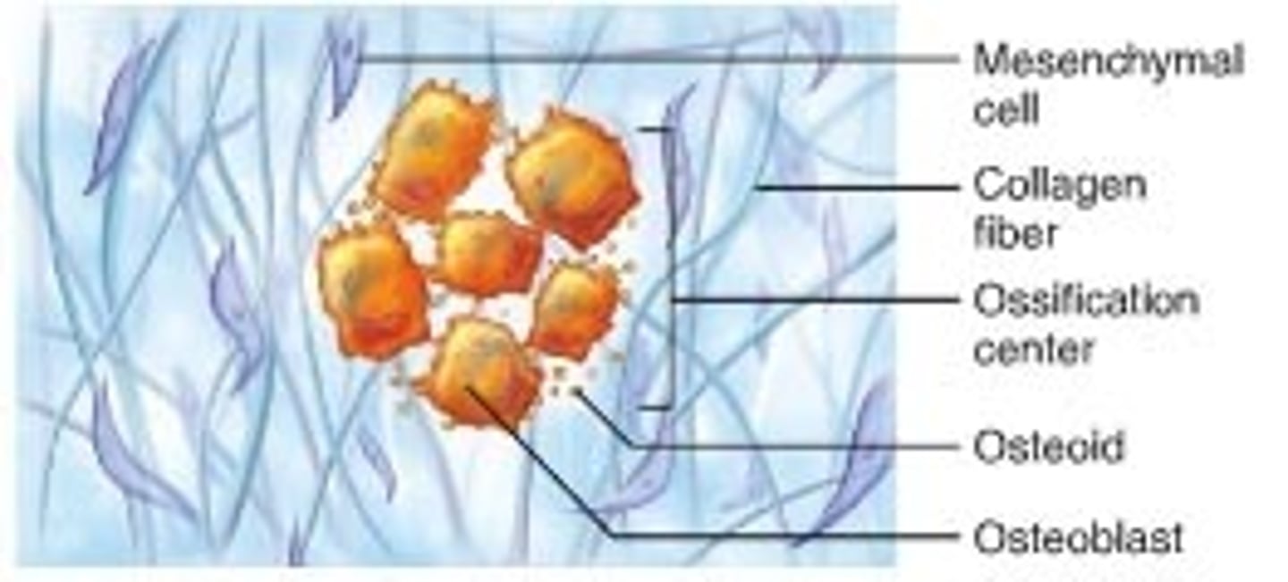
Intramembranous Ossification
-Bone grows within a membrane
-Forms many flat bones (bones of the skull) as well as maxillae, zygomatic, mandible & center of calvicle
Two types of Ossification
- Intramembranous Ossification
- Endochondral Bone Ossification
Endochondral Bone Ossification
-Most of the bones of the skeleton form this way
1) Skeleton begins as Hyaline Cartilage model
2) Bone replaces cartilage
3) Epiphyseal (growth) plates ossify eventually
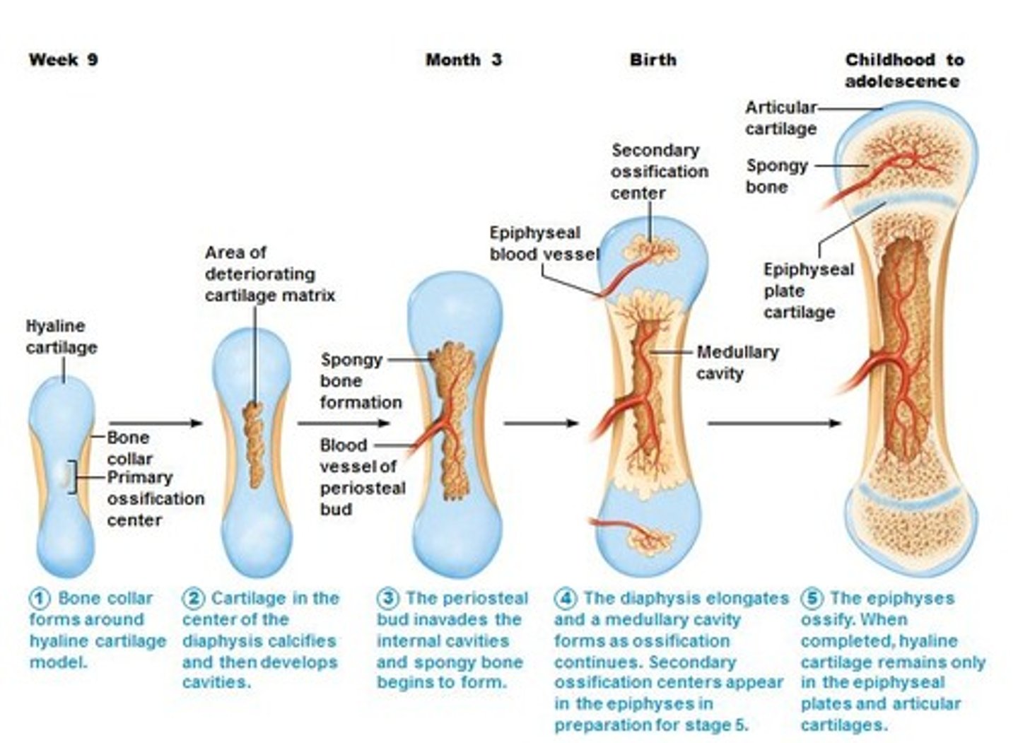
Closure of the Epiphyseal Plates
Cartilage is gradually replaced by bone tissue on both sides of the epiphyseal plate (primary center of ossification at diaphysis & secondary centers of ossification in epiphyses)
-When centers of ossification meets (a epiphyseal plate), growth stops
Skull
- 28 Bones - very complex
- Most are "flat" bones, formed via intramembranous ossification
- united by sutures (interlocking, immovable joints)
Major Structures of Skull
- Coronal Structure
- Squamous suture
- Lambdoid suture
- Sagittal Suture
- Parietal bone
- Frontal bone
- Temporal bone
- Occipital bone
- Sphenoid bone
- Ethmoid bone
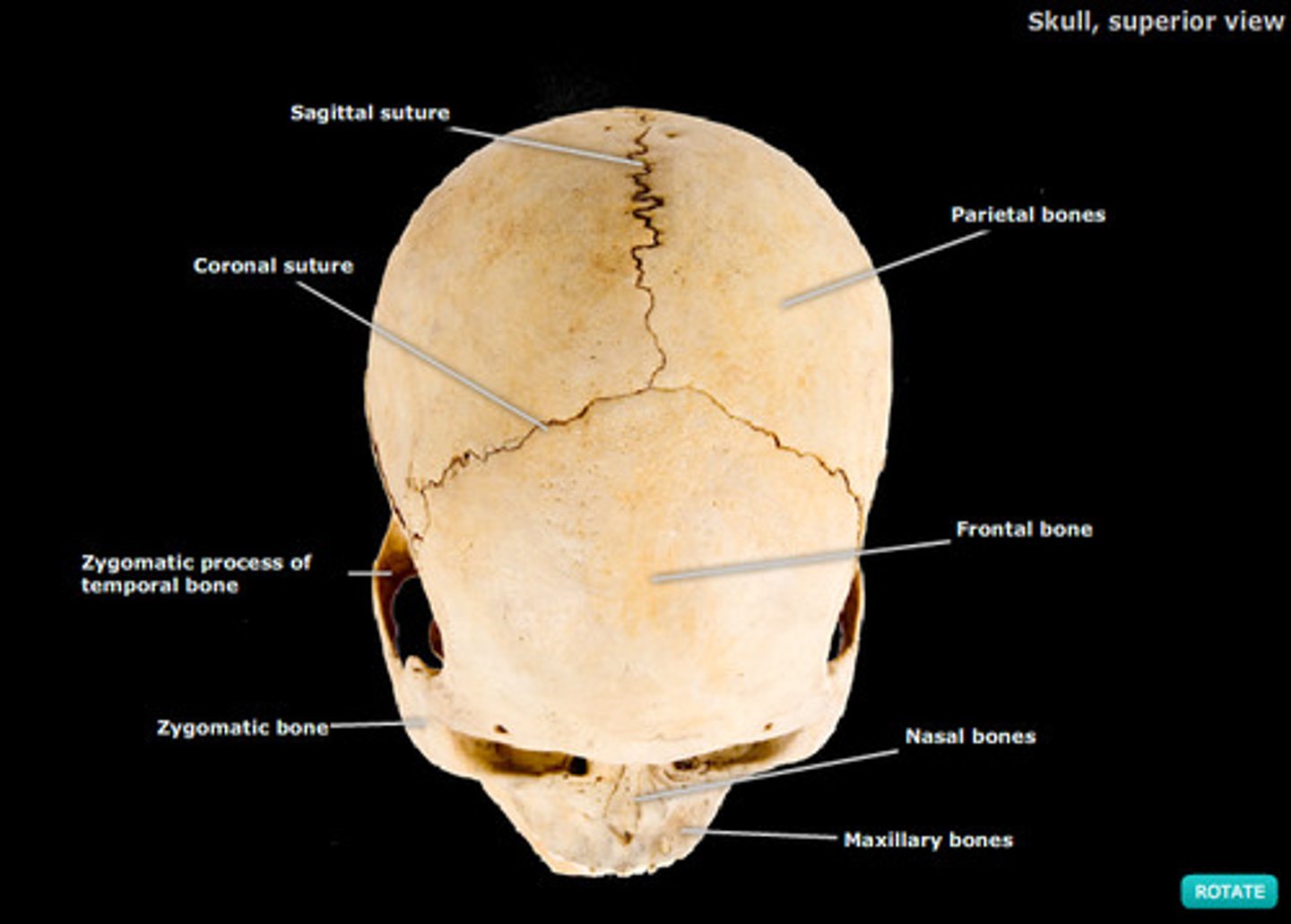
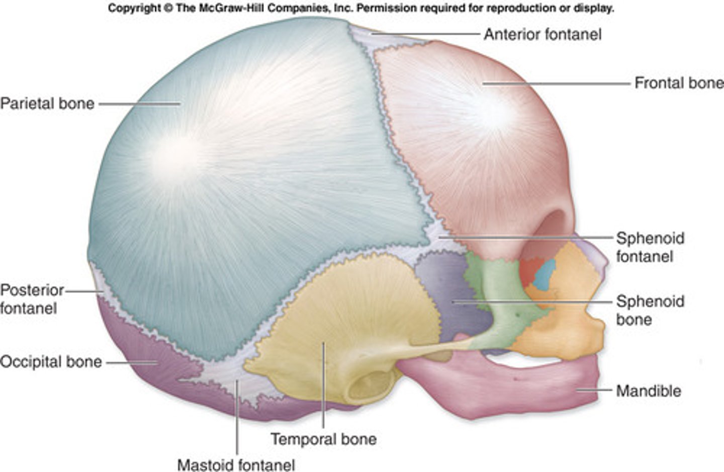
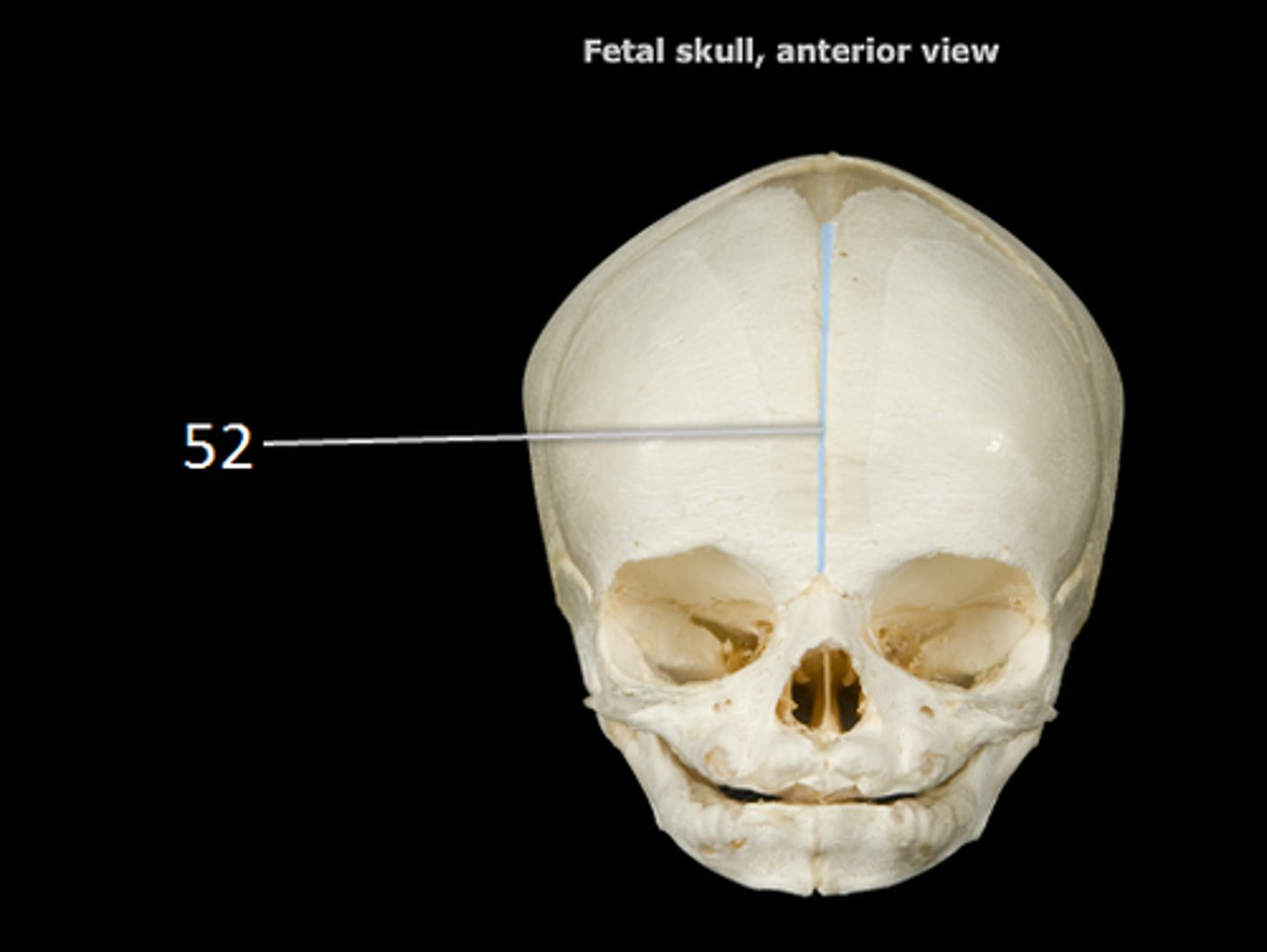
Fetal/Infant Skull & Structures
- Connected by flexible connective tissues to allow head to deform during birth & allow rapid brain growth
- Areas between bones are called fontanelles (soft spots) & fuses over times
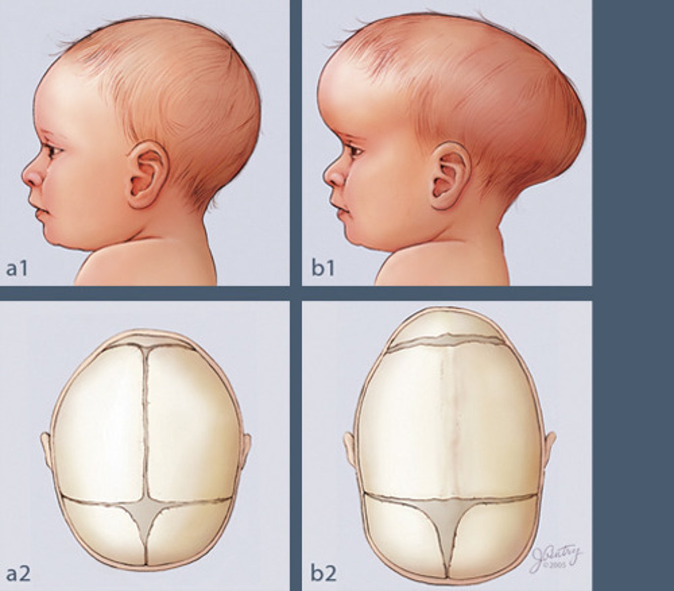
Craniosynostosis
pre-mature fusion of skull bones, leads to unusual cranial vault shape
The Skull
Subdivides into cranial & facial divisions
Cranial bone functions
Protect brain
provides attachments sites for some head/neck muscles
Facial bone functions
form the framework of face
opening the passage of air & food
Hold the teeth
anchor muscles of face
Cranium
Can be subdivided into vault (calvarium) & base
Vault = superior, lateral & posterior bones of skull (includes forehead)
Base = inferior part of cranium
Cranium is made up of ...
8 Bones
- Frontal (forehead)
- Occipital
- Sphenoid
- Ethmoid
- 2 Parientals (left & right)
- 2 Temporals (left & right)
Pariental Bones
Made up most of superior part of skull, extend posteriorly & laterally
**Note: Sagittal suture (between two pariental bones), lambdoid surure (between pariental & occipital bones)

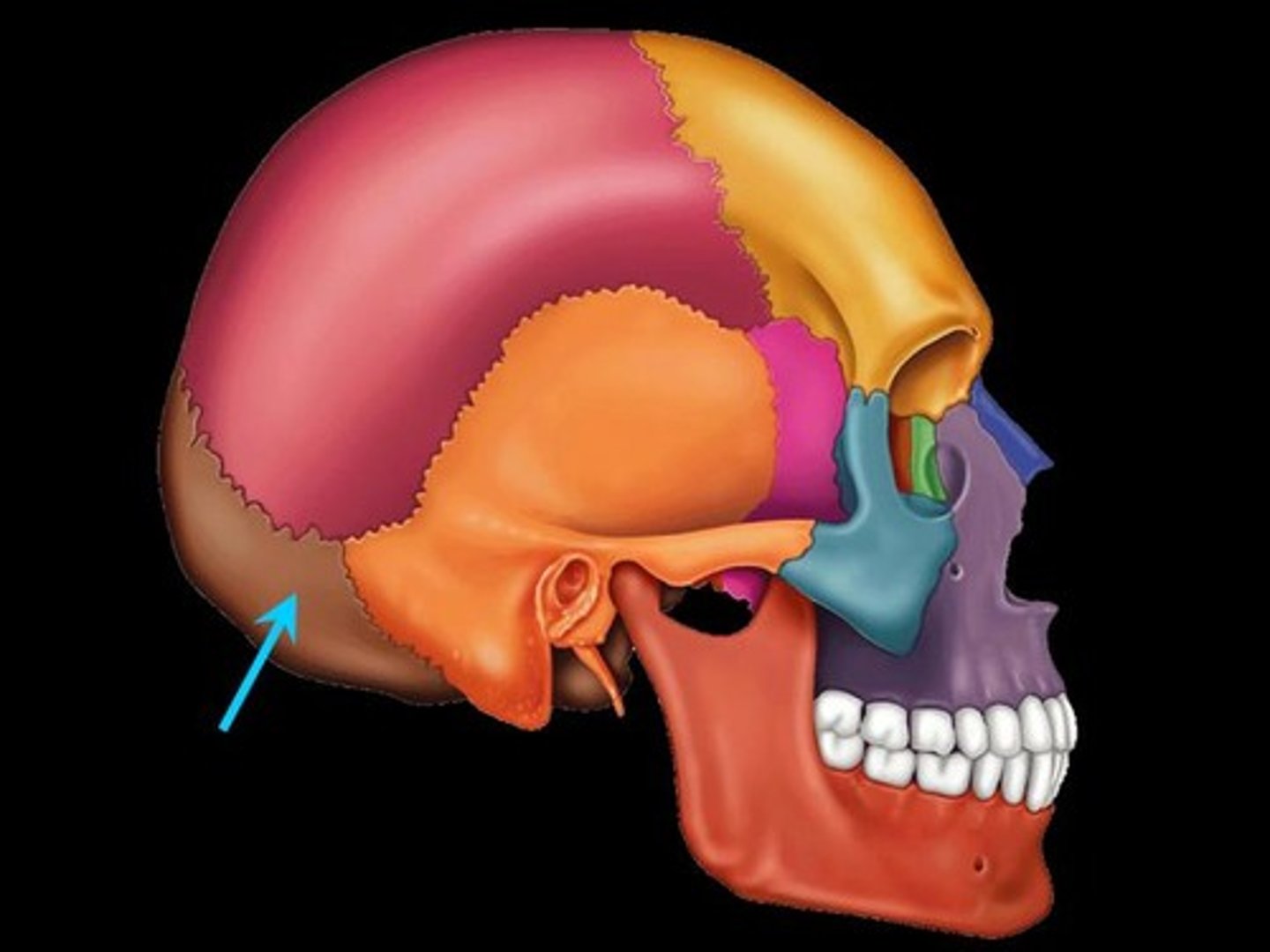
Occipital Bone
Posterior part of cranium & cranial base
Foramen magnum = large opening in occipital bone that allows the spinal cord to pass & connect the brain
Occupital condyles are region where the skull articulates with vertevral column (atlas or C1 vertebra)
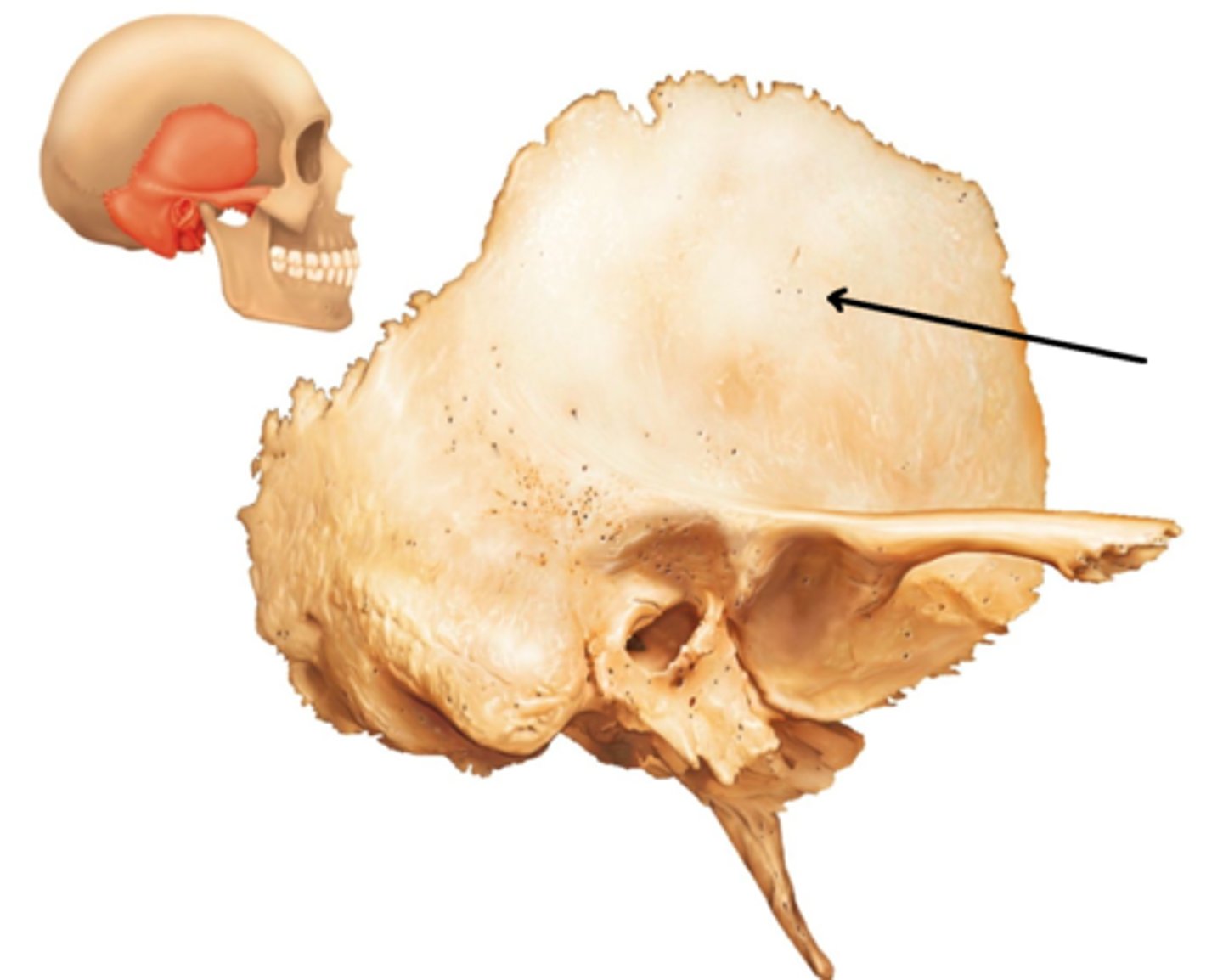
Temporal Bones
Paired temporal bones (left and right sides) house opening to ear, forms the base of cheekbone.
3 main regions: petrous, tympanic and squamous.
Temporal Bones Regions
Petrous is best seen internally, contains middle and inner ear cavities. Petrous=hard.
The external acoustic meatus (EAM): opening in the tympanic region leading to the middle & inner ear
Squamous portion is the vertical portion (part of cranial vault).
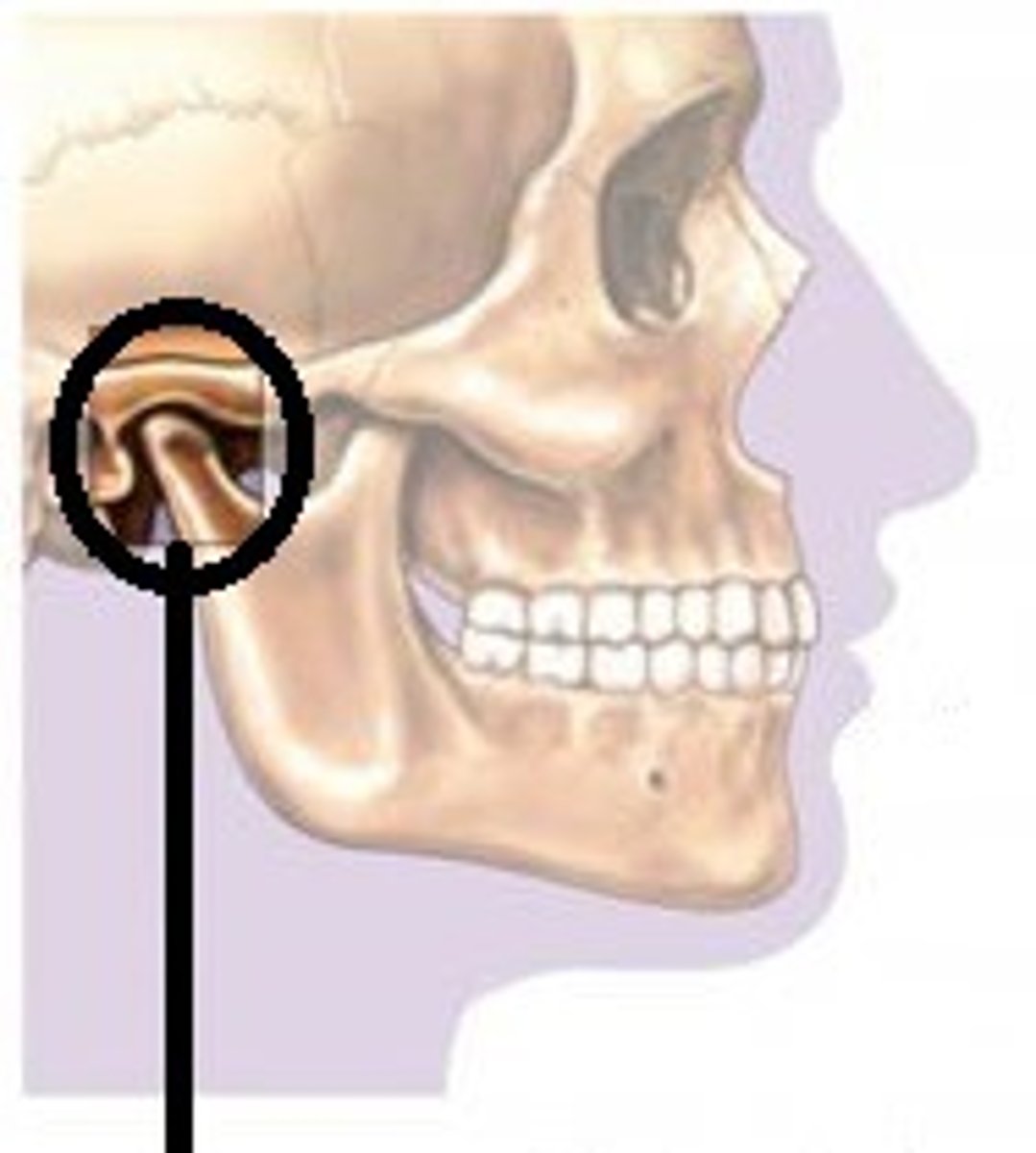
Temporomandibular Joint (jaw joint)
The condyle of the mandible articulates with temporal bone at the mandibular fossa.
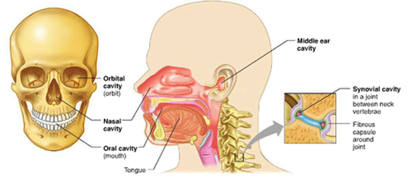
Middle Ear Cavity (inside temporal bone)
3 ear ossicles in the middle ear: malleus, incus and stapes. Bones are really small.
Sound waves cause vibrations of these bones that are transmitted to the inner ear (cochlea).
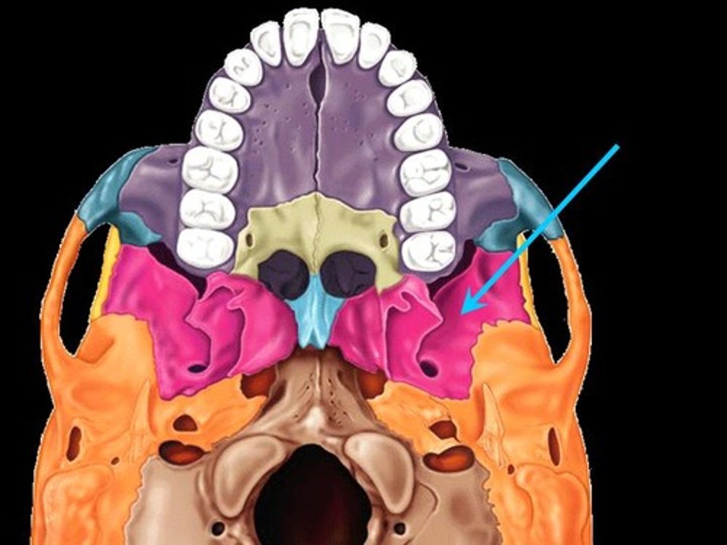
Sphenoid
Large and wing-shaped.
Landmark: Sella turcica - bony depression that holds the pituitary gland.
Sphenoid is the only cranial bone that articulates with every other cranial bone.
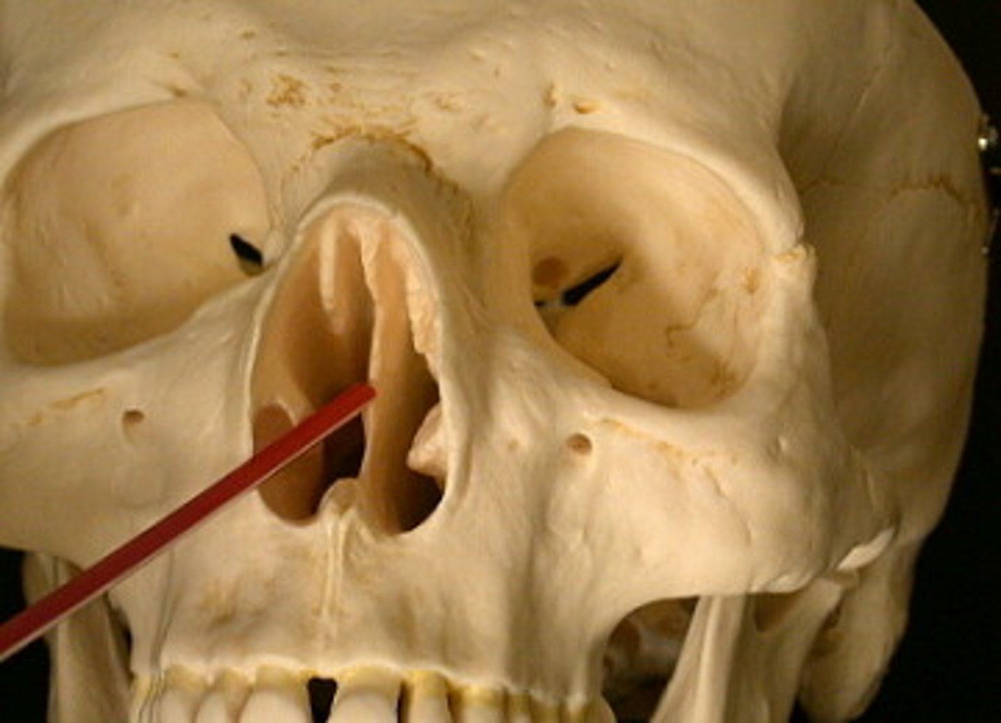
Ethmoid Bone
Just anterior to the sphenoid is the ethmoid bone.
Ethmoid takes up most of the area between the nasal cavity and the orbits.
Forms some boundaries of the nasal cavity, also separates nasal cavity from the brain.
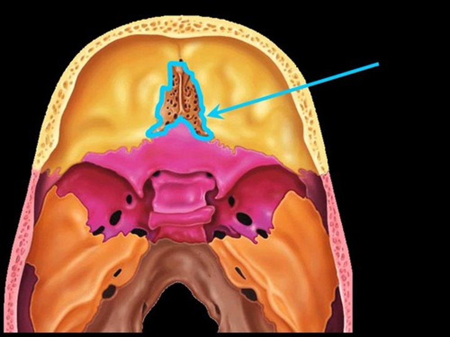
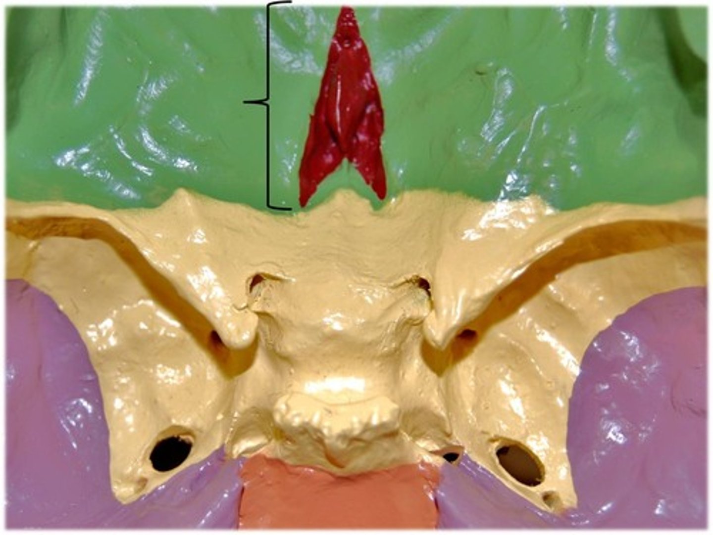
Ethmoid
- Crista galli attaches to cribriform plate; separates nasal cavity from brain, site of attachment for dura mater (membrane covering brain)
- Cribriform plate helps form the roof of nasal cavities; foramina allow passage of olfactory nerves into brain.
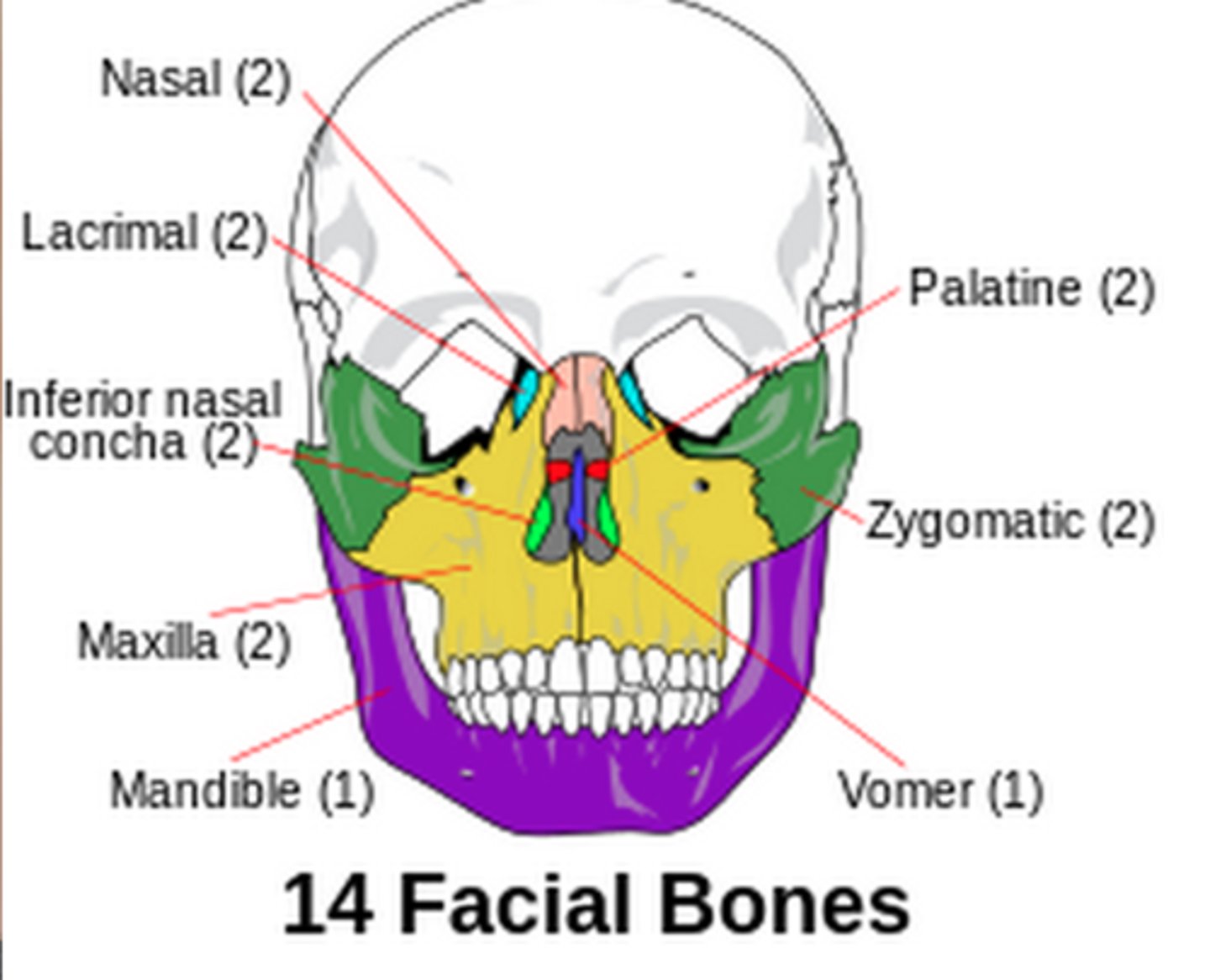
Facial Skeleton
14 Bones of the Face
Mandible (unpaired)
Vomer (unpaired)
2 Nasals
2 Lacrimals
2 Maxillae
2 Zygomatics
2 Palatine bones
2 Inferior nasal conchae
Bones of the Facial Skeleton
Lacrimal bones (paired):
Lacrimal groove allows tears to drain into nasal cavity
Inferior nasal concha
(paired)
Vomer
(unpaired)
Nasal bones (paired)
form bridge of nose
attach to cartilages
that form nose.
Zygomatic bones
(paired) form
cheekbones
Maxillae
(paired)
Mandible
(unpaired
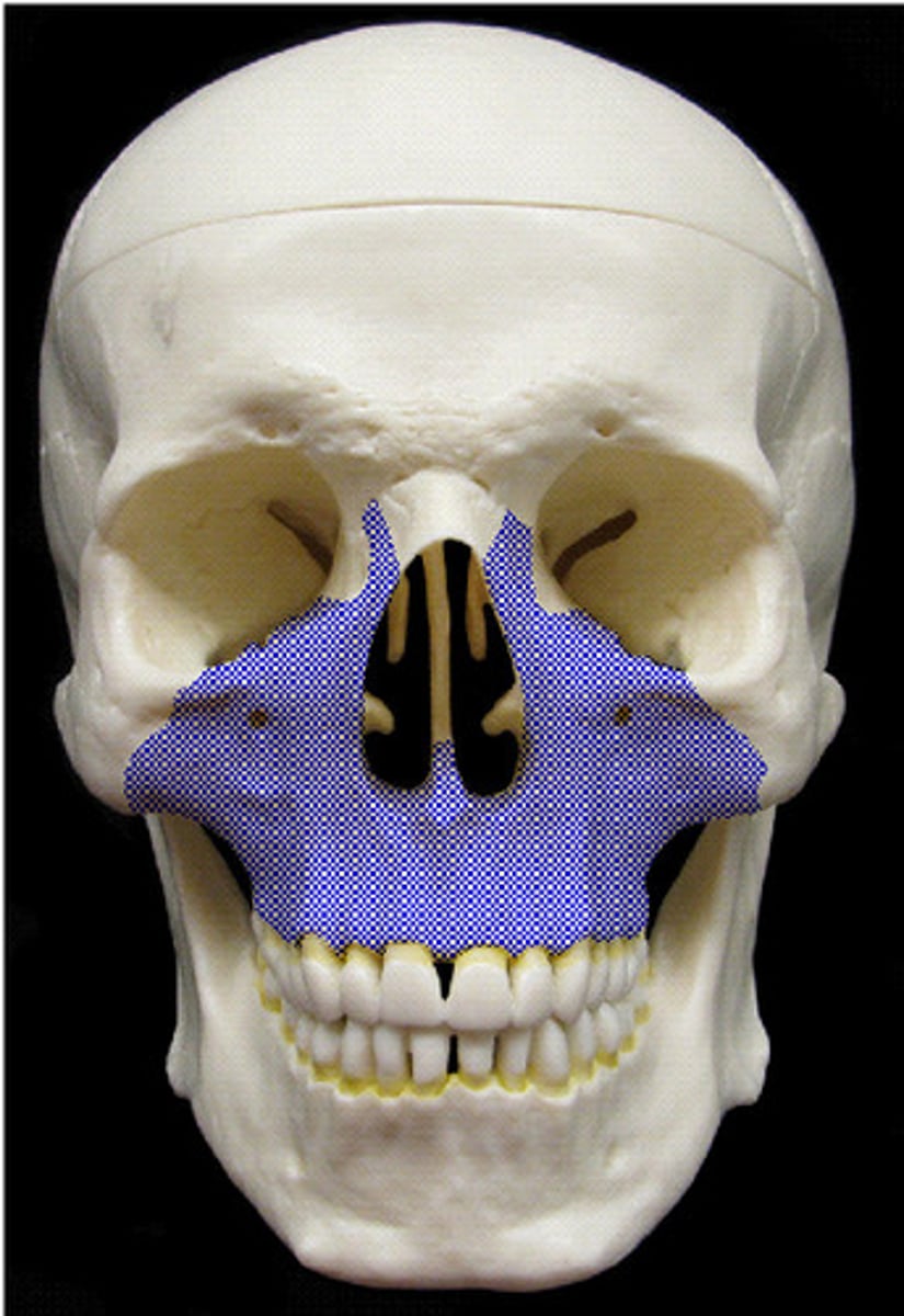
Maxilla(e)
Maxillary bones form upper jaw (paired, left & right).
Articulate with all other facial bones except mandible.
Alveolar processes contains teeth.
Frontal processes extend upward to reach frontal bone
Zygomatic processes of maxilla articulate with zygomatic bone
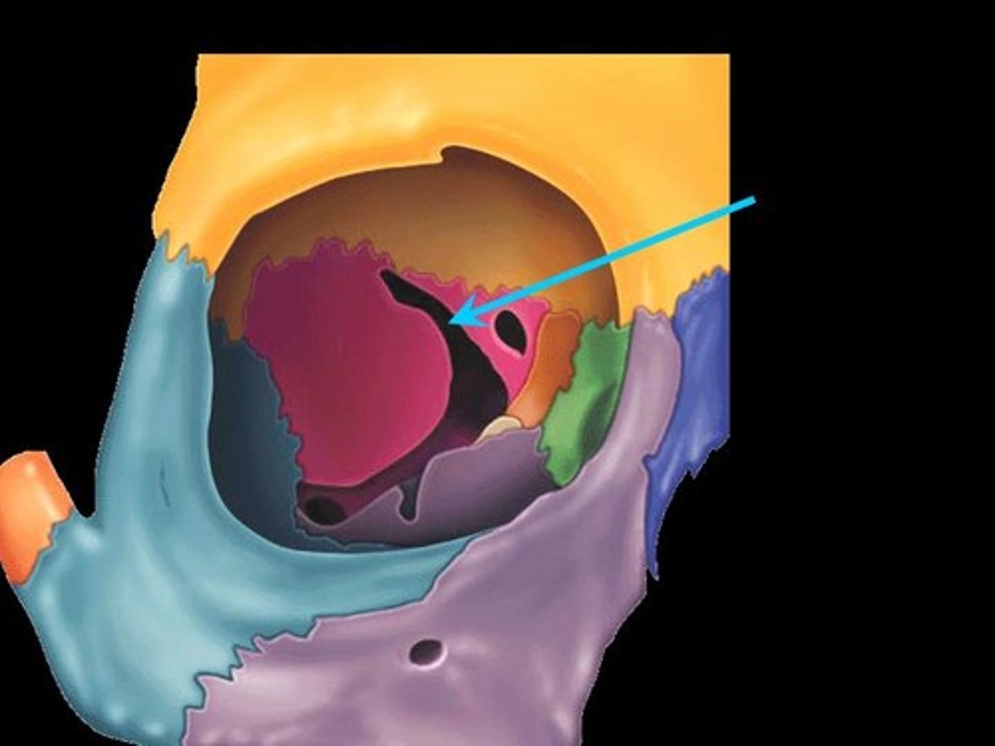
Orbit
Supports the eyes & muscles that move the eyes. Orbit also contains fat and lacrimal glands (tear producing glands).
Walls formed by frontal, sphenoid, zygomatic, maxillary, palatine, lacrimal & ethmoid bones.
Nasal Cavity in Sagittal Section
Lateral walls of nasal cavity: nasal bones, nasal conchae (superior, middle, inferior), maxillae, palatines.
Floor of nasal cavity = hard palate. Formed by palatine process of maxillae, horizontal plate of palatine.
Palate & Inferior Nasal Septum
Hard palate is composed of maxillary bones and the palatine bone.
Vomer forms inferior portion of nasal septum.
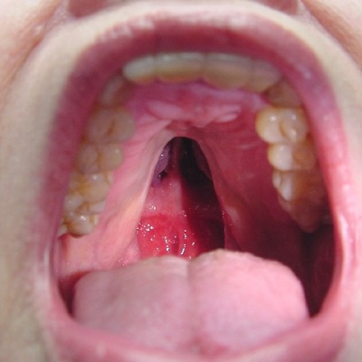
Cleft Palate
Failure of the 2 sides of the palate to join during development leads to cleft palate. Severity varies.
Opening between mouth and nasal cavity makes effective nursing difficult.
Can be repaired surgically with good outcomes.
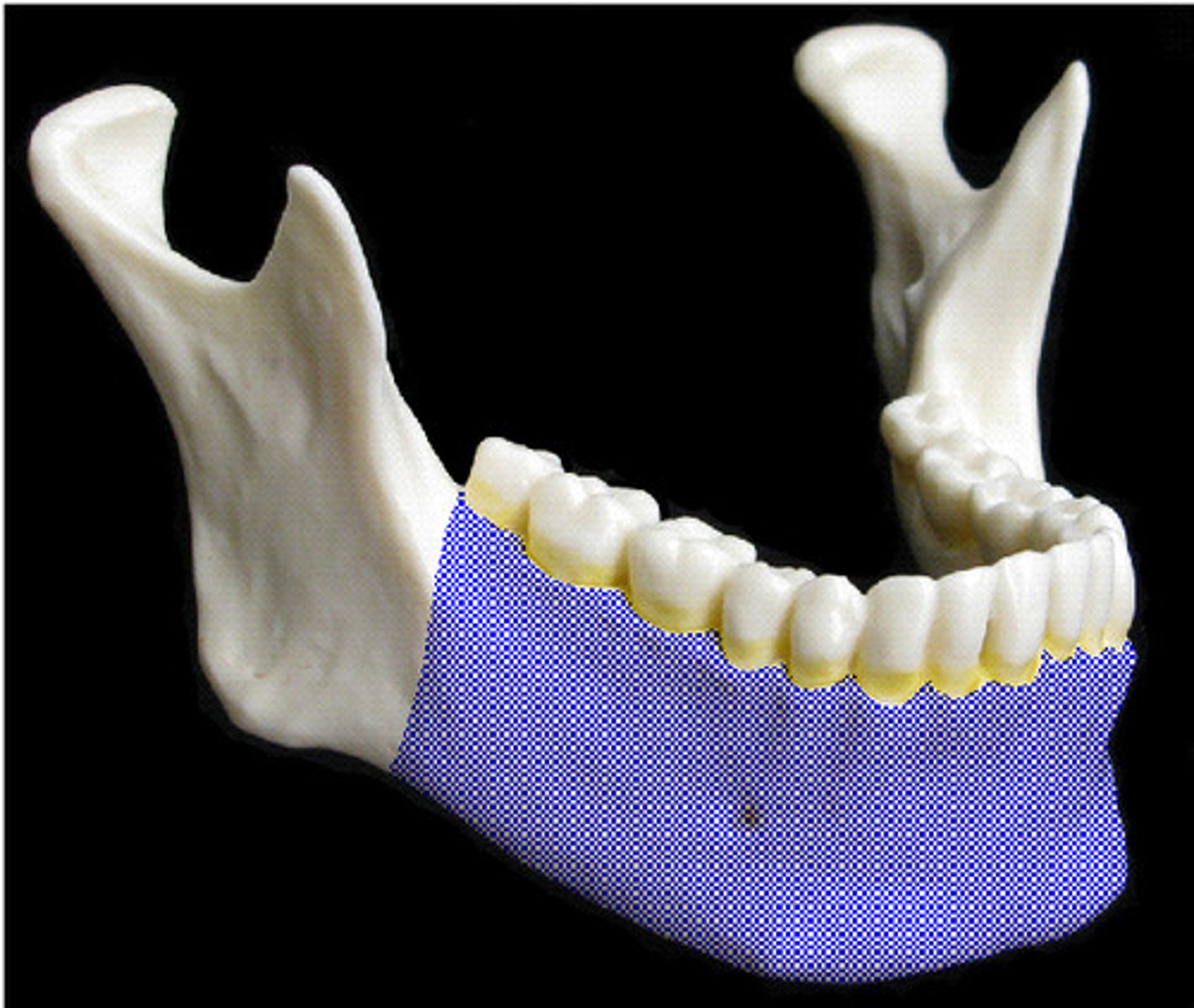
Mandible
Largest, strongest bone in the face.
Body of mandible contains lower teeth.
Tooth sockets are on the superior border (=alveolar processes)
Mandibular symphysis (not visible) is where the two halves of the body join to form the chin (=mental protuberance).
Vessels & nerves enter via mandibular foramen & exit via mental foramen inferior to teeth
Condyle articulates with the temporal bone to form the temporomandibular joint (TMJ; both sides of jaw).
Coronoid process serves as an attachment site for the temporalis muscle, a major chewing muscle.
Fetal/Infant Face
Cranium is proportionately huge relative to the face in infancy & early childhood.
By age 2, skull is 3/4 adult size.
Between ages 6-13, face grows outward & develops more "adult" proportions; body size begins to catch up with head.
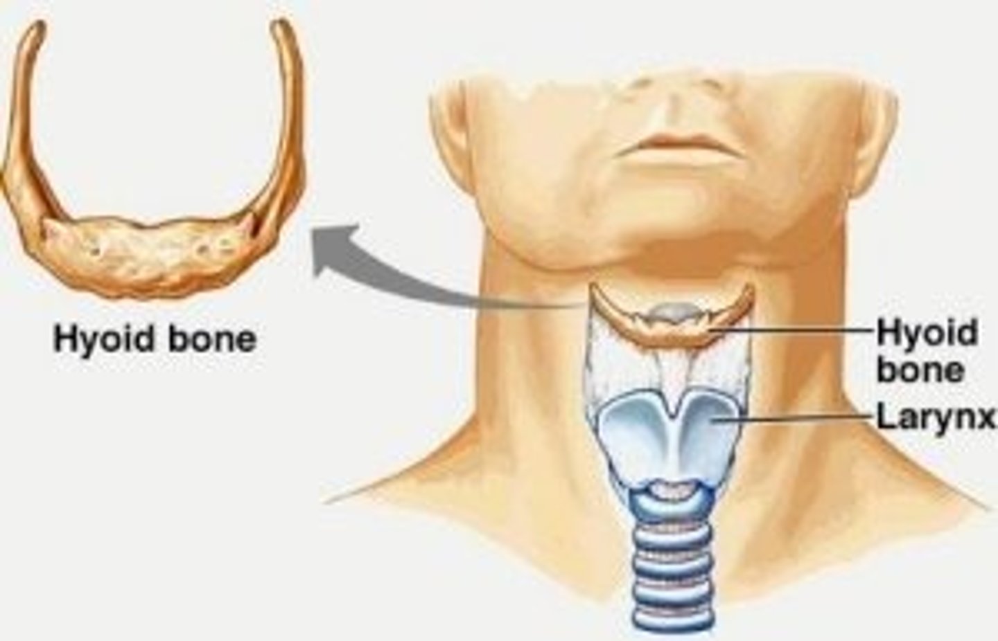
Hyoid
"Free floating" bone in neck inferior to mandible.
Only bone in skeleton that does not articulate with any other bone.
Acts as base for tongue, site of muscle attachments for muscles that move the larynx.
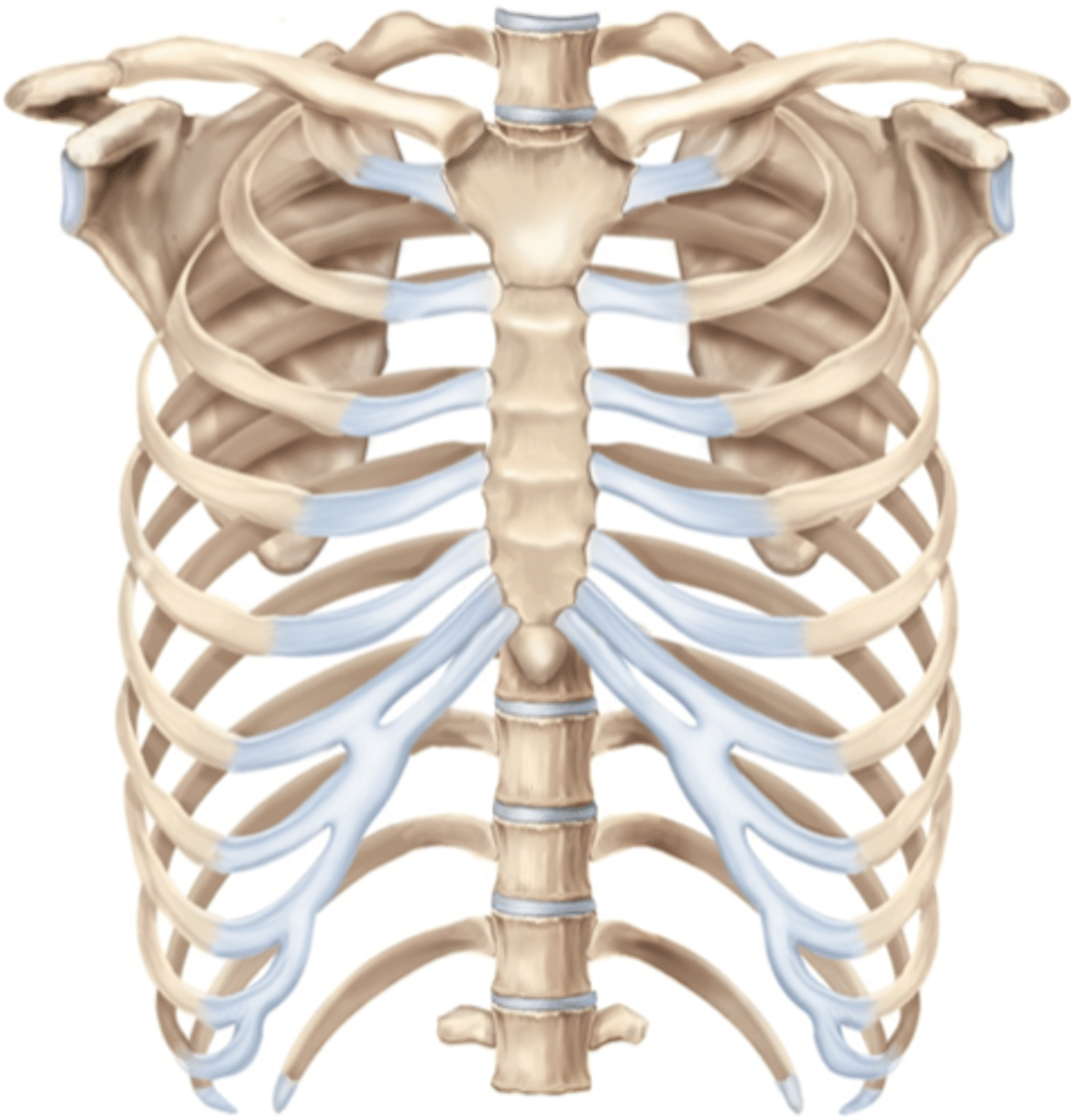
Thoracic Cage
Includes thoracic vertebrae, ribs, sternum and costal cartilages.
Protects heart, lungs, other organs.
Supports pectoral girdle and provides attachment points.
Intercostal spaces hold muscles that aid in breathing.
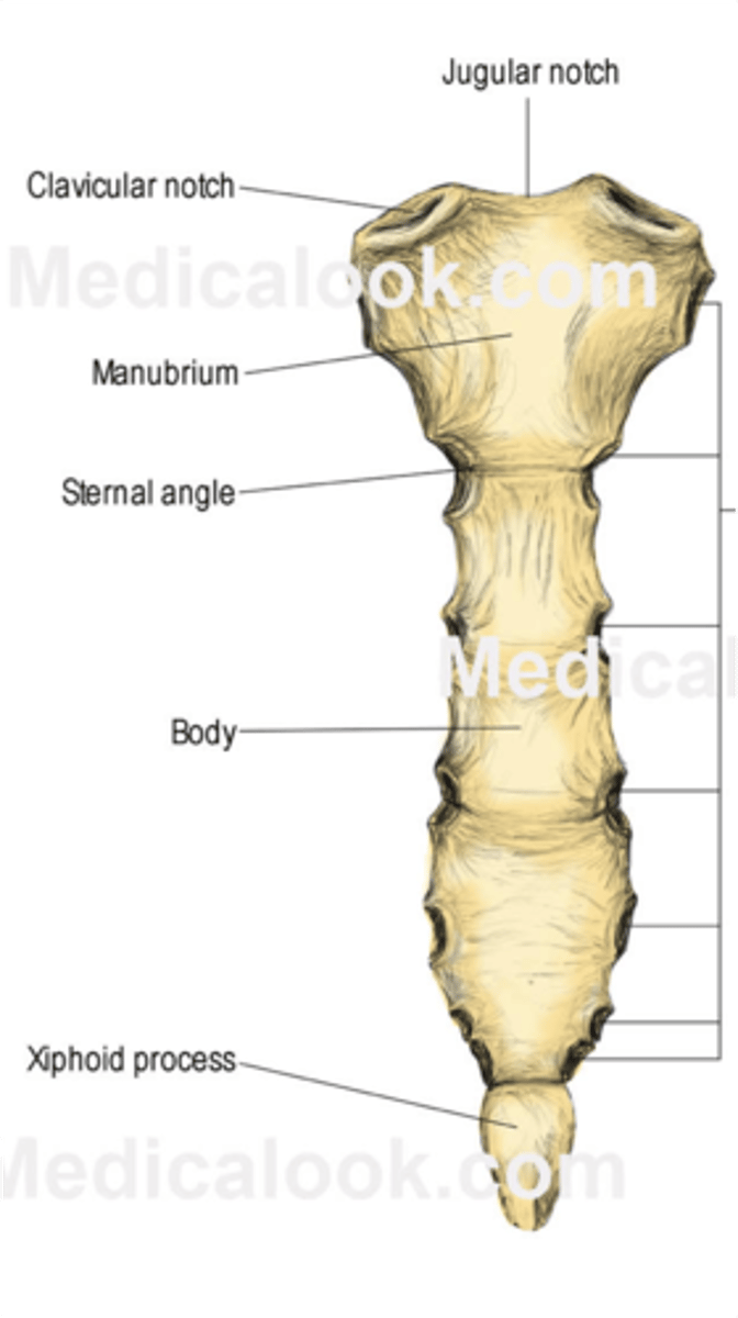
Sternum
3 bones
-- Manubrium
-- Body
-- Xiphoid Process
Articulates with:
-- Clavicles
-- Ribs & costal cartilages
Sternal angle is important landmark for thoracic anatomy.
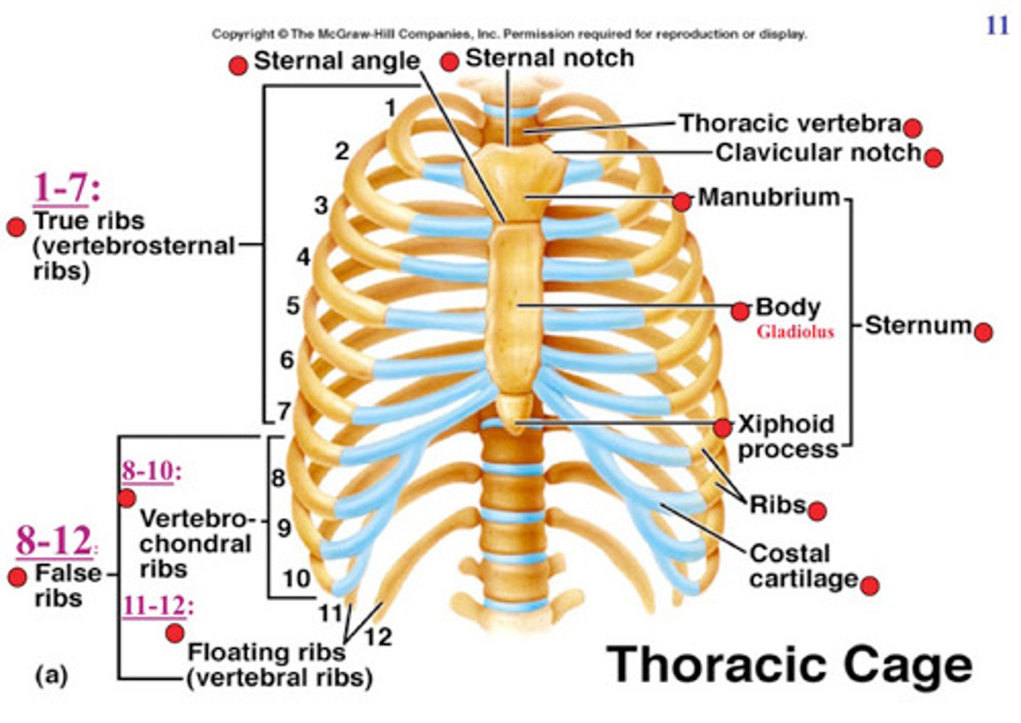
Rib Cage
Function: protect internal organs, aid in respiration.
12 pairs of ribs.
All ribs attached posteriorly to thoracic vertebrae
1st 7 attach to sternum by costal cartilages (true ribs).
8-10 are false ribs - do not have a direct attachment to the sternum. They attach via a shared costal cartilage.
11 and 12 are "floating ribs" - they do not attach anteriorly to the sternum.
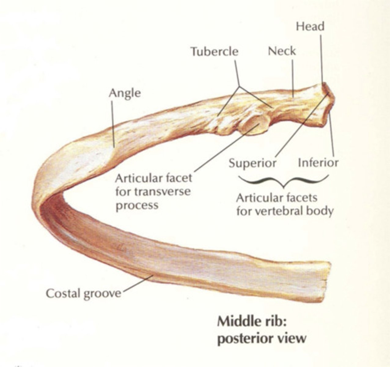
Rib Structure
Main part of rib is the shaft (body).
Rib articulates with the vertebra at the head and tubercle
"Neck" (not labeled) is a thinner region between the head and tubercle. Head has 2 "facets" - one facet articulates with the body of "its" vertebra; one articulates on the body of the vertebra superior to it.
Tubercle articulates with a facet on the transverse process of the vertebrae.
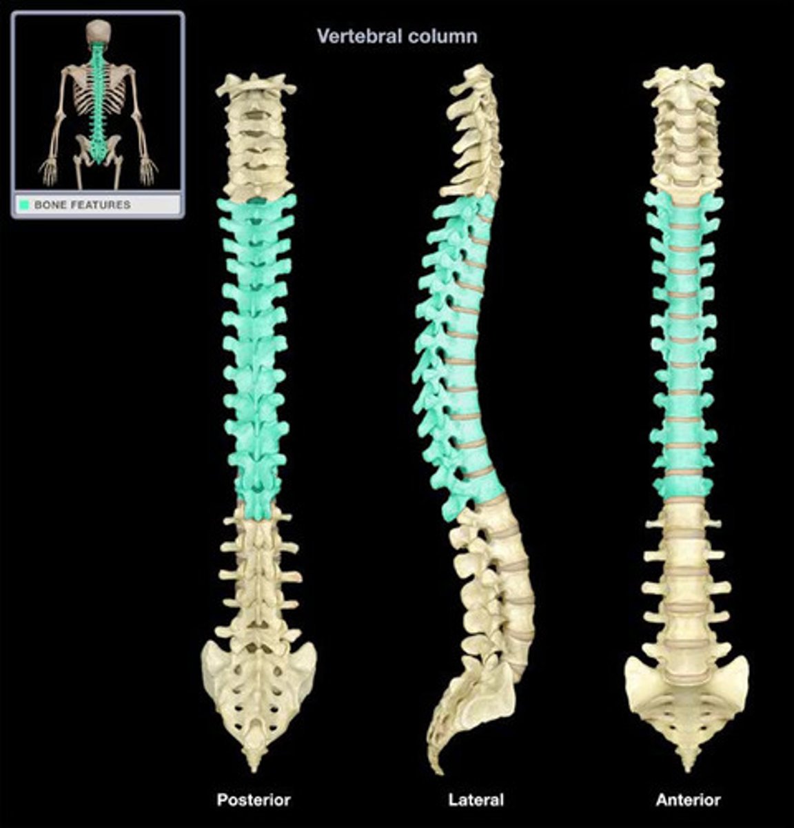
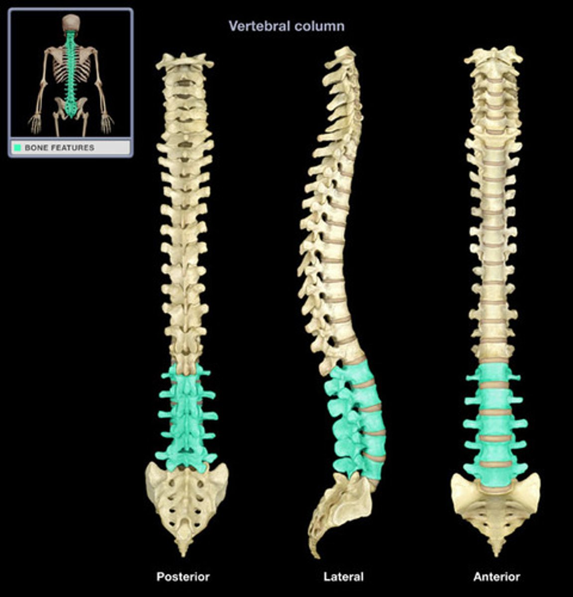
Vertebral Column
26 vertebrae
-- 7 Cervical
-- 12 Thoracic
-- 5 Lumbar
-- 5 Sacral (will fuse into 1)
-- 4 Coccyx (will fuse into 1)
Functions:
-- Protect spinal cord
-- Supports body axis
-- Attachment points for ribs & muscles of neck & back.
-- Anchor pectoral & pelvic girdles
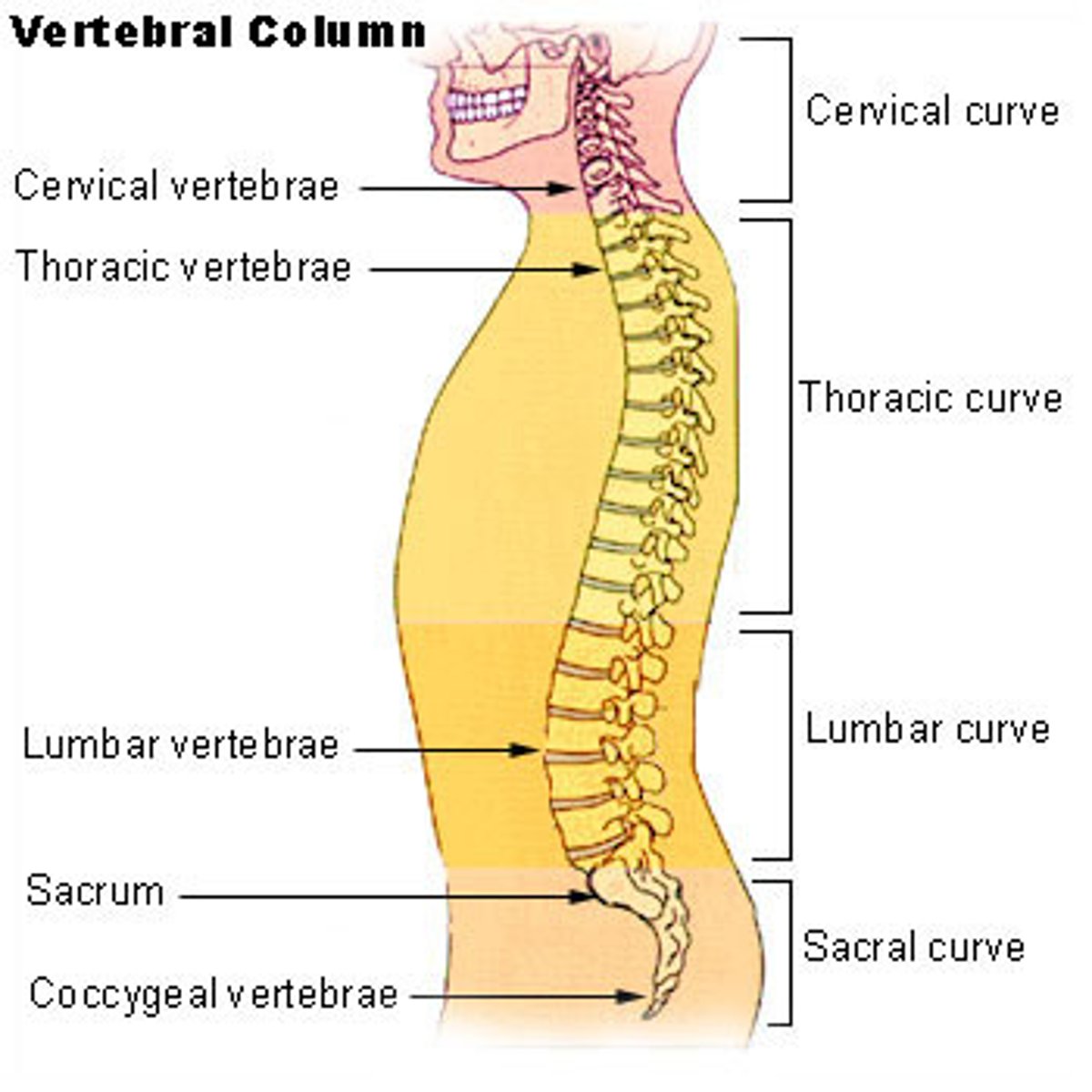
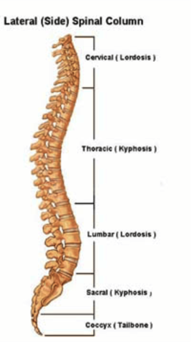
Curves of the Spine
Vertebrae become larger as move inferiorly to support weight
Sacrum articulates with hip bones of pelvis, passes weight to appendicular skeleton.
Curves increase flexibility; also position center of gravity over axis of body
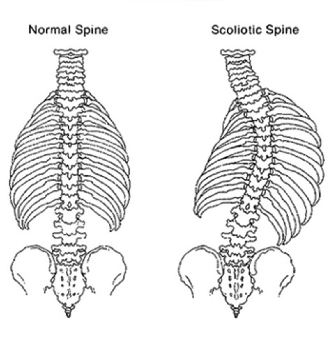
Curves of the Spine: Scoliosis
Scoliosis - lateral curvature of the spine.
Usually treated with body braces or surgery when young
Lateral curvature is abnormal!!.
Kryphosis (Dowager's Hump)
Excessive curvature of the thoracic spine.
Typically the result of vertebral body factures caused by osteoporosis.
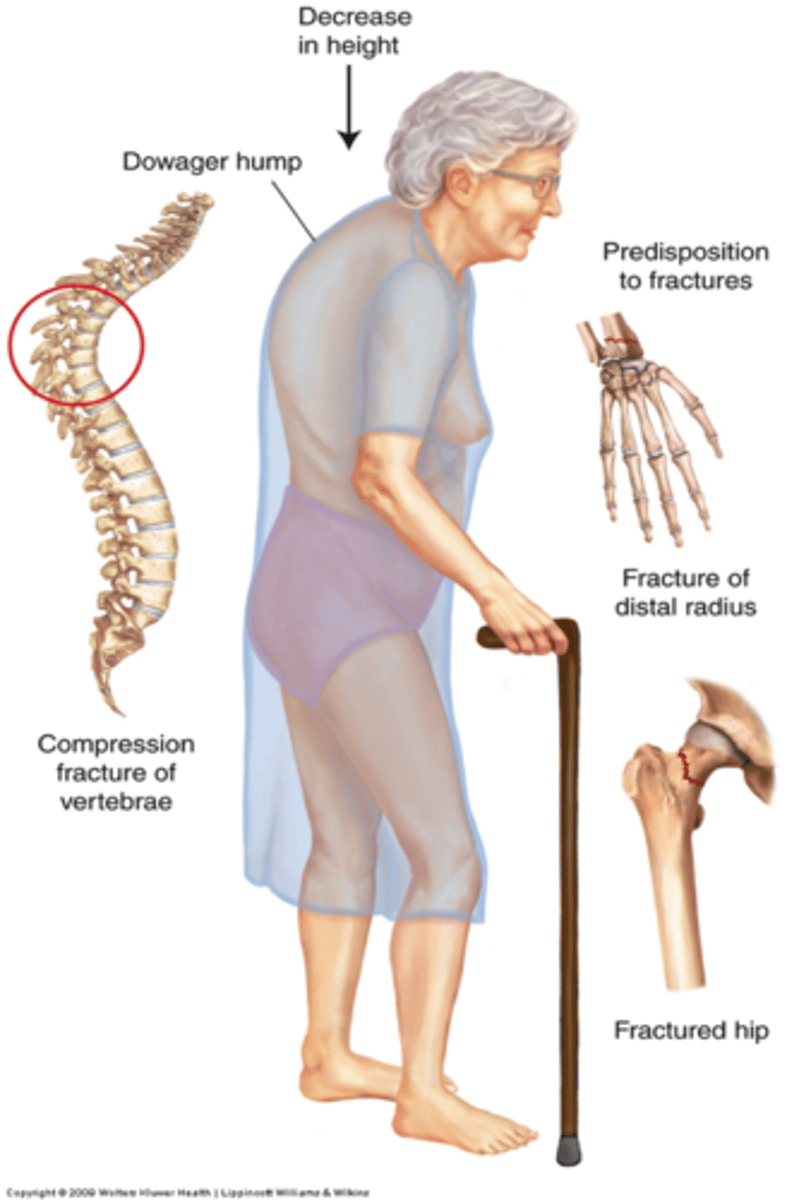
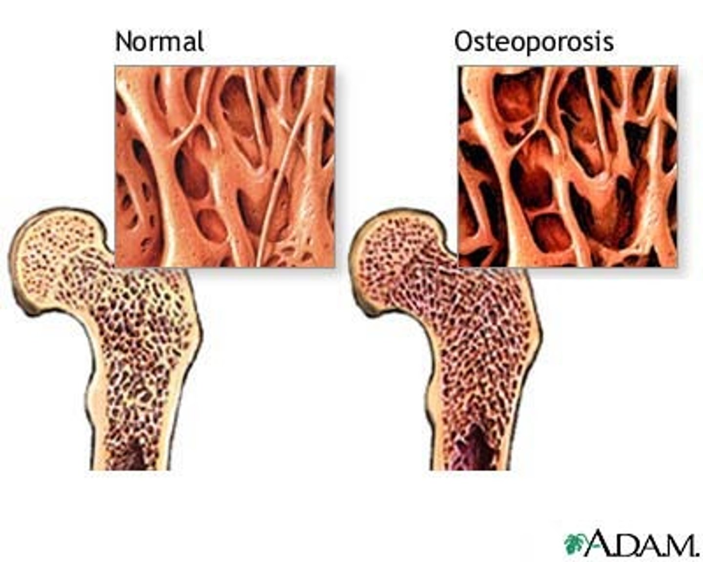
Osteoporosis
Results from imbalance in normal bone building & degradation cycle. Particularly after menopause, women don't absorb as much calcium & so osteoclasts break bone down to release the calcium into the bloodstream.
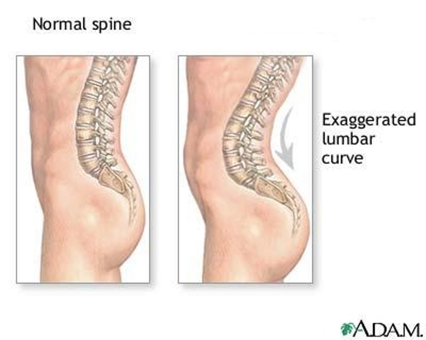
Lordosis
Lordosis is excessive curvature of the lumbar spine.
Usually temporary & resulting from shift for a larger front load (belly in men, pregnant women).
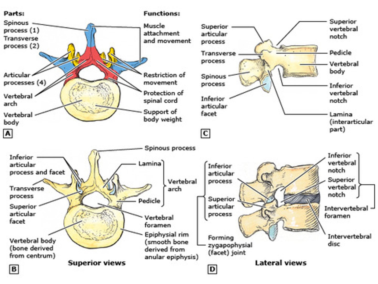
Basic Vertebral Anatomy
Body is located anteriorly.
Vertebral arch made of lamina & pedicles
-- Creates vertebral foramen
---- Surrounds spinal cord
Stacked vertebrae result in vertebral canal.
-- Spinous processes and transverse process are ligament & muscle attachment sites
Vertebrae articulate at superior and inferior articular processes and facets
-- Each articular process has an articular facet.
-- Individual vertebrae articulate with the vertebrae just superior and inferior to it.
-- This articulation also makes intervertebral foramina between two vertebrae.
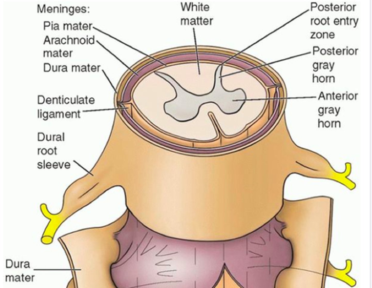
Vertebral Column & Spinal Cord
Notice how the spinal cord passes through, & is protected within, the vertebral (spinal) canal.
Spinal nerves exit between the body of the vertebra & the vertebral arch via intervertebral foramina
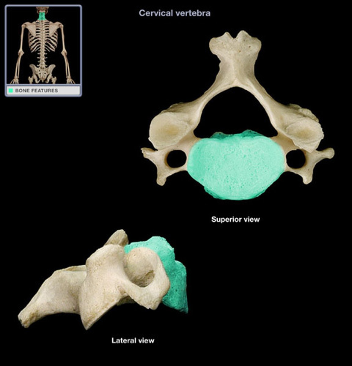
Seven Cervical Vertebrae (C1-C7)
Cervical vertebrae features:
-- Have transverse foramina
-- Articular facets face superior/inferior
-- Bifid (split) spinous processes
Atlas (C1) and Axis (C2) are unusual (see the next slides
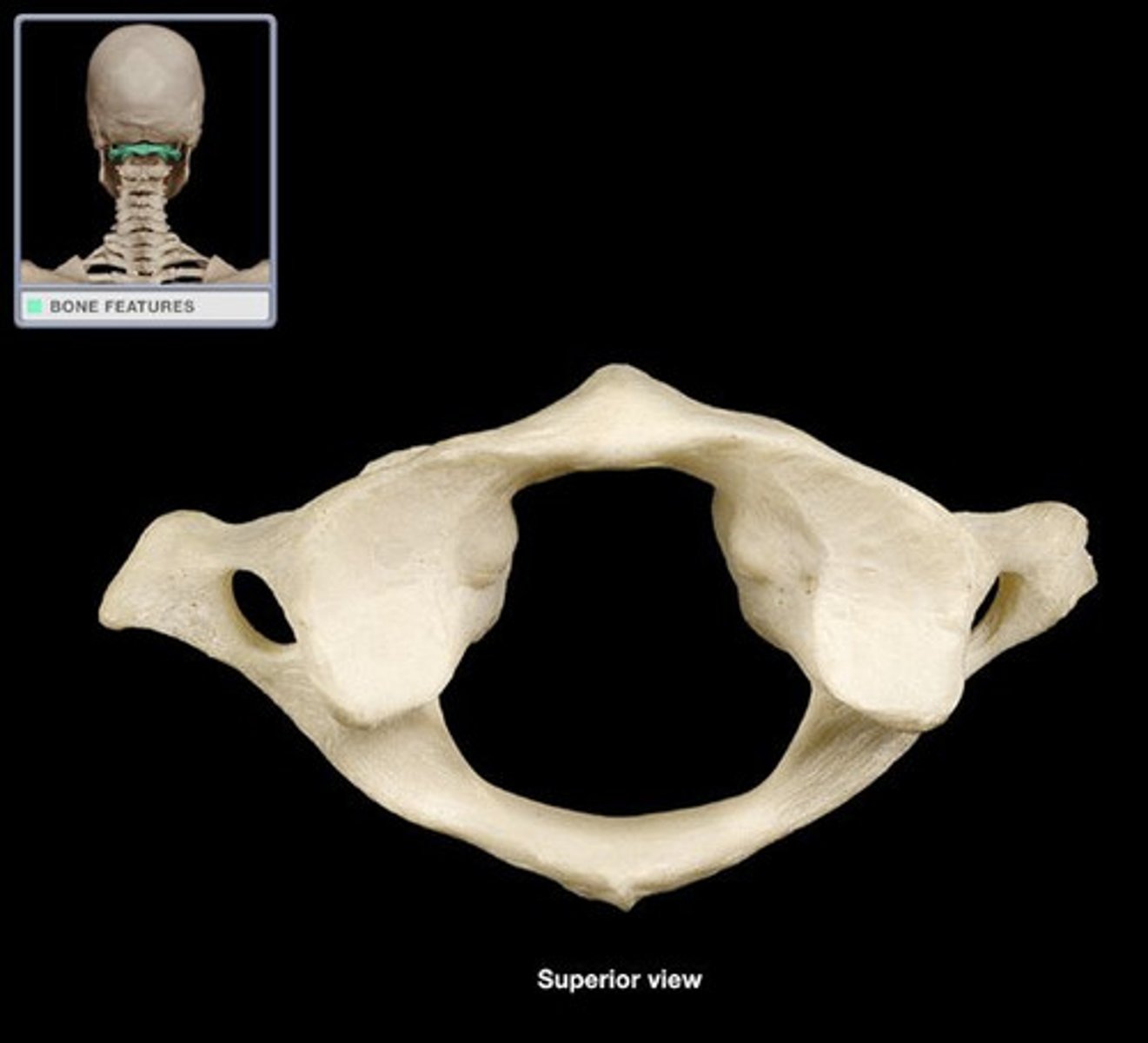
Atlas (C1)
Atlas does not have a body or a spinous process.
Articulates with occipital condyles, allows flexion/extension of
head (nodding "yes").
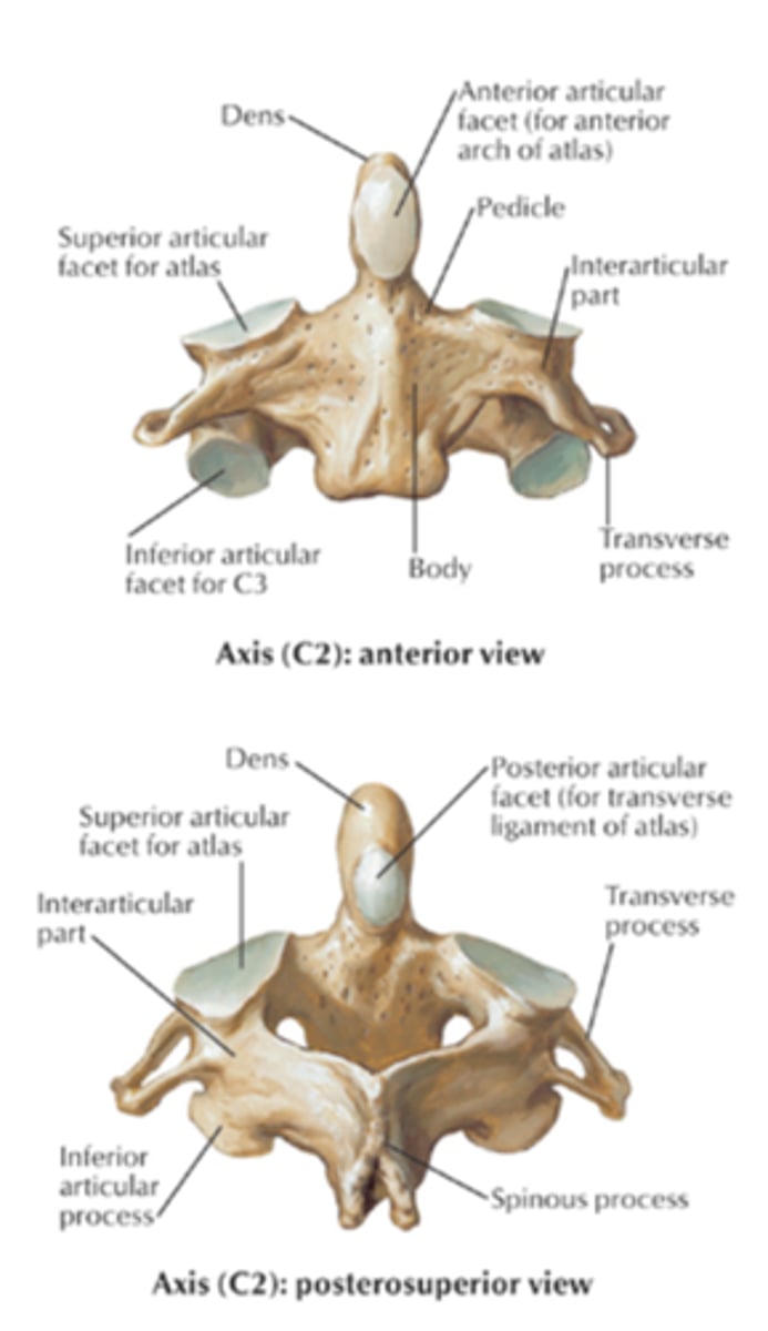
Axis (C2)
Dens articulates with atlas, allows rotational movement (shaking head "no"). Dens used to be the body of the atlas, but not part of axis.
Twelve Thoracic Vertebrae (T1-T12)
Thoracic vertebrae:
Costal facets for ribs, located on each body of the thoracic vertebrae.
Articular facets face anterior/posterior
Spinous processes are long and project inferiorly
Five Lumbar Vertebrae (L1-L5)
Lumbar vertebrae:
-- Large bodies
-- Articular facets face medial/lateral
-- Short, flat spinous processes
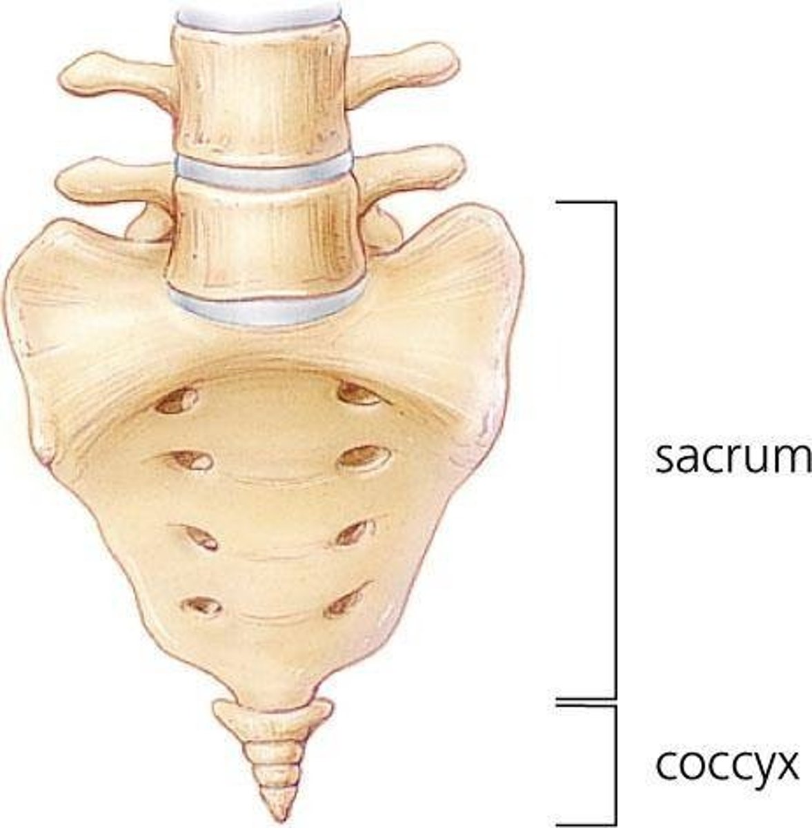
Sacrum & Coccyx
Sacrum
-- 5 Fused Vertebrae
-- Forms posterior wall of pelvis.
-- Ala on lateral sacrum articulate with hip bones to form sacroiliac joints.
Coccyx
-- 3-5 fused vertebrae
-- "Tail bones"
Appendicular Skeleton
Pectoral Gridle & Uppper Limb
Pectoral Girdle, Arm, forearm, hand
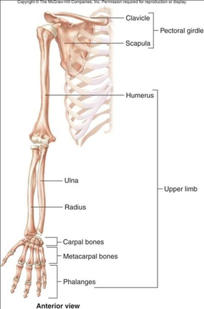
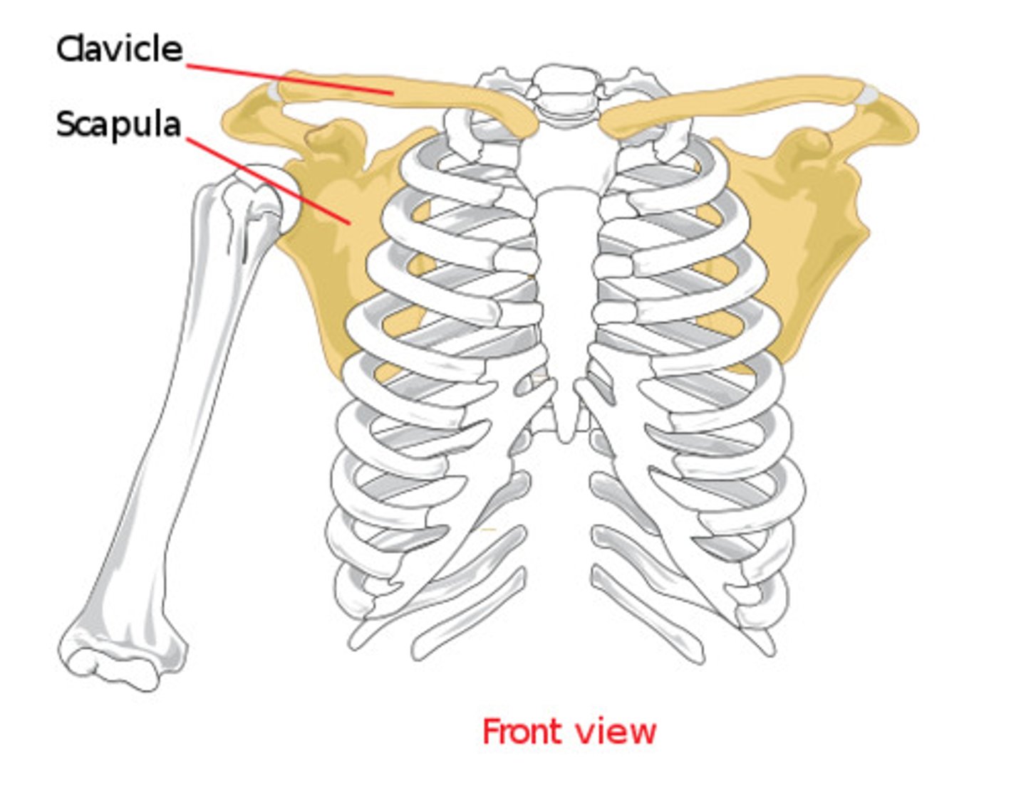
Pectoral Girdle: Clavicles & Scapulae
Includes the left and right scapulae & left & right clavicles.
Note that the scapulae do NOT join to the axial skeleton at all, and their articulation with the clavicle is very loose.
Attached to the axial skeleton by way of associated muscles & ligaments.
Provides a highly flexible system (lots of movement allowed), but not very stable.