Introduction of Protein
 Call Kai
 Learn
Learn Practice Test
 Spaced Repetition
 Match
Match Flashcards
 Knowt Play
1/55

There's no tags or description
Looks like no tags are added yet.
Name | Mastery | Learn | Test | Matching | Spaced | Call with Kai | Chat |
|---|
No analytics yet
Send a link to your students to track their progress
56 Terms
(Catalysis) (Structure & Support) (Movement) (Transport & Regulation) (Immune Defense)
(________): Enzymes drive all biochemical reactions within the cel
‣ (___________): Collagens form cellular and extracellular frameworks, making them the most abundant body proteir
◦(_________): Contractile proteins power muscle movement and cellular
locomotion
• (____________): Specialized proteins carry body fluid materials (transferrin), receive signals (receptors), and control gene expression (transcription factors
• (__________): Antibodies serve as critical defense components in
the immune system
(peptides) (Polypeptide) (Amino acids) (condensation/dehydration synthesis) (dipeptide) (DIPEPTIDE) (OLIGOPEPTIDE) (POLYPEPTIDE) (PROTEIN)
Peptide and Peptide Bonds
● A typical protein contains 200–300 amino acids, but some are much smaller, such as (________).
● (__________): large amounts of amino acids
● Large polypeptide constitutes a protein
● (__________) are bound together through peptide bonds
● A peptide bond forms when amino group of one amino acid combines or links with the carboxyl group of another amino acid
○ This causes the loss of one molecule of water called (________________________)
● When one amino acid joins another amino acid, this is termed a (________).
● one amino acid joins another amino acid ● Example: Aspartame
○ Modified dipeptide ○ It is an artificial sweetener seen in Coke Zero. ○ It is modified because aspartic acid and phenylalanine are the amino acids, and it is modified with the addition of methanol. | ||
approximately a combination of 2–20 amino acids. | ||
If many more amino acids are joined together |
(____________) If one or more large polypeptides are bound together
(Amino acids) (Proteins) (peptide bonds)
(CARBOXYL GROUP) (AMINO GROUP) (R- GROUP)
The "building block of life." (_______) are monomers. (________) are a polymer of amino acids. (1) (__________) are joined from head-to-tail via the formation of (________)
C - terminal (yellow box) | ||
N - terminal (green box) | ||
● Side chain (red box) ● The R group or side chain is the only thing that changes between the different types of amino acids. ● Amino acids differ from one another only in their R group. ○ Phenylalanine ○ Tyrosine |
(R group) (polypeptide)
Amino acids differ from one another only in their (______). A chain of amino acids is a (_________); a large polypeptide constitutes a protein
(amphoteric) (low pH (acidic)) (high pH (alkaline)) (isoelectric point (pl)) (zero)
Amino Acids are (__________) they act as both acid and base
At (__________) -> the molecule gains H —> net positive charge
At (__________) -> the molecule loses H* —> net negative charge
At the (__________) > positive and negative charges are equal > net charge = (______)
(20) (-Phenylalanine, valine, tryptophan, threonine, isoleucine, methionine, histidine leucine, lysine; Arginine) (-Alanine, asparagine, aspartic acid, cysteine, glutamic acid, glutamine, glycine proline, serine, tyrosine)
Essential Amino Acids
About half of the (___) amino acids cannot be synthesised at a rate fast enough to support growth, and must be supplied by the diet in the form of proteins
Essential: (_____, _____, ______, ______, _____, ____, ______, _______, _________)
Nonessential: (_____, ______, _____, ______, _____, _____, _____, _____, _____, _______,)
(Protein Synthesis) (hepatocytes:) (immunoglobulins)
(__________)
Most plasma proteins are synthesized in the liver by (________) (_________) are the exception- they are synthesized in plasma cells.
Transcription
DNA unfolds in the nucleus; one strand serves as a template for a complementary strand of mRNA.
Translation
mRNA moves to the cytoplasm and attaches to ribosomes; the mRNA is read three nucleotides (one codon) at a time
1. .In the cytoplasm, an amino acid is activated in an energy-requiring reaction (ATP).
2. The amino acid is activated by ATP using a specific enzyme.
3. The activated amino acid attaches to a specific transfer RNA (tRNA). Each amino acid has a specific tRNA.
4. The activating enzyme and adenosine monophosphate are released.
5. The anticodon matches the corresponding codon on the mRNA.
(ribosome. ) (codon) (nucleotides) (peptide bond) (synthesized.)
Initiation
1. Translation begins at the (_________)
2. The mRNA loads onto the ribosome at an initiation (______).
Elongation
1. The mRNA is read three (_________) at a time.
2. Matching tRNA brings the corresponding amino acid to the ribosome.
3. Codon and anticodon base pairing allows them to bind together.
4. Ribosomal enzymes form a (_________).
5. The preceding amino acid is transferred onto the amino group of the newly arrived amino acid.
6. This is peptide bond formation.
7. The peptide continues to be (_________)
Termination
When the ribosome reaches a stop or termination codon:
● The complete polypeptide chain detaches.
● The mRNA-ribosome complex dissociates.
● The now-uncharged tRNA is released back into the cytoplasm for recycling.
● It can continue to another synthesis.
tRNA
Each amino acid is carried by a specific transfer RNA whose anticodon base- pairs with the mRNA codon; the ribosome forms the peptide bond as each new amino acid arrives
(Rate) (2) (6)
(_____)
Protein synthesis occurs at approximately (_) to (_) peptide bonds per second. Intracellular proteins are made on free ribosomes; proteins made for secretion are made on ribosomes attached to the rough endoplasmic reticulum
(Protein Metabolism) (Biological Value) (Protein Turnover) (Rapid turnover) (Stable turnover:) (○ Thyroxine ○ Growth Hormone ○ Insulin ○ Testosterone) (Glucagon, Cortisol)
(_________)
Unlike fats and carbohydrates, the body has no designated storage form for nitrogen
(_________)
Dietary protein quality depends on providing all necessary amino acids; lacking just one limits essential protein synthesis
(___________)
(___________) Approximately 125 to 220g per day
● This can be larger if there is a larger requirement,
such as bodybuilding and training for something
● It can also be lower if the person is in a decreased
state or in a deficit.
● (_________): Plasma proteins (albumin) and
most intracellular proteins
● Stable turnover: Structural proteins, such as
collagen
Hormones Regulating Protein Metabolism
Anabolic: (___________, _____________, ____________, ________)
Catabolic: (________, _______)
(Nitrogen Balance) (16%) (Positive Balance:) (Negative Balance) (Liver (primary), digestive tract, and kidneys) (Lysosomal Pathway)
(Cytosolic Pathway) (Ammonia ) (Keto acid )
(_____________)
The nitrogen content of serum protein is, on average approximately (__)
(______________)
Intake > Excretion e.g. Growing children, pregnant women, recovery from illness
(_____________)
Excretion > Intake e.g. Starvation, severe burns, wastina diseases, high fevers
Sites of Disintegration: (______________)
Intracellular Degradation Routes:
(_______________): Extracellular proteins
(_____________): Intracellular proteins
■ (_______)
➢ Converted to urea in the
urea cycle through the
liver
➢ Urea is then excreted in
the urine
■ (_______)
➢ Recycled
➢ Oxidized through the
krebs cycle
➢ Converted to glucose
➢ Converted to fat
(PROTEIN STRUCTURE) (Heat) (Extreme pH) (Strong alkali) (Strong acid) (Organic solvent) (Detergents) (Heavy metals) (Mechanical agitation) (UV light)
(__________)
● Primary, secondary, tertiary, and quaternary
● Levels determine the protein’s shape, charge, and
solubility
● Disruption of these structures can cause
(___________)
○ Loss of the native folded structure.
○ The protein loses its biological activity
causes includes:
■ (____)
■ (______)
➢ (______)
➢ (______)
○ (________)
○ (________)
○ (________)
○ (___________)
○ (________)
(Primary) (peptide bonds)
Protein Structure
(_______________)
● The number and types of amino acids in the specific sequence.
● The primary structure is the amino acid sequence itself.
● It is a linear sequence of amino acids.
○ Amino acids are joined by (______)
● E.g. valine substituted for glutamic acid in the alpha-chain of hemoglobin A produces hemoglobin S (sickle cell disease).
● Primary structure determines the genetic code and determines every higher level of structure.
(Secondary) (Alpha helix ) (Beta-pleated sheet)
Regularly repeating structures stabilized by hydrogen bonds; the alpha- helix, beta-pleated sheet, and turns. Most serum proteins form a helix
● (_______)
○ Coiled or spiral arrangement.
○ Most serum proteins are formed from an alpha helix.
● (_________)
○ An extended strand lying side by side.
○ Amyloid is an example of a beta-pleated sheet configuration.
■ Protein abnormality
■ Aggregate into insoluble fibrils
■ These can infiltrate organs and cause organ damage.
Tertiary
The overall three-dimensional shape, stabilised by hydrophobic effect, ionic attraction, hydrogen bonds and disulfide bonds. Determines a protein's function. One chain folding on itself.
Quaternary
Interaction of more than one protein subunit, held by noncovalent forces, forming a larger complex.
● Interaction of more than one
protein subunit, held by
noncovalent
forces(hydrogen bonds and
electrostatic interactions),
forming a larger complex
● One or more separate
chains come together.
● Example is hgb
○ Four subunits
○ Each subunit
carries a heme
group
(Isoelectric point (pl):) (pH > pl) (pH < pl)
(_______________)
The pH where net surface charge equals zero
(_______): net Negative charge
(_______): net Positive charge
Henderson-Hasselbalch equation:
pH=pKa+log [conjugate base] / [conjugate acid]
(Surface Charge & Hydration) (Lowest Solubility at pl) (Physiological Imperative) (7.35) (7.45) (Methodological Significance)
(________________): Net surface charges make proteins hydrophilic, creating a hydration shell that keeps them dissolved .
(________________): With zero net charge at its pl, repulsive forces vanish- increasing protein-protein interactions and causing precipitation. .
(_______________): Maintaining normal blood protein solubility requires tight regulation of systemic pH between (___) and (____).
(________________): Differences in protein solubility under varying pH and salt conditions serve as the foundation for laboratory protein fractionation and purification.
(energy) (Osmotic force) (Acid-base balance) (Transport) (Antibodies) (Hormones) (Structure) (Enzymes) (Hemostasis)
tissue nutrition, Plasma proteins can act as a reserve energy source for tissues during starvation and severe stress | ||
Maintenance of water distribution between cells and tissues, interstitial compartments, and the vascular system of the body ● Plasma proteins generate colloid osmotic pressure and oncotic pressure ● This draws fluid back into the capillaries. ● A decrease in plasma protein levels: ○ Reduces oncotic pressure. ○ Drives fluid into the interstitial spaces. ○ Causes edema, or accumulation of water in tissues. ● This can signify disease, including renal disease or proteinuria in renal disease. | ||
participation as buffers to maintain pH to maintain the overall pH of the body. |
metabolic substances ● Transport proteins move ions, small molecules, and macromolecules across membrane or in circulation ● Examples include albumin, hgb (transports oxygen in blood), and transferrin (transports iron) | ||
part of immune defense system Antibodies or immunoglobulins such as IgG, IgM, and Ig are synthesized by B lymphocytes and identify and neutralize foreign antigens. | ||
hormones and receptors are chemical messengers that regulate metabolism, growth, reproduction, and behavior ● Examples are insulin, growth hormone, cortisol, follicle-stimulating hormone ● Hormones and their receptors can be proteins |
connective tissue ● Structural or fibrous proteins form cell and tissue frameworks. ● Examples: Collagen, elastin, and keratin in hair ● They also form connective tissues and participate in | ||
catalysts ● They can be released into the bloodstream during tissue damage and can serve as diagnostic markers. ● Transaminases, dehydrogenase, and phosphatases | ||
participation in coagulation of blood ● Fibrinogen is a protein: ○ Synthesized in the liver. ○ One of the largest plasma proteins. ○ Participates in clot formation. |
(Simple Proteins) (Conjugated Proteins) (Derived proteins)
Classification by Structure Proteins
-Protamine
-Histone
-Albumin
-Globulin
-Epidermal
Simple Proteins:
-Nucleoproteins
-Chromoproteins
-Phosphoproteins
-Glycoproteins and mucoproteins
-Lipoproteins
-Metalloprotein
Conjugated Proteins
(Plasma Proteins) (Prealbumin) (Albumin)
(____________)
are divided into albumin and globulins
(_____________)
Migrates before albumin on electrophoresis. Transports thyroxine, triiodothyronine and (with retinol-binding protein) vitamin A. Short half-life (~2 days), so it decreases rapidly in malnutrition and hepatic damage - a sensitive marker of nutritional status. Increased in steroid use, alcoholism, chronic renal failure.
(_______________)
The most abundant plasma protein; synthesised in the liver (9-12 g/day, no reserve). Responsible for ~80% of colloid osmotic pressure. Binds bilirubin steroids, fatty acids, calcium, magnesium, and many drugs. A negative acute phase reactant.
malnutrition, liver disease, protein-losing enteropathy, nephrotic syndrome, burns hypothyroidism, overhydration, sepsis. Increased only with dehydration
Plasma Proteins
Decreased in: (_________, _________, ________, _______, _______, ___________)
(ALPHA-1 GLOBULINS) (ALPHA-2 GLOBULINS)
Globulins I:
(alpha1-Antitrypsin) (alpha1-Fetoprotein (AFP)) (AFP-L3%) (alpha1-Acid Glycoprotein) ()
ALPHA-1 GLOBULINS
(_________________)
Inhibits neutrophil elastase; deficiency (SERPINA1 mutations, allows destruction of alveoli (emphysema) and can cause cirrhosis. Acute-phase reactant. Seen on electrophoresis as a missing alpha1-globulin band.
(____________)
Principal fetal protein; used in maternal screening (elevated: neural tube defects; low: Down syndrome risk), (_______) is a tumour marker for hepatocellular carcinoma
(_________________)
Acute-phase reactant;, important in drug binding, affecting drug action and distribution.
(Haptoglobin (Hp) (hemolytic anemia) (Hp1-1, Hp2-1, Hp2-2.) (Ceruloplasmin) (alpha2-Macroglobulin)
ALPHA-2 GLOBULINS
(____________)
Binds free hemoglobin, preventing its loss (and iron) into urine Decreased in (__________); used to distinguish it from other anemias.
Three phenotypes: (_____, _____, _____)
(___________)
Copper-containing acute-phase reactant. Decreased in Wilson's disease, with excess copper storage in liver, brain, cornea (Kayser-Fleischerrings).
(____________)
Inhibits proteases; increases up to 10x in nephrotic syndrome due to its large size aiding retention.
(BETA GLOBULINS) (GAMMA GLOBULINS)
Globulins II:
(Transferrin Transports iron) (Fibrinogen) (C-Reactive Proteln) (High-sensitivity CRP (hsCRP))
Globulins II
BETA GLOBULINS
(________________); only 20-50% of binding sites normally occupied. Increased in iron deficiency; decreased in inflammation and hereditary hemochromatosis (excess iron deposition).
(_________)
Forms the fibrin clot when activated by thrombin; virtually absent-from serum since it is consumed in clotting. An acute" phase reactant.
(______________) (CRP One of the first acute-phase proteins to rise in inflammation, promotes opsonization. (______________) is used to assess cardiovascular risk
(IgG) (lgA/lgM ) (IgẠ) (IgMA) (IgD/lgE) ( IgD)
GAMMA GLOBULINS
(______) Most abundant antibody; crosses the placenta (the only one that does). Acts by agglutination, opsonization, complement activation, toxin neutralization.
(________)
(_____) is the main immunoglobulin in secretions (tears, saliva) (_____) is the first antibody in response to a new antigen and is the only one made by the neonate
(________)
(_____) may help regulate B-cell function, IgE mediates allergic and anaphylactic reactions. Monoclonal spikes on electrophoresis signal myeloma.
(Oncotic pressure; transport buffering) (Thyroid hormone) (vitamin A) (Protease inhibition) (Binds free haemoglobin ) (Copper transport) (Iron transport) (Clot formation) (Opsonisation; acute-phase marker)
(Immune defense)
Protein: Albumin
Function: (_____________________)
made by: Liver
Protein: Prealbumin
Function: (___________) and (________) transport; early nutritional marker
Made by: Liver
Protein: al-Antitrypsin
Function: (___________)
made by: Liver
Protein: Haptoglobin
Function: (_____________)
Made by Liver
Protein: Ceruloplasmin
Function: (__________)
Made by: Liver
Protein: Transferrin
Function: (___________)
Made by Liver
Protein: Fibrinogen
Function: (__________)
Made by: Liver
Protein: CRP
Function: (_________________)
Made by: Liver
Protein: Immunoglobulins
Functions: (___________)
Made by: Plasma cells
(Myoglobin) (8-12) (18-30) (Cardiac Troponin) (cTnl ) (cTnT) (BNP / NT-proBNP) (NT-proBNP) (Cystatin C) (Amyloid) (Congo red) (Amyloidosis)
Other Proteins of Importance
(______________)
Oxygen-carrying protein in striated and cardiac muscle. Rises within 2-3 hours of an AMI peaks at (_____) hours; small size means it clears in (_____) hours - useful for monitoring reperfusion success but not cardiac-specific
(_________)
(_____) and (____) are the gold standard for diagnosing acute coronary syndrome - specific for heart muscle and elevated longer than CK-MB Decision limit: 99th percentile of the. reference population.
(_____________)
Natriuretic peptides released from myocytes under ventricular stress; a popular marker for congestive heart failure. (_______) is the biologically inert precursor fragment.
(_________)
A sensitive endogenous marker of glomerular filtration rate, unaffected by muscle mass, gender, age or race - an alternative to creatinine in patients with reduced muscle mass
(_________)
Insoluble fibrous protein aggregates from altered secondary structure (beta-pleated sheets); stains with (______), (_________) can infiltrate the heart, kidneys, liver and nerves, causing organ failure
(Serum) (Kjeldahl) (16%) (Biuret) (Dye-Binding ) () () () ()
Total Protein Methods of Analysis
(______) is the specimen most often used, a fasting sample is not required. Hemolysis falsely elevates total protein by releasing RBC proteins into the serum
(_________)
The reference method - measures total nitrogen and assumes an average (___) nitrogen content. Too time-consuming for routine clinical use.
(________)
The most widely used routine method. Cu2+ ions complex with peptide bonds in alkaline solution, forming a violet coloured chelate read at 540 nm. Requires at least two peptide bonds - free amino acids and dipeptides do not react
(_________)
Coomassle brilliant blue shifts absorbance from 465 to 595 nm on binding protein, Fast and simple, but unequal dye binding between individual protelns limits accuracy for complex serum mixtures
(Albumin) (Albumin) (BCP (bromocresol purple)) (BCG (bromocresol green)) () (3.5-5.0 g/dl)
(_________): Dye-Binding Methods
(_________) is positively charged at the working pH and binds an anionic dye, shifting the dye's absorption maximum.
(__________): specific, sensitive and precise: today's preferred dye, though it underestimates albumin in renal insufficiency.
(_________): the most commonly used dye; sensitive but overestimates low albumin levels because alpha-globulins also react
Reference intervals: Albumin (_______) (53-65% of total protein); Globulins by subtraction (Total protein - Albumin]
(6.5 to 8.3 g/dL) (6.0 to 7.8 g/dL.) (HYPOPROTEINEMIA) (Excessive loss) (Decreased intake) (Accelerated catabolism)
Total Protein Abnormalities
Reference interval (ambulatory adults): (______). Recumbent: (______) Lower at birth, reaching adult levels by age 3; slightly lower in pregnancy,
(______________)
Total protein below the reference interval - occurs whenever a negative nitrogen balance exists,
(_____________): renal disease (urine), Gl inflammation, open wounds, internal bleeding, burns
(_____________): malnutrition, malabsorption (sprue Decreased synthesis: liver disease, inherited immunodeficiency
(___________): burns, trauma, other injuries
(HYPERPROTEINEMIA) (Dehydration) (Excessive production)
(__________________)
Not a true disease state - it is the result of an underlying cause, most often dehydration.
(__________): vomiting, diarrhea, excessive sweating diabetic acidosis, hypoaldosteronism - absolute protein quantity unchanged, but concentration rises as water volume falls
(____________): primarily gamma-globulins - monoclonal (multiple myeloma, Waldenstrom's) or polyclonal (chronic inflammatory states)

SERUM PROTEIN ELECTROPHORESIS
● At pH 8.6 all major serum proteins carry a net negative charge and migrate toward the anode. Albumin travels farthest, followed by albumin, alpha1-, alpha2-, beta-, and gamma-globulins.
● Samples are applied near the Cathode end of a support medium saturated with an alkaline buffer at pH 8.6
○ At this pH, all major serum proteins have a net negative charge.
○ Therefore, they migrate toward the opposite pole positive pole / anode
(Albumin) (alpha1-Globulin) (alpha2-Globulin) (beta-Globulin) (gamma-Globulin)
THE FIVE ZONES (REFERENCE %) | DISEASE PATTERNS | |
● (______): 53-65% (3.5-5.0 g/dL) ● (________): 2.5-5% (0.1-0.3 g/dL) ● (________): 7-13% (0.6-1.0 g/dL) ● (______): 8-14% (0.7-1.1 g/dL) ● (________): 12-22% (0.8-1.6 g/dL) A reference serum control is run with each electrophoretic run; densitometric scanning computes each band as a percentage of total protein. | ● Nephrotic syndrome: sharp albumin decrease, marked alpha2 and beta increase ● (_______________): decreased albumin; increased alpha1, alpha2, and beta bands | |
(Monoclonal Gammopathy) (alpha1-Antitrypsin
deficiency) (Nephrotic syndrome) (Cirrhosis:)
DISEASE PATTERNS
● (________): sharp spike in gamma (or beta/alpha2) - suggests multiple myeloma | ||
● (_______): absent or faint or flat alpha1 band | ||
● (_________): sharp albumin decrease, marked alpha2 and beta increase |
Inflammation (acute-phase pattern) | ||
(__________) decreased albumin, increased broad gamma band overlapping beta—the classic beta-gamma bridge (beta-gamma cirrhosis bridge) | ||
(○ Increased loss of protein in the urine. The proteinuria exceeds 3.5 g every 24 hours. This is the nephrotic threshold) (○ Too large to pass when there is
glomerular damage. Retained because
of filtration damage. Smaller proteins
are lost and filtered. The retained
alpha-2 macroglobulin becomes more
concentrated.) (○ Nephrotic syndrome; The tetrad of nephrotic syndrome are heavy proteinuria, hypoalbuminemia, edema, and hyperlipidaemia) (IgG) (Transferrin)
CASE 1: A 34-year-old man presents with progressive facial and leg swelling over three weeks.
Questions:
1. Which mechanism explains the low albumin?
2. Why has the a2 fraction increased?
3. What is the diagnosis?
● Other consequences:
○ (_____): Leads to increased susceptibility to
infection.
○ (______): May lead to anemia because
iron transport is affected

(○ Haptoglobin is consumed because it
binds free hemoglobin released by
lysed cells, and the complexes are
cleared from the circulation. It is not a
synthesis problem. It is low because it is
binding to the large amount of free
hemoglobin.)
(○ Both come from the ruptured red cells.
LDH is released from the cell interior.
Heme is catabolized to unconjugated
bilirubin.) (○ Drug-induced metabolism because
the symptoms started after a new
medication and the anti-globulin/DAT
test is positive; the case can point
toward drug-induced immune
hemolysis. Dark urine is due to free
hemoglobin exceeding haptoglobin's
binding capacity. The excess is filtered
into the urine. This produces
hemoglobinuria.)
CASE 2:
A 27-year-old woman develops fatigue and dark urine two
days after starting a new medication.
Questions:
1. Why is the haptoglobin so low?
2. What do the raised LDH and bilirubin indicate?
3. What is the mechanism?
(INTRAVASCULAR HEMOLYSIS)
(Extravascular hemolysis)
(___________________)
● Mechanism: red cells lyse within the circulation,
releasing free hemoglobin into plasma. Free Hb
released → haptoglobin binds it → the complex is
cleared by the reticuloendothelial system →
haptoglobin is consumed
● (___________) usually occurs in the
spleen and liver. There is much less free
hemoglobin. Haptoglobin decrease is not as drastic
as in intravascular hemolysis.
(○ Multiple myeloma, the globulins are massively increased with a sharp gamma-region spike. In multiple myeloma globulins increase because of M protein. M protein is produced by a malignant plasma-cell clone. Albumin is)
○ A narrow, sharp spike indicates a single clone. The clone produces identical molecules that migrate together. This is a neoplastic process. Broad increase indicates many clones and polyclonal reactive increase. Broad increases are usually seen in chronic infection and liver disease rather than a neoplastic monoclonal process
○ The urine dipstick detects albumin. It does not detect free light chains well. Bence Jones protein is a hallmark associated with multiple myeloma.
CASE 3:
A 68-year-old man presents with back pain, fatigue and
recurrent infections
1. Why is total protein high while albumin is low?
2. What does the sharp spike indicate, and how does it differ from a broad increase?
3. Why is the urine dipstick negative despite renal
involvement?

(MULTIPLE MYELOMA) (Calcium) (Renal impairment) (Anaemia) (Bone lesions) ()
malignant proliferation of a single
plasma cell clone, producing one identical
immunoglobulin—the M-protein (paraprotein).
● Clinical features - CRAB:
○ (_______)
○ (_______)
○ (_______)
○ (________)
● Confirm with immunofixation to identify the heavy
and light chain type
● (1) (________) shows sharp, narrow spikes in
the gamma region.
● Urine may be a positive protein dipstick due to
Bence Jones protein.
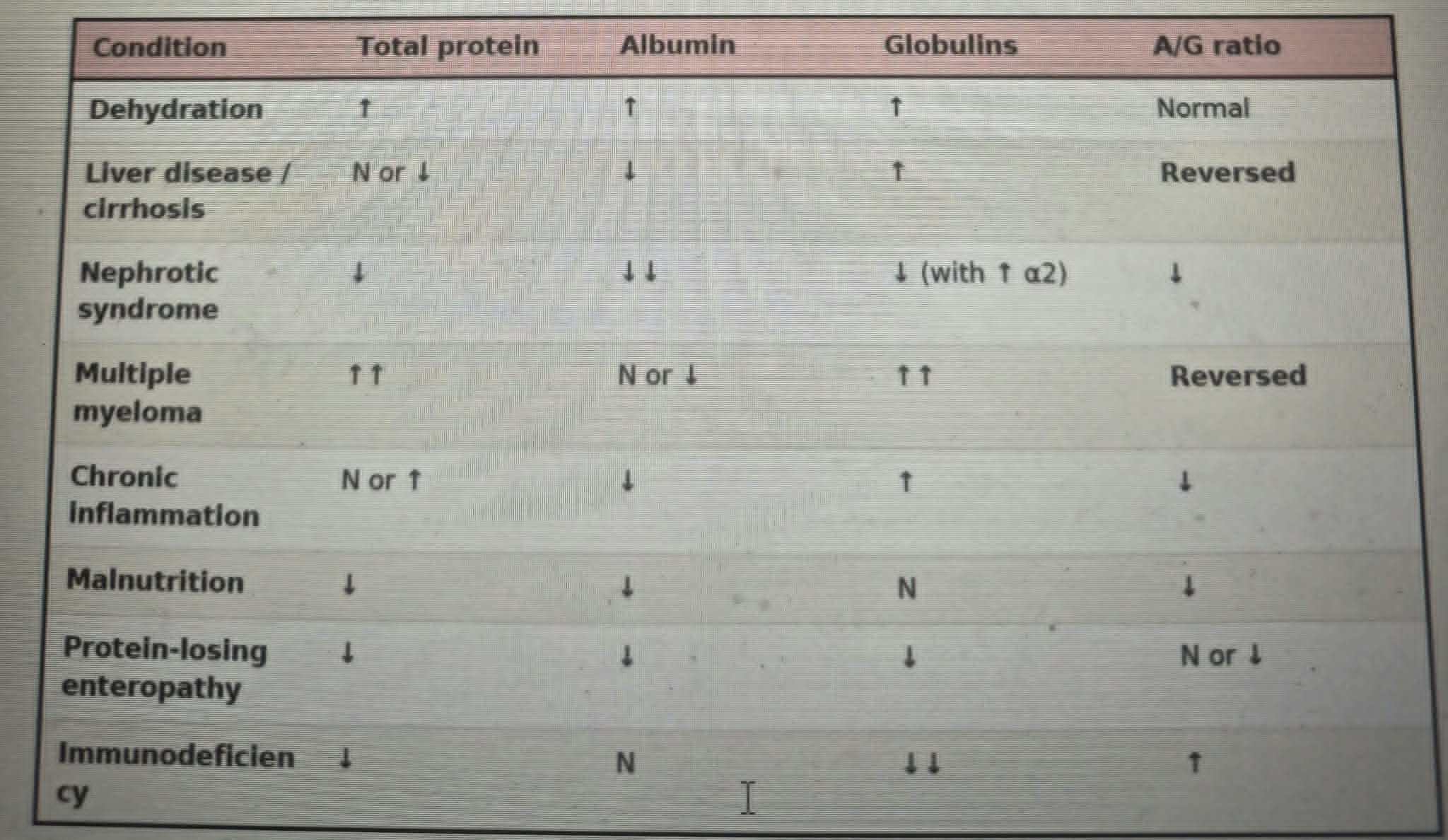
○ Positive acute phase reactants: CRP and Haptoglobin. During inflammation or infection positive acute phase reactants increase. CRP and haptoglobin increases. During inflammation or infection, negative acute phase reactants decrease. Examples: Albumin and Prealbumin
○ No. The low albumin in this case does not indicate malnutrition. Albumin decreases because it is a negative acute phase reactant.
○ The rapid fall is not due simply to reduced synthesis. Possible mechanisms include:
1. Redistribution
○ Increased capillary permeability moves albumin into the interstitium.
2. Dilution
○ IV fluids can dilute albumin. If CRP rises in just 3 days after surgery:
■ This can be assumed/considered most probably a post-operative infection.
■ There may be infection at the operation site.
CASE 4: A 55-year-old woman is 3 days postoperative with fever and rising white cell count.
Questions:
1. Identify the positive and negative acute-phase reactants here.
2. Does the low albumin indicate malnutrition?
3. Could albumin have fallen this fast through
reduced synthesis alone?

(inflammation) (mask)
● Mechanism: cytokine-driven (IL-6) reprioritisation of
hepatic protein synthesis - the acute-phase
response
● UP ↑ CRP (first and largest) • ↑ fibrinogen • ↑
haptoglobin • α1-antitrypsin • ↑ ceruloplasmin
● Down ↓ albumin • ↓ prealbumin • ↓ transferrin
● Electrophoresis: ↓ albumin with ↑ α1 and α2 bands
● Two traps to avoid:
○ Low albumin/prealbumin here means
(_________), not necessarily malnutrition -
check CRP
○ A raised acute-phase protein can (____) an
underlying deficiency (α1-antitrypsin,
ceruloplasmin, haptoglobin)
Globulin rises and offsets
the fall in albumin. ↓ Albumin, ↑ Globulin, Total
protein may remain normal. Dysproteinemia
abnormal distribution with a normal sum. This is
why albumin/globulin ratio and serum protein
electrophoresis
Immunoglobulins are produced by plasma cells. They are not produced by the liver. Therefore, liver failure does not affect immunoglobulin production in the same way and gamma globulins can increase.
The liver synthesizes
clotting factors. Liver failure causes reduced
synthesis of clotting factors and prolonged
PT/INR. This causes easy bruising and bleeding
CASE 5:
A 58-year-old man with a long history of alcohol use
presents with abdominal distension and easy bruising.
Questions:
1. Why is the total protein normal despite a
markedly low albumin?
2. Why are the globulins raised if the liver is failing?
3. What causes the bruising?
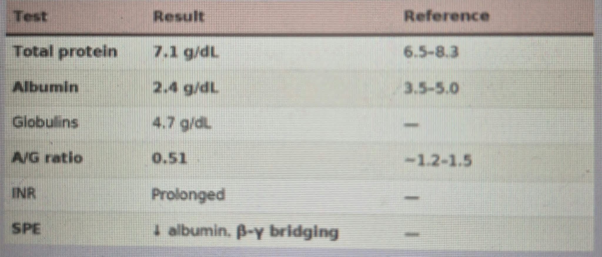
LIVER DISEASE
● Mechanism: the liver synthesizes albumin, clotting
factors, and most transport proteins. Hepatocellular
failure reduces synthesis of all of them
Clinical features: edema (swelling of the legs), ascites
(accumulation of fluid and causes abdominal distension),
bruising, bleeding, and jaundice (yellowing of the skin due to
accumulation of bilirubin because of decreased transport
proteins).
(Wilson’s disease) ( Alpha-1 antitrypsin deficiency)
● (________)
○ Genetic disorder.
○ Due to failure of a transport protein.
○ Specifically involves transport of copper.
○ Results in extra copper in the body.
●(_______________________)
○ Absent or faint alpha-1 band on electrophoresis.
○ Leads to emphysema, alveolar dysfunction, and liver disease