Biology: The Cardiovascular System
1/78
Earn XP
Description and Tags
Chapter 10
Name | Mastery | Learn | Test | Matching | Spaced | Call with Kai |
|---|
No analytics yet
Send a link to your students to track their progress
79 Terms
Corinary Andiogram (Aka. Cardiac Catheteristation)
A procedure where a special dye is injected into someone to see how well the dye moves through your arteries (Heart veins). X-ray images are then taken to observe the dye moving through the arteries.
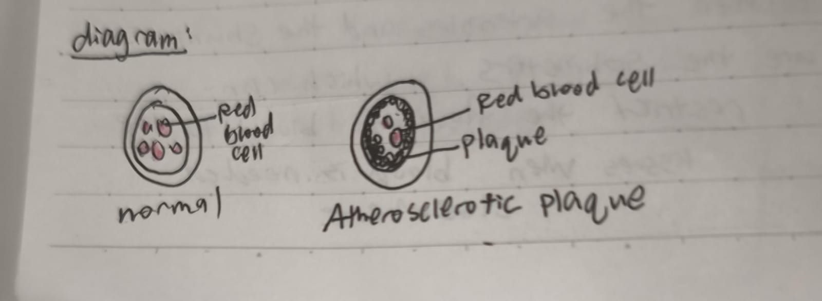
Atherosclerosis
The build-up of plaque (Hardened fats). It could cause a blockage in the artery. (Dangerous if present in the coronary artery, which leads to a heart attack.)
What is the purpose of a cardiovascular system?
To move blood around the body to help tissues get enough oxygen and nutrients (Glucose) and to get rid of waste products (CO2)
3 types of blood vessels
In order:
Arteries
Capillaries
Veins
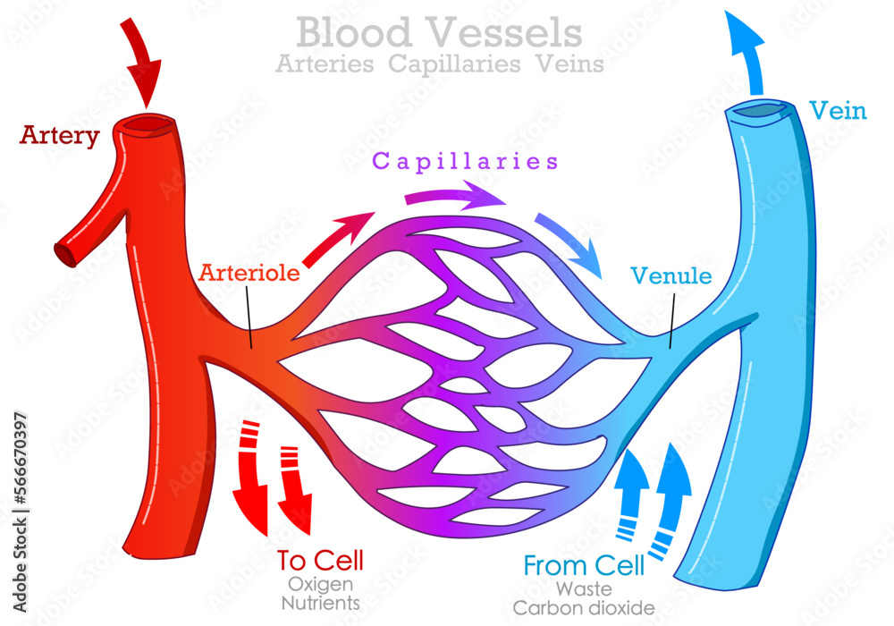
What is the cycle of blood flow?
Heart → Arteries → Capillaries → Veins → Heart
Flow from the heart:
Carry blood AWAY from the heart. Then it brings it to smaller ARTERIOLES.
In tiny CAPILLARIES, blood exchanges gases with the surrounding tissues.
Flow to the heart:
After dropping off O2 and picking up CO2, the capillaries collect blood into larger VENULES.
Venules collect into larger VEINS, which return the blood to the heart.
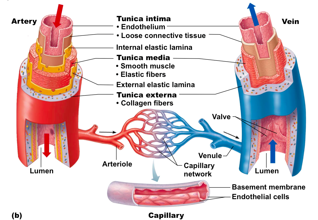
What are the structural differences between veins, arteries, and capillaries?
Capillaries only have ONE CELL LAYER, allowing for EASY GAS EXCHANGE.
Arteries have thick, muscular walls to handle high pressure
Veins have thinner walls with valves to prevent backflow
Each type of blood vessel is structurally adapted to its specific role in the circulatory system.
Arteries and Veins both have 3 layers. What are they?
Tunica Intima - Slippery, THIN layer
Tunica Media - THICKER layer of muscle.
Tunica Externa - Aka. Tunica adventitia, fibrous CONNECTIVE TISSUE to SUPPORT and PROTECT.
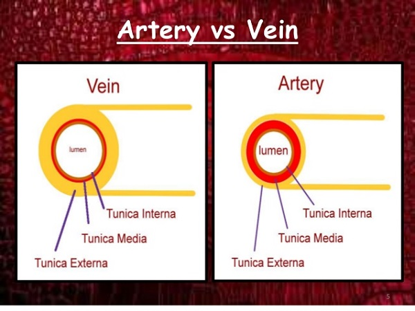
Artery vs. Vein (What are their differences?)
The artery’s Lumen (Hollow cavity inside a vein) is smaller than the lumen in the vein.
The walls of the artery is thicker than the walls of the vein.
ARTERY: THICC WALLS, especially in TUNIVA MEDIA LAYER (MOOSCULAR LAYER). Receive blood from the heart under HIGH PRESSURE, so they NEED TO BE FLEXIBLE!
VEIN: THIN walls, receive blood FROM TISSUES under LOW PRESSURE.
Veins: 3 ways of maintaining blood flow even under low pressure:
Muscular pumping: As muscles contract, blood is squeezed through the veins.
Respiratory pumping: a mechanism to pump blood back to the heart using inspiration.
Valves: LARGER veins have VALVES to PREVENT BACKFLOW of blood. (Blood only flows in one direction.)
How do valves open / close? Also, why is the backflow of blood a bad thing?
Valves open when muscles CONTRACT, close when muscles relax. (Open = contract, Close = relax)
Backflow prevents blood from flowing forward as it should.
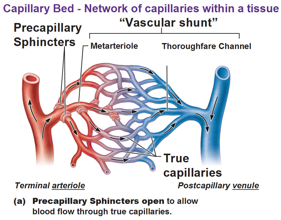
What is the primary function of the precapillary sphincters? Using this info, explain why it is beneficial for it to close during cold temperatures.
Precapillary sphincters: Tiny rings of smooth muscle located at the entrance to the capillaries
Control blood flow into true capillaries.
If open → blood flows INTO the true capillaries.
Closed → blood bypasses the capillaries.
When it’s cold, precapillary sphincters close to reduce blood flow to the skin’s surface. This prevents heat loss by keeping warm blood deeper in the body, helping you conserve body heat and maintain core temperature.
What is the shunt (Thoroughfare channel)? (Hint: In the capillary)
Direct pathway connecting the arteriole to the venule.
Allows blood to bypass the true capillaries.
Used when tissues don’t need much blood supply (During rest or in cold temps to conserve heat.)
No exchange happens here — it’s just a shortcut for blood when tissues don’t need much supply.
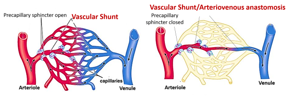
What are the true capillaries
Small blood vessels where exchange of gases, nutrients, and wastes occurs between blood and tissues.
Blood flows through them when the precapillary sphincters are OPEN.
How do precapillary sphincters help regulate the delivery of blood through the capillaries?
Precapillary sphincters control which capillaries receive blood.
When open, blood flows into true capillaries ➝ tissues get oxygen and nutrients.
When closed, blood bypasses the area through the shunt ➝ less blood goes to that tissue.
This helps the body prioritize blood flow to where it’s most needed (e.g., muscles during exercise, digestive system after eating).
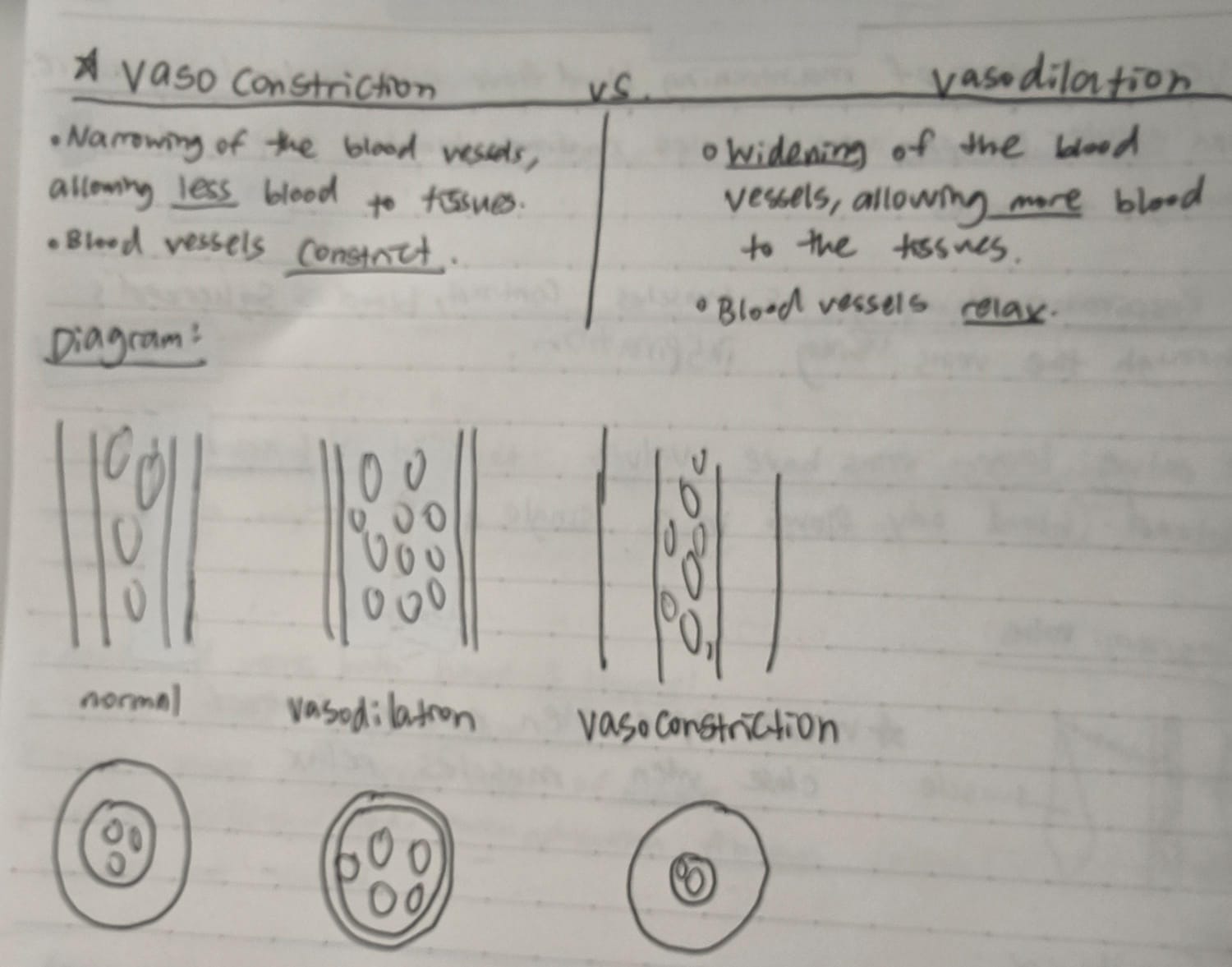
Vasoconstriction Vs. Vasodilation
Vasoconstriction: NARROWING of the blood vessels, allows for LESS blood TO TISSUES. Blood vessels CONSTRICT.
Vasodilation: WIDENING of the blood vessels, allows for MORE blood to the tissues. Blood vessels RELAX.
Do vasoconstriction and vasodilation cause problems? How?
Pros:
Vasoconstriction: Normal when used appropriately like to CONSERVE HEAT IN COLD WEATHER, or REDIRECT BLOOD to important organs DURING STRESS (Fight or flight)
Vasodilation: normal after eating (Increases blood flow to digestive organs), during exercise, to release excess heat (Cool the body)
Cons:
Vasoconstriction: High blood pressure (Narrow vessels increase resistance to blood flow.), Reduced oxygen delivery, less blood reaching the fingers and toes (Causing coldness), organ damage.
Vasodilation: Low blood pressure, dizziness or fainting, dangerous drop in blood pressure, blood pooling in legs (Making it harder to return blood to the heart)
Describe the dangers of Atherosclerosis.
Can lead to high blood pressure, blood clots in the blood vessels, and heart attacks.
Medication for Atherosclerosis
Oral antiplatelet therapy (To prevent platelets from sticking together, beta blockers (Helps lower your heart rate and blood pressure).
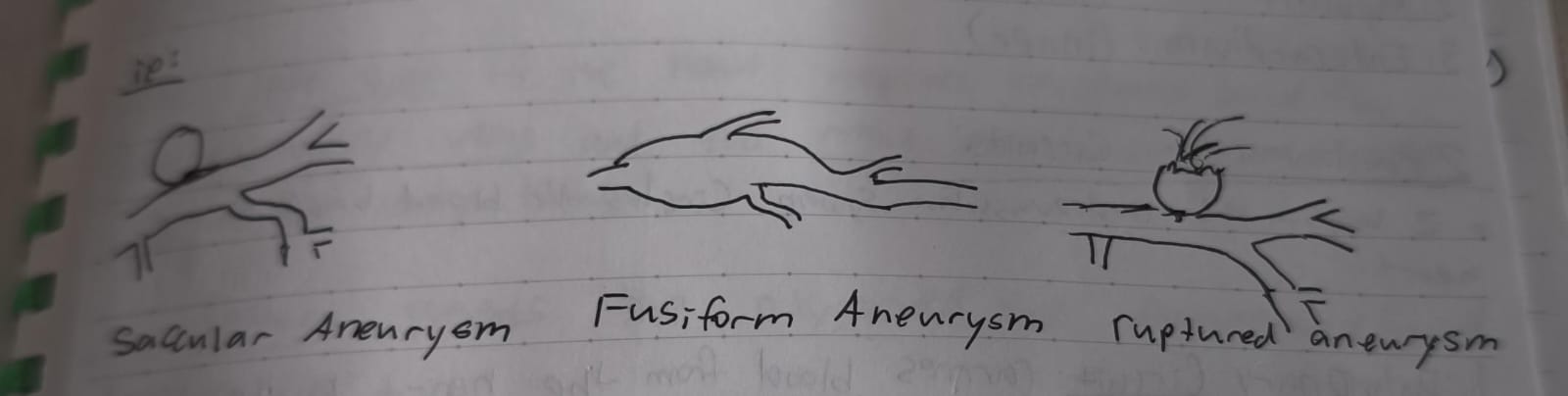
Aneurysm
A bulge in the weakened wall of a blood vessel (Usually happens in an artery).
The thinner wall offers less support and the tissue eventually ruptures. Less oxygen and nutrients are delivered to the tissues.
What is the difference between saccular, fusiform, and ruptured aneurysm?
Saccular: Aka. Berry aneurysm. Small, rounded bulge on one side of the blood vessel. Often found in the BRAIN. Could lead to bleeding or a stroke.
Fusiform: Uniform widening of the entire circumference of the blood vessel. Found in larger ARTERIES like the aorta. May weaken / rupture the vessel over time.
Ruptured: When ANY TIME OF ANNEURYSM breaks open, causing internal bleeding. Causes strokes, hemorrhage (Internal bleeding), organ failure, or death depending on the location.
Ways to treat an aneurysm?
Doctors could put a special cord into the aneurysm then a type of string is pushed into the aneurism to clog it and stop it, OR they could clip the aneurysm to stop the blood flow to it.
What is the apex of the heart?
The “tip” of the heart that is pointed towards the left hip.
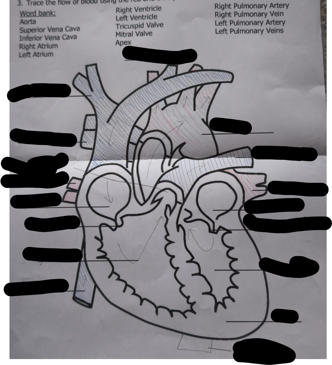
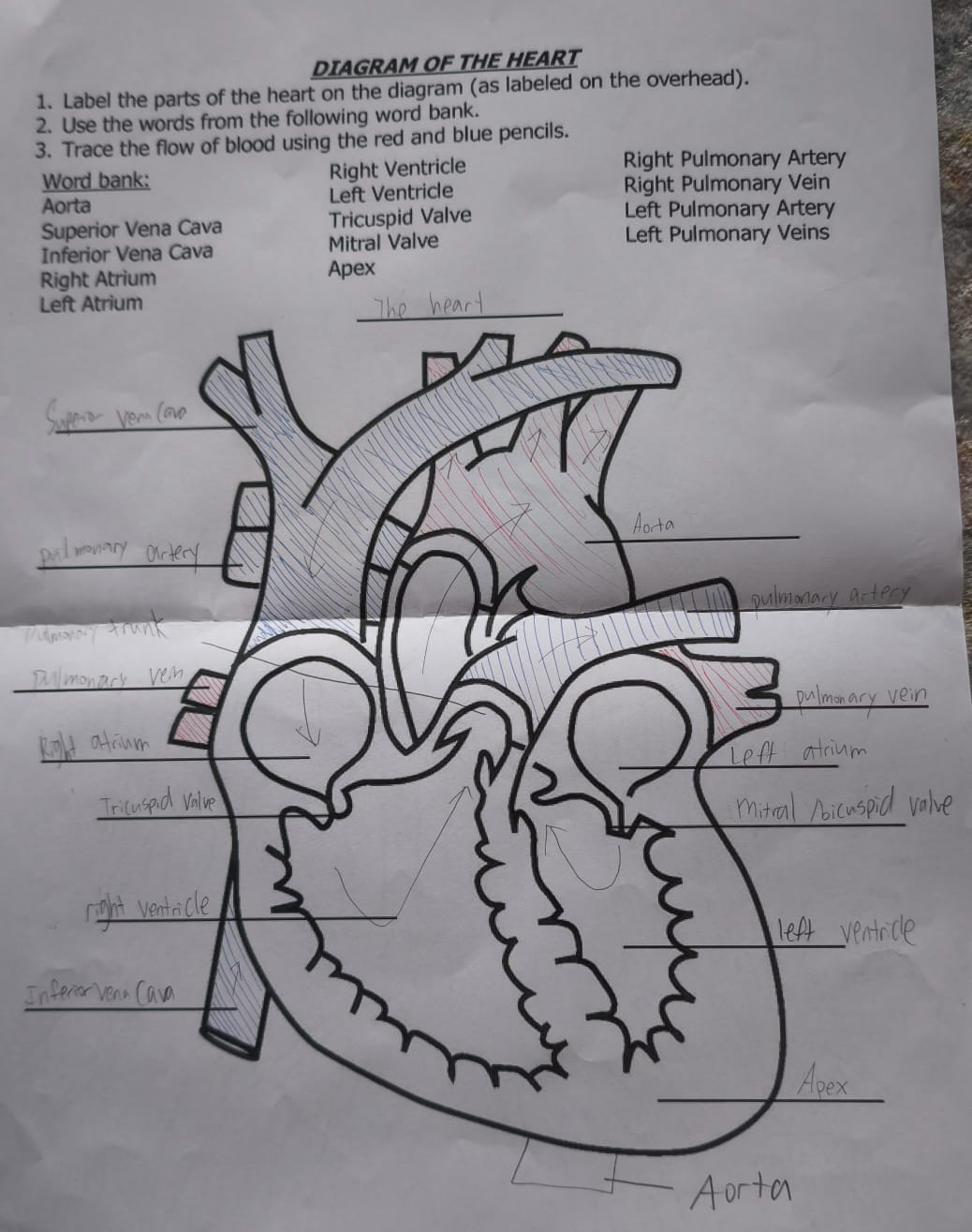
Label the parts of the heart.
Answer:
What’s the difference between the superior vena cava and inferior vena cava? Where do they bring blood, and what kind of blood do they carry?
Both carry deoxygenated blood into the right atrium, but from different parts of the body.
Superior vena cava: from the upper body (head, arms)
Inferior vena cava: from the lower body (legs, abdomen)
The right atrium and left atrium are both upper chambers of the heart. What kind of blood does each receive, and where does it come from?
Right atrium receives deoxygenated blood from the body via the vena cavae
Left atrium receives oxygenated blood from the lungs via the pulmonary veins
Compare the right ventricle and left ventricle in terms of function and strength. Why is one thicker than the other?
Right ventricle pumps deoxygenated blood to the lungs (short distance)
Left ventricle pumps oxygenated blood to the entire body (long distance)
→ The left ventricle has a thicker muscular wall because it needs more force to push blood all the way around the body.
What do the tricuspid valve and mitral valve have in common, and how do they differ in position and function?
Both are atrioventricular valves that prevent backflow of blood.
Tricuspid valve is on the right side, between right atrium and right ventricle
Mitral (bicuspid) valve is on the left side, between left atrium and left ventricle
Pulmonary arteries vs. pulmonary veins — what’s the key difference, and what’s unique about these vessels compared to most arteries and veins?
Pulmonary arteries (left and right) carry deoxygenated blood from the right ventricle to the lungs
Pulmonary veins (left and right) carry oxygenated blood from the lungs to the left atrium
➤ Unlike most arteries and veins, the pulmonary arteries carry deoxygenated blood, and the pulmonary veins carry oxygenated blood — opposite of the usual rule!
What is the function of the aorta, and how does it differ from the pulmonary trunk?
The aorta carries oxygenated blood from the left ventricle to the body.
The pulmonary trunk carries deoxygenated blood from the right ventricle to the lungs.
➤ They are both major arteries, but serve opposite sides and functions in circulation.
What is the apex of the heart, and what chamber forms most of it? Why is its position important?
The apex is the pointed tip at the bottom of the heart, mostly made by the left ventricle.
It points down and to the left — this helps orient doctors when placing stethoscopes or interpreting heart sounds.
What are the 3 layers of the heart?
Muscle tissues:
Epicardium (outer)
Myocardium (middle)
Endocardium (inner)
There are 2 cardiovascular circuits. What are they and what do they do?
The two loops in the cardiovascular system
Pulmonary circuit: carries blood from the HEART to LUNGS and BACK. Picks up O2 and releases CO2.
Systemic circuit: carries blood from the HEART to the TISSUES and back. Drops off O2 and picks up CO2.
Which blood vessels play a role in the pulmonary circuit?
Major blood vessels attached to the heart: Pulmonary trunk, pulmonary veins (pulmonary veins collect oxygen-rich blood from your lungs and carry it to your heart.)
Which blood vessels play a role in the systemic circuit?
Major blood vessels attached to the heart are: Vena cavae (Superior and inferior) (Right side of the heart), and Aorta (Left side of the heart.)
Aorta → Arteries → Arterioles → Capillaries → Venules → Veins → Vena Cavae → Right Atrium
Tricuspid valve vs Bicuspid / mitral valve?
“Tri” = 3 flaps → Tricuspid = Right side
“Bi” = 2 flaps → Bicuspid/Mitral = Left side
→ Think: "Try to do what's right" (Tricuspid = Right side)
Why do arteries carry deoxygenated blood in the heart, but oxygenated blood in the rest of the body?
Arteries = carry blood away from the heart
Veins = carry blood toward the heart
It’s not about oxygen content — it’s about direction of blood flow.
Flow of blood (Deoxygenated ←→ Oxygenated)
Deoxygenated Blood (low oxygen)
Comes from body
→ Superior & Inferior Vena Cava
→ Right Atrium
→ Tricuspid Valve
→ Right Ventricle
→ Pulmonary Valve
→ Pulmonary Arteries
→ Lungs (to pick up oxygen)
Oxygenated Blood (rich in oxygen)
Comes from lungs
→ Pulmonary Veins
→ Left Atrium
→ Mitral (Bicuspid) Valve
→ Left Ventricle
→ Aortic Valve
→ Aorta
→ Body (to deliver oxygen)
What do the 4 chambers of the human heart consist of?
2 atria (Aka atrium) on top to receive blood
2 ventricles on the bottom to pump blood back out.
The left side of the heart receives oxygenated blood from the pulmonary veins, and the right side receives deoxygenated blood from the body tissues.
What separates deoxygenated / oxygenated blood in the heart?
The septum prevents the oxygenated and deoxygenated blood from mixing.
Why is the left side of the heart bigger than the right?
Because the left side of the heart pumps blood to your entire body, while the right side only pumps blood to your lungs.
How does the heart make sure blood is pumped in one direction?
Valves. They are flaps of tissue that prevent blood from flowing backwards (Like a set of swinging doors).
What is the direction of blood flow in the heart?
Top → bottom
Right → left
What is the atrioventricular valves?
Valves between the atria and ventricles
They prevent backflow of blood when the ventricles contract
2 AV valves: Tricuspid valve + mitral (bicuspid) valve
How they open and close:
Open when atria contract, allowing blood into the ventricles
Close when ventricles contract, stopping blood from flowing back into the atria
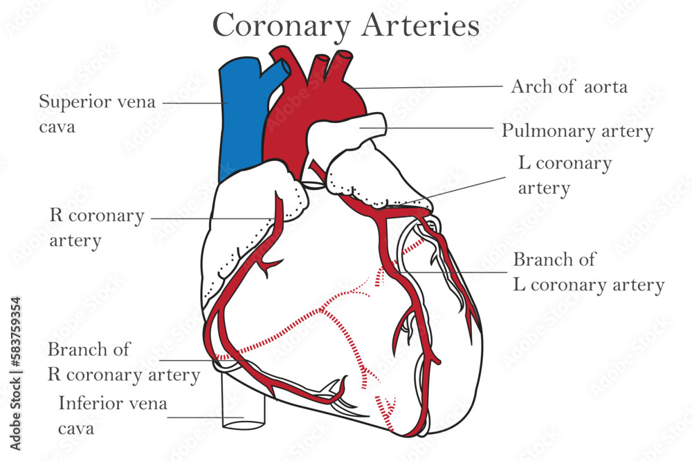
How does the heart itself get an oxygen supply?
Through the coronary artery, an artery that supplies the cardiac muscle with oxygen and nutrients.
If a coronary artery gets blocked, the heart muscle doesn’t get oxygen → can cause a heart attack (myocardial infarction)
That's why they're often checked during heart disease diagnosis or bypass surgery
“Coronary = crown” — these arteries wrap around the top of the heart like a crown to keep it nourished.
often embedded in a layer of fat called epicardial fat (found on the outer layer of the heart).
What happens when the heart does not receive enough oxygen?
Angina (Chest pain) could occur when too little oxygen reaches the heart due to blocked coronary arteries. (Plaque buildup inside the artery is a bad sign.)
SOLUTION: Coronary bypass: Surgery for increasing blood flow to the heart.
Why is there fat around the heart?
This fat cushions and protects the coronary arteries
the heart can use fatty acids as fuel — in fact, fatty acids are the heart’s main energy source at rest!
But the fat around the heart is not stored “for emergencies” like body fat — it’s more protective and structural
Too much fat around the heart (especially in obesity) can actually be harmful, increasing the risk of heart disease
How does deoxygenated blood flow through the heart? (Hint: right)
From body tissues → superior vena cava and inferior vena cava → right atrium → right ventricle → pulmonary arteries → lungs (to pick up more O2 and drop off CO2)
How does oxygenated blood flow through the heart? (Hint: left)
From lungs → pulmonary veins → left atrium → left ventricle → aorta (Drop off oxygen to the rest of the body)
How does the intrinsic conduction system regulate the heart?
It sets the pace for heart contractions. It is composed of special tissues able to generate their own impulse.
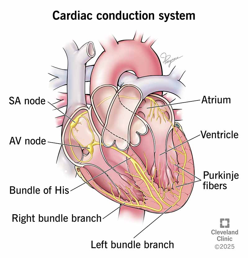
What is the intrinsic conduction system composed of? (In order)
The sinoatrial (SA) node: begins the impulse, causing the contraction of the atria. The SA node is AKA. the CARDIAC PACEMAKER.
The impulse then reaches the atrioventricular node (AV). This node PAUSES, letting the atria empty to the ventricles.
The impulse continues into the AV bundle and the bundle branches to the PURKINJE FIBERS, which contract and the ventricles sending blood out of the heart.
AV bundle VS. Purkinje Fibers
Purkinje Fibers
Tiny nerve-like fibers in the inner walls of the ventricles
Spread out from the bottom of the heart (apex) upward along the ventricle walls
Function:
Deliver the electrical impulse to the ventricular muscle cells
Cause the ventricles to contract from the bottom up ➝ pushing blood up into the arteries
Think of them as the final messengers that say “now squeeze!”
AV Bundle (Bundle of His)
A bundle of conducting fibers that carries the electrical signal from the AV node
Travels through the septum (wall between left and right ventricles)
Function:
Acts as a highway for the signal to move from the atria ➝ ventricles
Splits into right and left bundle branches, which lead to the Purkinje fibers
What is a diastole and a systole?
The heartbeat is one contraction and relaxation of the heart muscle.
Diastole: Relaxation of the heart muscle + FILLING of blood
Systole: Contraction of heart + pumping blood out. (Aortic valve = open)
Our heart rate is usually about 75 times per minute
Stages of Diastole
Atrial contraction (Ventricles fil with blood)
AV valves close, making the first heart sound “lub”
Stages of Systole
ISOVOLUMETRIC CONTRACTION: ventricles are contracting with all valves closed.
Ejection phase: Pressure in the ventricles build until the semilunar valves open and blood is released into the blood vessels. BLOOD is also refilling the atria in this phase.
ISOVOLUMETRIC relaxation: Ventricles relax with all the valves closed.
Diastole begins again and the cycle repeats.
What does blood pressure measure?
Measures the amount of pressure in artery.
Two parts (120/80)
Systolic Vs Diastolic pressure
Systolic: High number (120 mmHg), pressure in artery when ventricles CONTRACT.
Diastolic: Low number (80 mmHg), pressure in artery when ventricles relax. MOST IMPORTANT IN DIAGNOSIS OF HEART HEALTH.
How is blood pressure measured?
Measured using Sphygmomanometer.
How can you tell if blood pressure is too high?
Any # (Systolic / Diastolic) over 120/80 is a sign of high blood pressure (Bad)
Cardiac output
Amount of blood pumped by each ventricle in ONE MINUTE.
Calculated by this formula: CARDIAC OUTPUT = HEART RATE x STROKE VOLUME (Stroke volume: volume of blood pumped from the ventricle per beat.)
Stroke volume
The volume of blood PUMPED OUT by a ventricle in a heartbeat.
Heart rate
# of times a heart beats per minute.
How can the stroke volume change?
Exercise (Additional blood return from muscles brings in more volume to the ventricles.
Rapid blood loss: Decreases blood returning to the heart.
Heart rate can change by?
Stress, hormones (Epinephrine and thyroxine), ions (electrolyte imbalances affect the heart’s ability to contract), physical factors (Age, overall health, body temp)
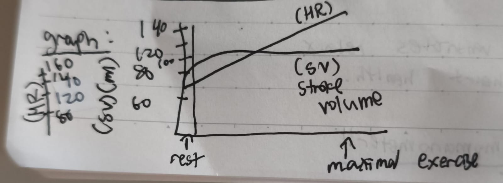
How do heart rate and stroke volume change with exercise?
Heart rate steadily increases while stroke volume increases but plateau after a certain point.
Blood pressure depends on 2 factors. What are they?
Cardiac output (Increase in cardiac output → increase in blood pressure)
Arteriolar resistance (Constriction → higher blood pressure.) (Increased blood volume → higher blood pressure) (Vasoconstriction / Vasodilation)
Hypertension
High blood pressure
Cause: Increased resistance to blood flow
Hypertension leads to…
Weakened / ruptured blood cells.
Body compensates by increasing support to connective tissues which leads to hardened arteries. (Heart pumps harder, blood does not easily flow through vessels)
Preventions for hypertension?
Reduce salt to less than 5g daily.
Eat fruits and vegetables regularly.
Avoid saturated fats and trans fats.
Avoid tobacco
Reduce alcohol
How does your body maintain a stable temperature? (Hint: Thermoregulation)
High temp → hypothalamus turns on cooling systems → Skin blood vessels dilate via arterioles (Increased blood flow to skin) / sweat glands initiate sweating; evaporation of sweat → body temp decreases
OR
Low temp → hypothalamus turns on warming systems → skeletal muscles contract; shivering generates heat (Uses atp) → body hair erects to conserve heat. → body temp increases: Hypothalamus turns off warming systems.
Arterioles
Arterioles are small blood vessels that branch off from arteries
They are the last small branches before capillaries
Control blood flow into capillary beds
Act like "pressure valves" — they regulate blood pressure by tightening or relaxing
Can vasoconstrict (narrow) or vasodilate (widen) to direct blood where it’s needed
Increased # of red blood cells = ???
Increase efficiency in oxygen delivery.
Capillaries
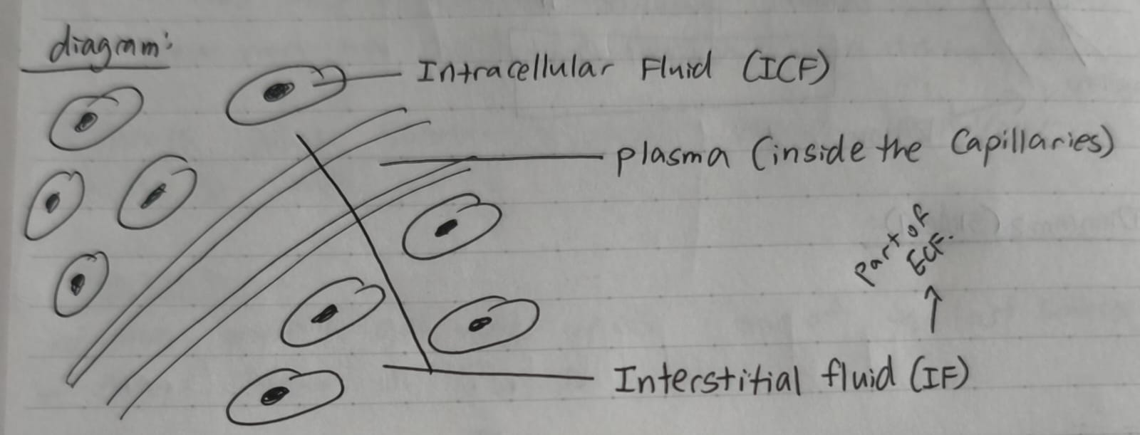
Allow for the exchange of nutrients as well as wastes (Between the blood and the SURROUNDING extracellular fluid (ECF/IF) (The IF is a type of ECF)
Oxygen, nutrients, ions, water into body cells.
Metabolic wastes like CO2 LEAVE the ECF and ENTER the CAPILLARIES.
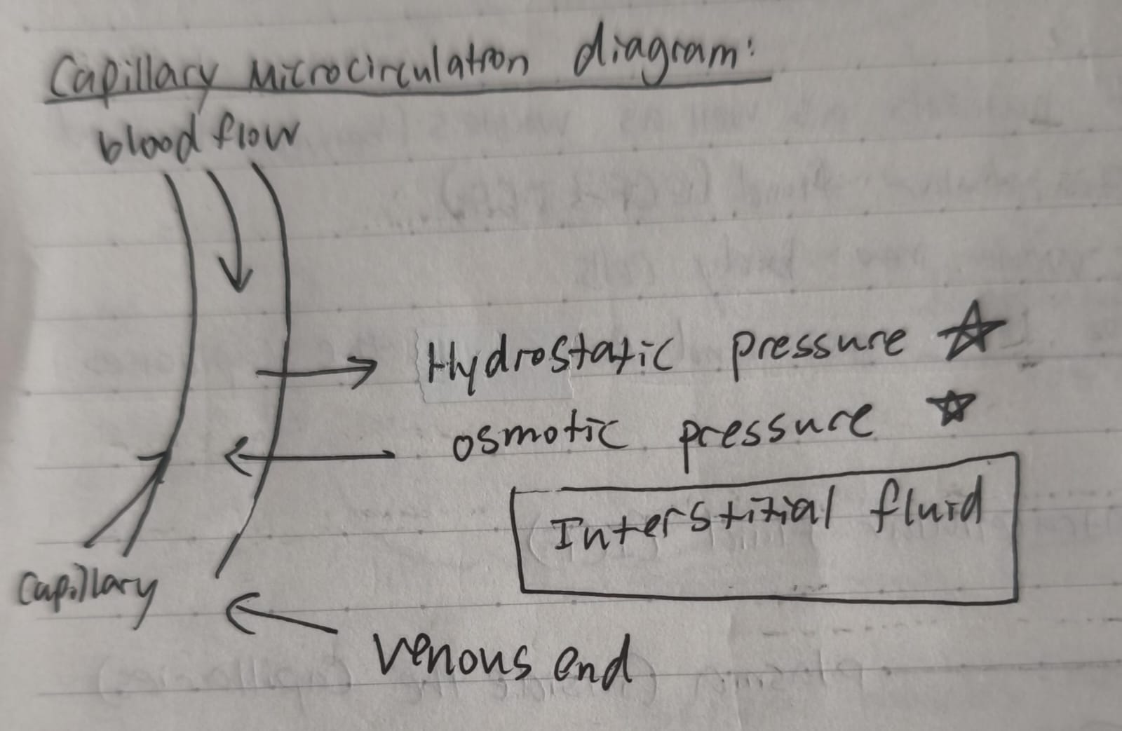
2 forces that regulate the movement of water (Between blood and ECF) (Hint: Capillaries)
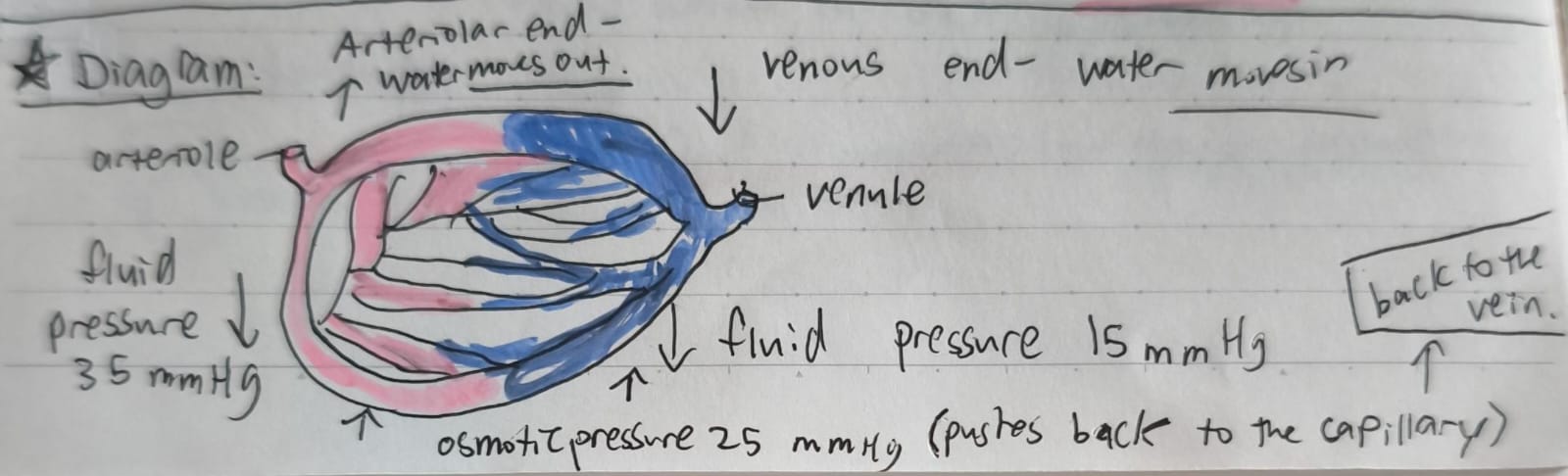
Fluid pressure (In the body, it’s mostly the blood pressure inside capillaries. It pushes water and small solutes out of the capillaries into the surrounding tissue. Higher at the arteriole end, lower at the venule end)
Osmotic pressure (Water moves from low solute to high solute areas to balance concentration. Pulls water back into the capillaries from the surrounding tissue.)
THIS IS DONE SO THAT TISSUES WON’T SWELL.
Summary:
Water moves from high fluid pressure (capillary) to an area of low fluid pressure (ECF)
Proteins and dissolved minerals in blood cause fluid from the ECF to move into the blood by osmosis.
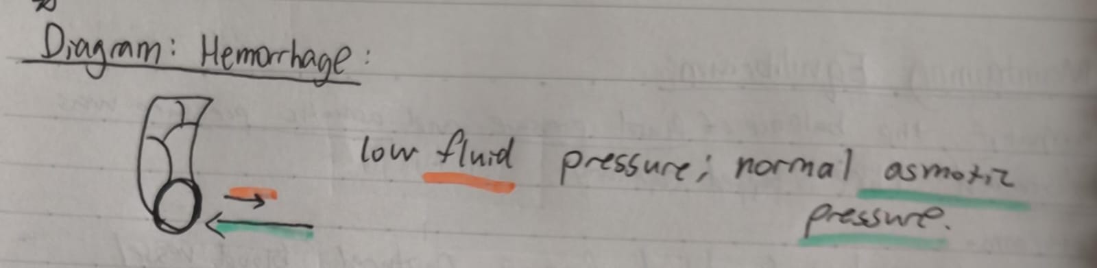
What happens if the balance of fluid pressure and osmotic pressure was disturbed? (Like a popped capillary)
Hemorrhage: Blood loss from a ruptured blood vessel.
ie. (Decrease in blood volume = decrease of fluids in blood vessels. → force that drives fluids from capillaries diminishes but osmotic pressure remains the same. → force drawing water into the capillaries is greater! As water moves into capillaries, fluid volume is eventually restored.)
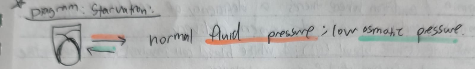
Why does starving yourself sometimes lead your belly to swell?
Plasma proteins get used up as one of the last sources of energy = decrease in plasma proteins → decrease in osmotic pressure = decrease in reabsorption of fluids into blood. → more water in ECF = swelling
lymphatic system
Lymphatic system: network of vessels, nodes, and organs that helps drain excess fluid (called lymph)
Filter pathogens and waste through lymph nodes
Support the immune system by housing white blood cells (like lymphocytes)
What is the lymphatic system made of?
Lymph (Clear fluid that carries waste, immune cells, and fats, comes from excess interstitial fluid).
Lymphatic Vessels (Veins, but carry lymph instead of blood to transport lymph from tissues to blood stream).
Lymph nodes (Small bean shaped filters that destroy pathogens and cancer cells and house white blood cells.
Organs:
Structure | Function |
|---|---|
Spleen | Filters blood, removes old red blood cells, stores white blood cells |
Thymus | Where T cells mature (especially active in children) |
Tonsils & Adenoids | Trap pathogens from food and air |
Bone Marrow | Makes lymphocytes (WBCs) |
Lymphoid organs
Spleen: stores and purifies blood
Thymus gland: Where lymphocytes mature.
Lymphocytes: Help the body’s immune system fight cancer and other foreign viruses and bacteria. (A type of white blood cell.)
Papillary muscles
Their primary function is to anchor the chordae tendineae (heart strings) and prevent the inversion or prolapse of the atrioventricular (AV) valves (tricuspid and mitral/bicuspid valves) during ventricular contraction (systole).
Valve Stability: Contract synchronously with the ventricles to keep the AV valves tightly closed, preventing backflow of blood into the atria.
Force Distribution: Transmit tension from the chordae tendineae to the valve leaflets, ensuring efficient sealing.
If papillary muscles are damaged (e.g., by a heart attack), the valves may leak (regurgitation), leading to inefficient blood flow and potential heart failure.