A&P/2 LAB 1: Anatomy of the Eye & Ear
 Call Kai
 Learn
Learn Practice Test
 Spaced Repetition
 Match
Match Flashcards
 Knowt Play
1/55

There's no tags or description
Looks like no tags are added yet.
Name | Mastery | Learn | Test | Matching | Spaced | Call with Kai | Chat |
|---|
No analytics yet
Send a link to your students to track their progress
56 Terms
Fibrous Tunic
Outer layer of the eye, consisting of the sclera and cornea. Their main functions are to provide shape to the eye and support the deeper structures
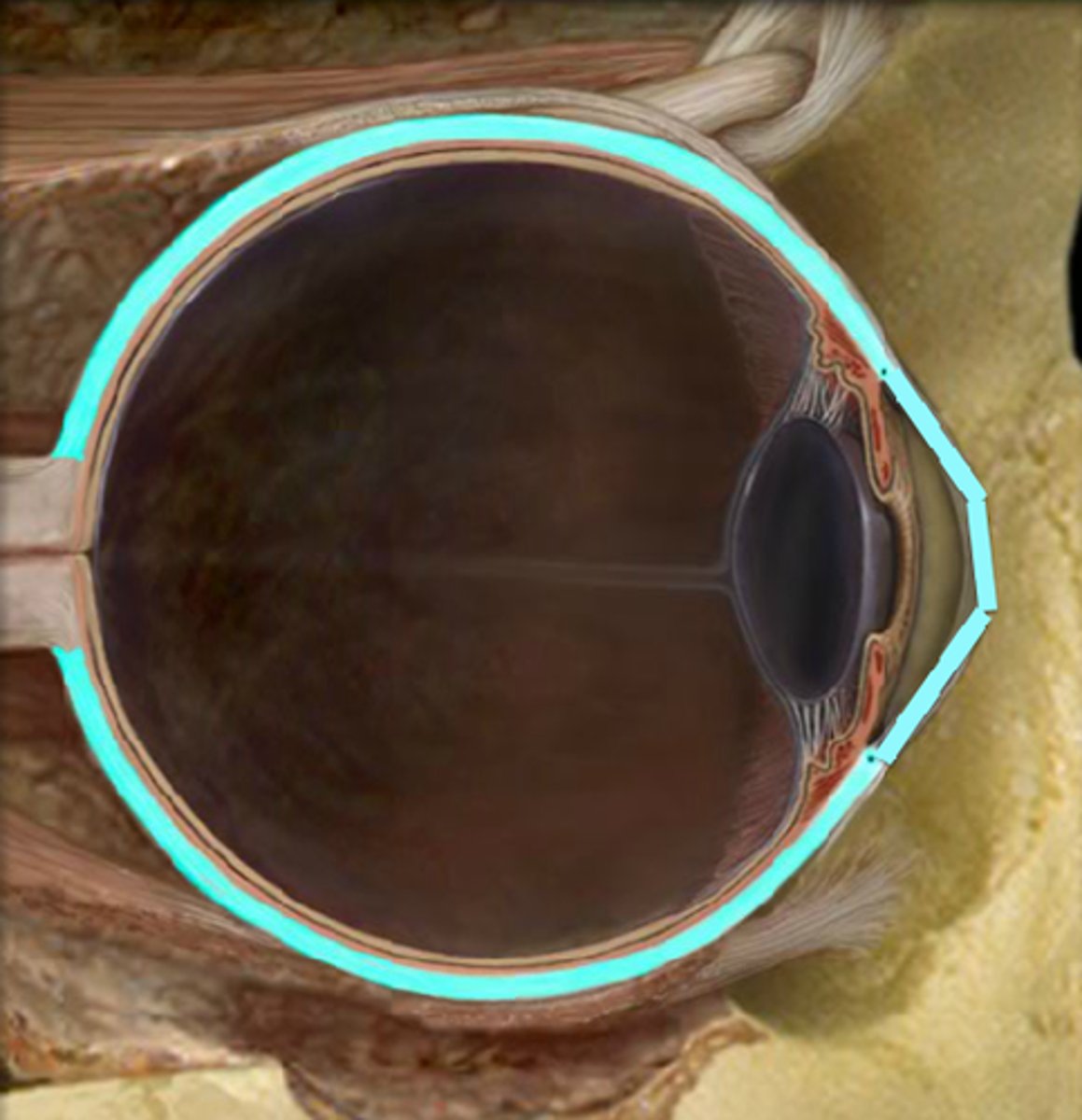
Sclera
White of the eye. Provides protection and form.
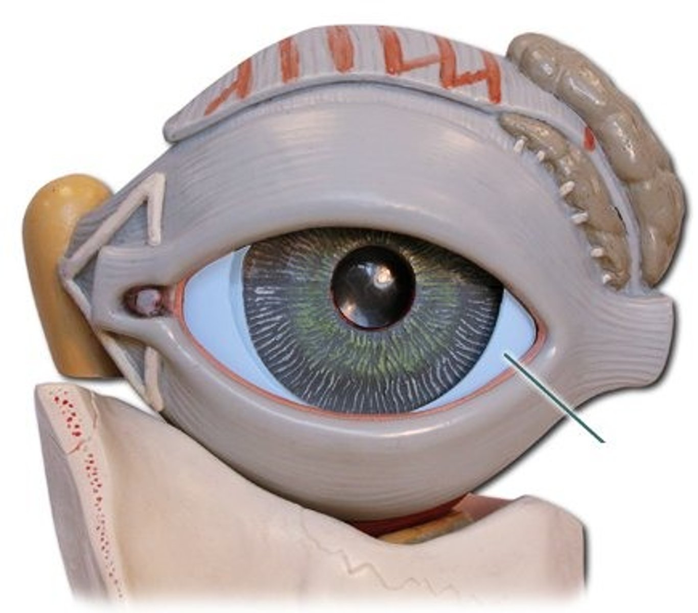
Cornea
The transparent layer forming the front of the eye. It functions like a window that controls and focuses the entry of light into the eye.
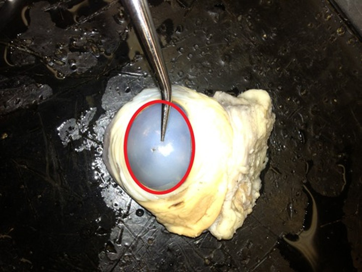
Vascular tunic
Middle layer consisting of choroid, ciliary body, iris. Its function is to provide nourishment to the outer layers of the retina through blood vessels. It is part of the uveal tract.
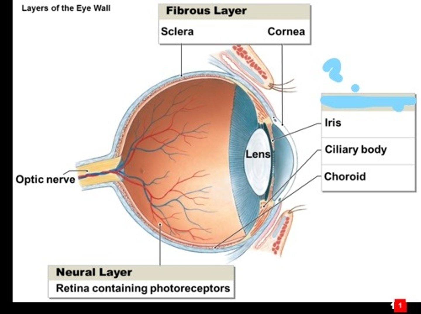
Choroid
middle, vascular layer of the eye, between the retina and the sclera. provides oxygen and nourishment to the outer layers of the retina.
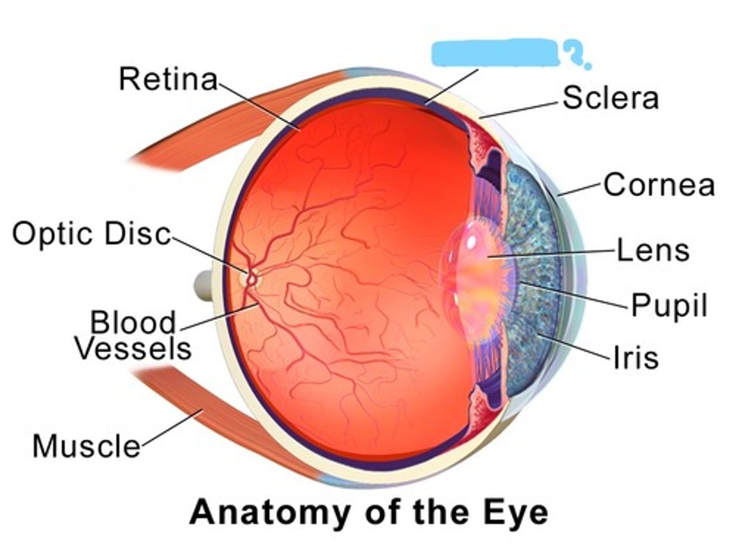
Iris
A ring of muscle tissue that forms the colored portion of the eye around the pupil and controls the size of the pupil opening
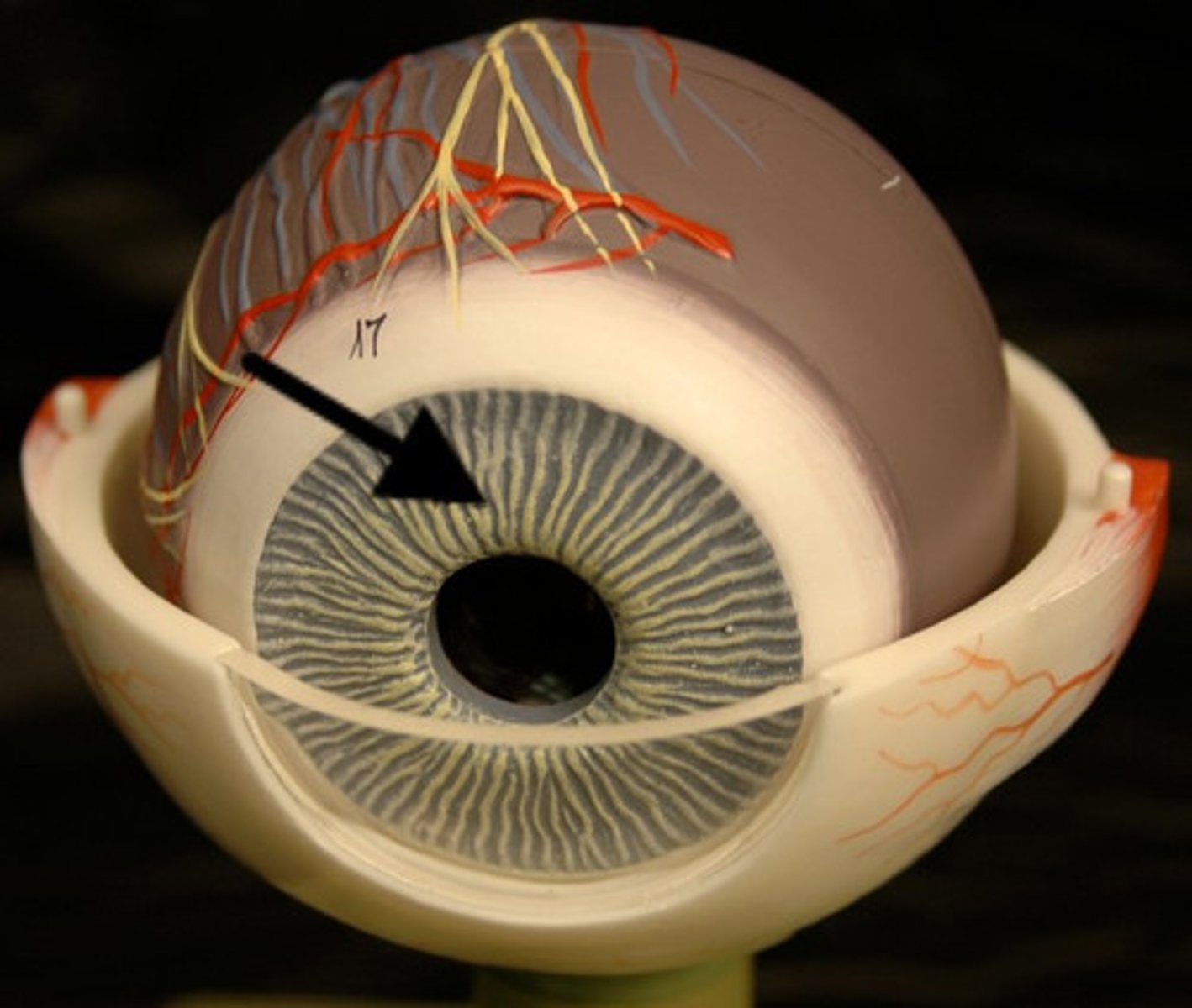
Ciliary Body
Structure surrounding the lens that connects the choroid and iris. It contains ciliary muscles, which control the shape of the lens, and it secretes aqueous humor.
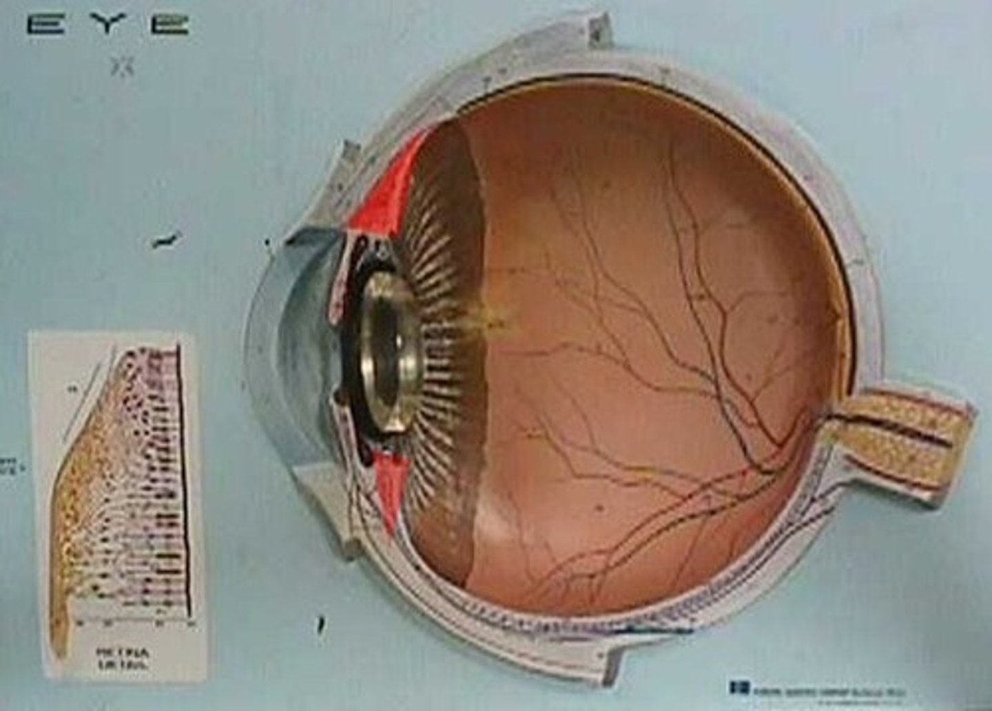
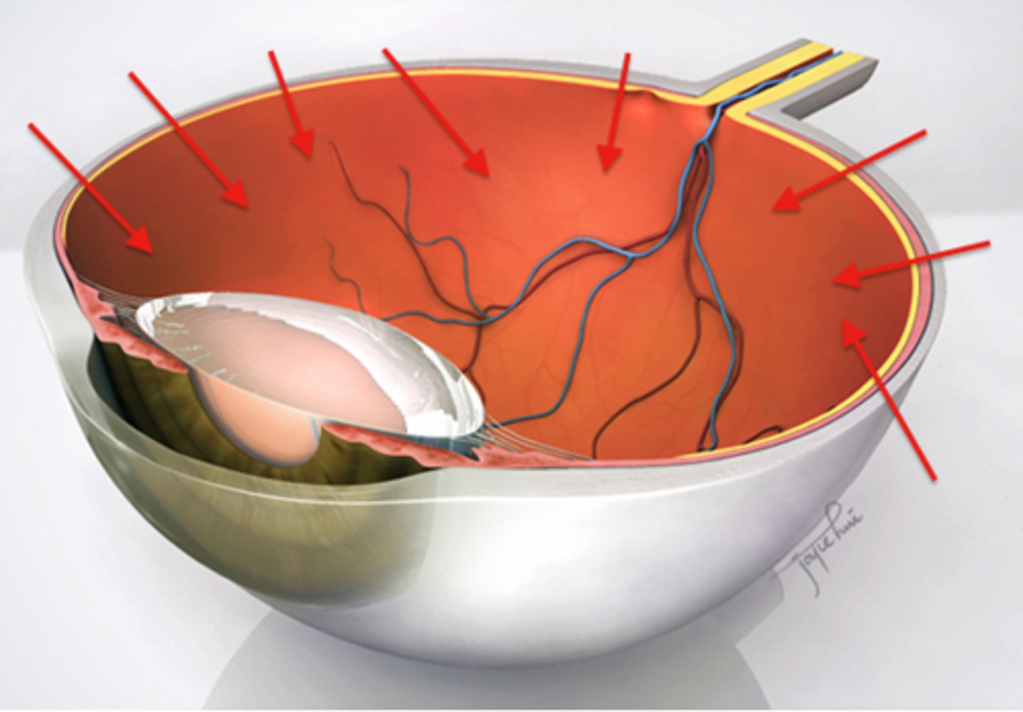
Sensory Tunic (retina)
(two- layered) Outer layer stores vitamin A for photoreceptor cells, Inner layer contains all the photoreceptor cells
Rods
Retinal receptors that detect black, white, and gray; necessary for peripheral and twilight vision, when cones don't respond
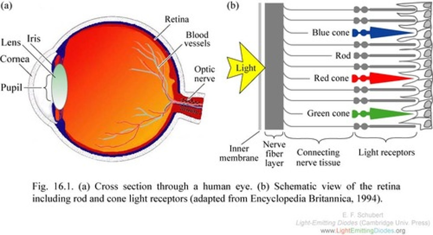
Cones
Retinal receptor cells that are concentrated near the center of the retina and that function in daylight or in well-lit conditions. Detects fine detail and give rise to color sensations.
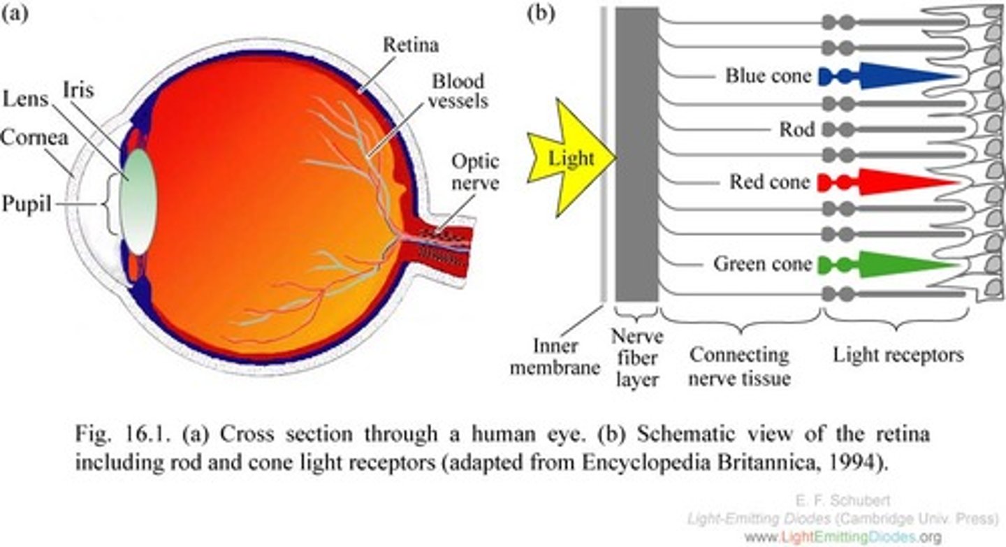
Bipolar Cells
In the retina, the specialized neurons that connect the rods and cones with the ganglion cells.
Ganglion Cells
In the retina, the specialized neurons that connect to the bipolar cells; form the optic nerve.
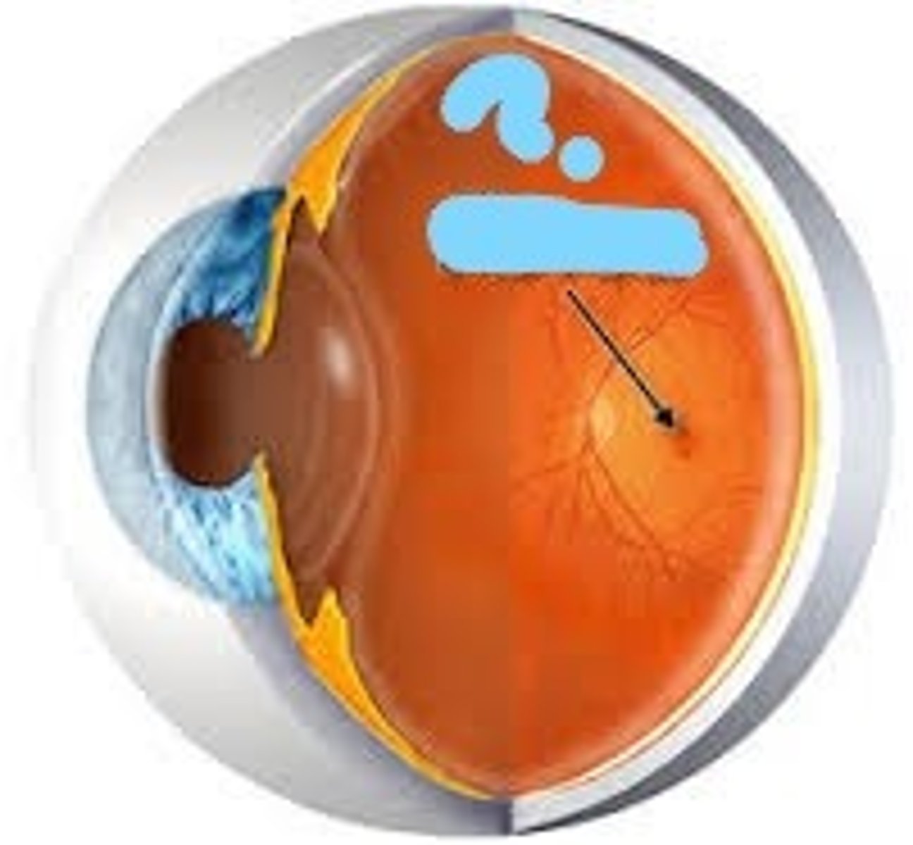
Fovea Centralis
Pinpoint depression in the center of the macula lutea that is the site of sharpest vision
Optic Disc
Region at the back of the eye where the optic nerve meets the retina. It is the blind spot of the eye because it contains only nerve fibers, no rods or cones, and is thus insensitive to light.
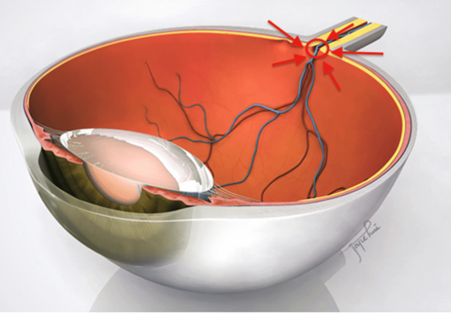
Central Artery & vein
Supply retina with nutrients, only area in body where blood vessels can be directly observed
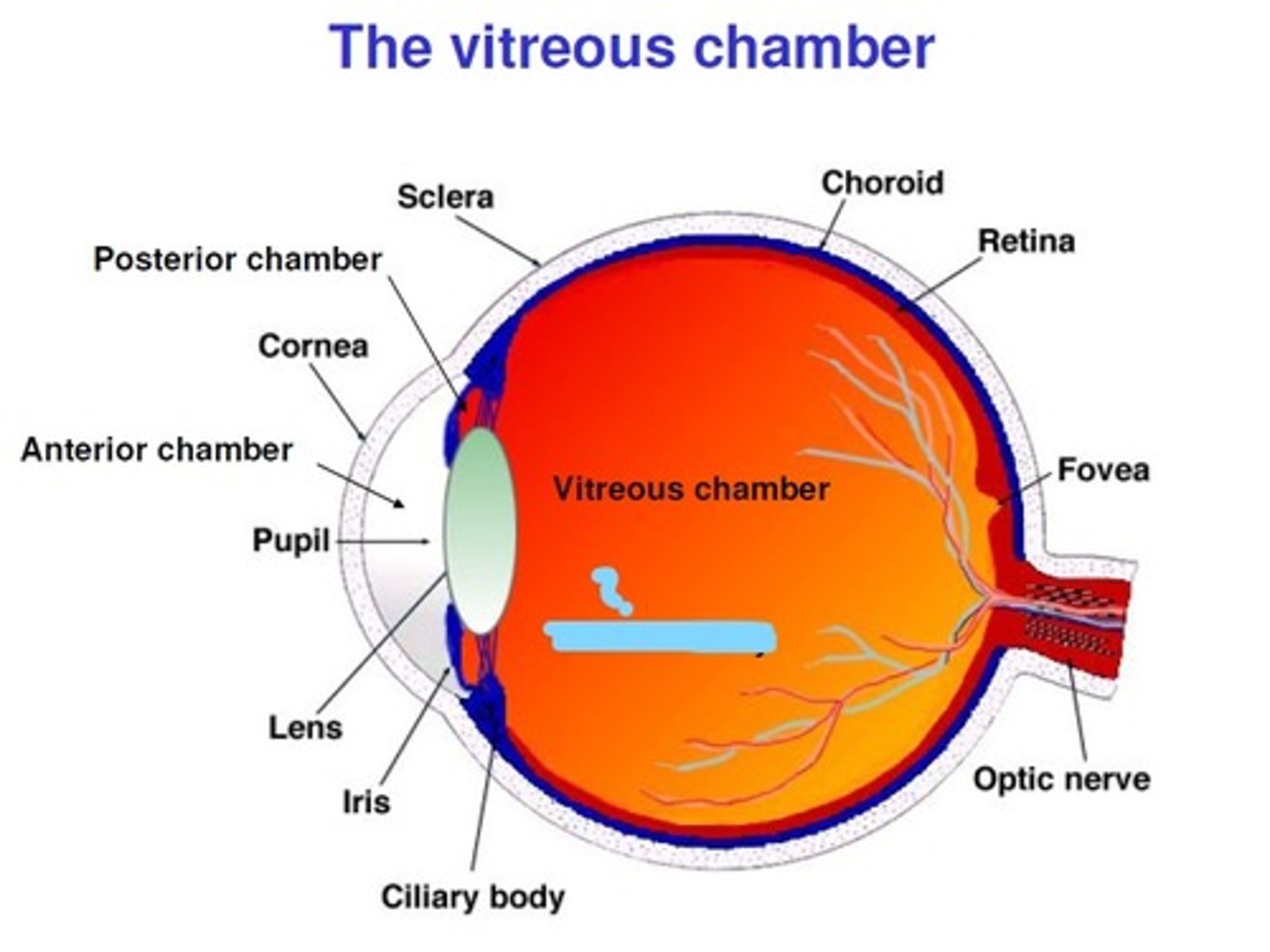
Vitreous Body / Vitreous Humor
Is the clear gel that fills the space between the lens and the retina of the eye. It doesn't replace once lost. Gel-like fluids inside the eye help it maintain its shape, which plays an important role in overall eye health.
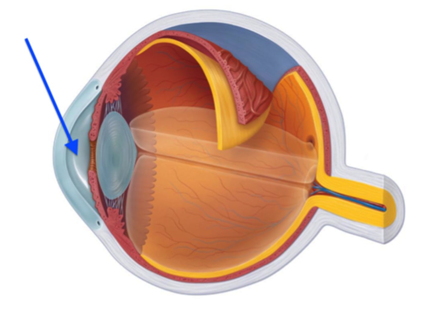
Aqueous humor
Fluid produced by the ciliary body and found in the anterior chamber. Replaces when lost. As well as nourishing the cornea and the lens by supplying nutrition such as amino acids and glucose, the aqueous humour will: Maintain intraocular pressure. Transport vitamin C in the front segment to act as an antioxidant agent
Lens
The transparent structure behind the pupil that changes shape to help focus images on the retina
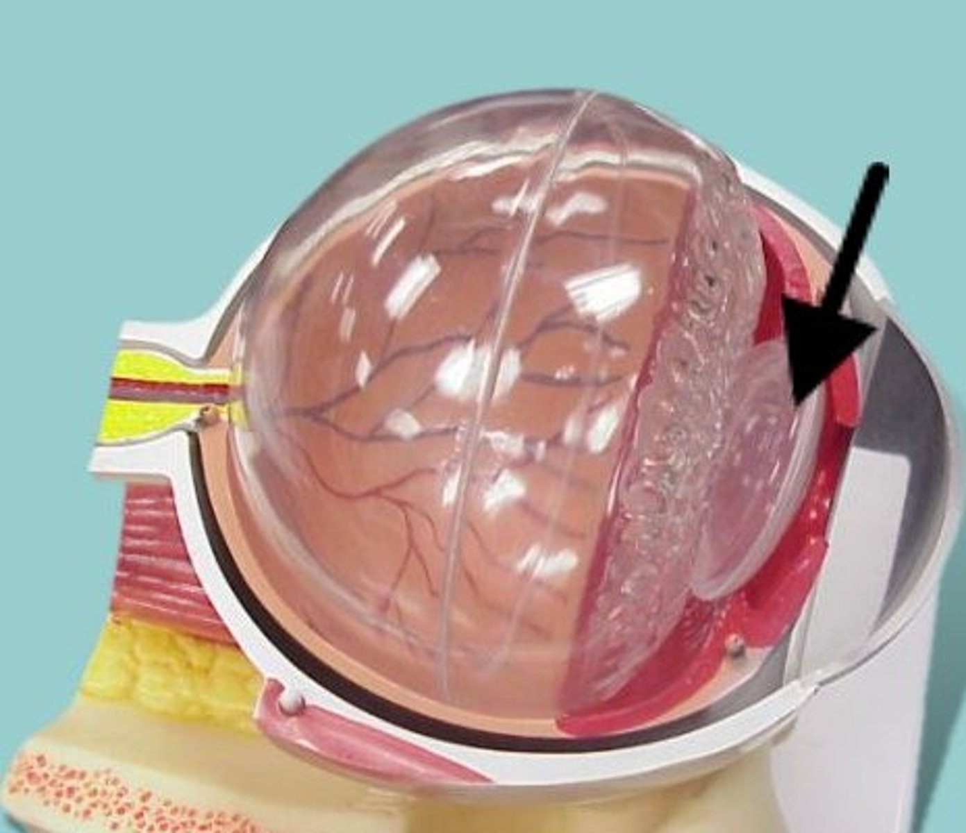
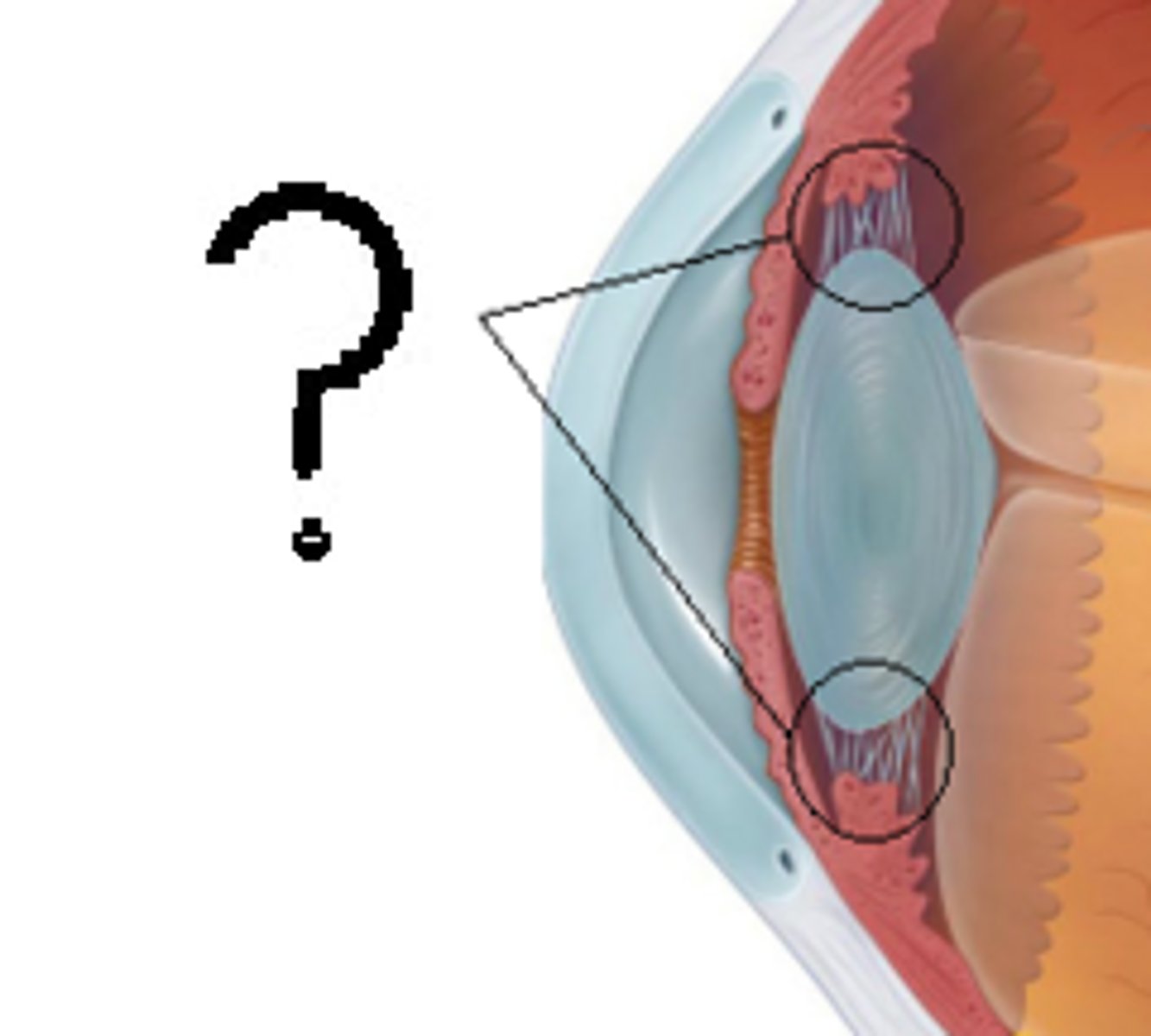
Suspensory Ligaments
A fibrous membrane that holds the lens of the eye in place
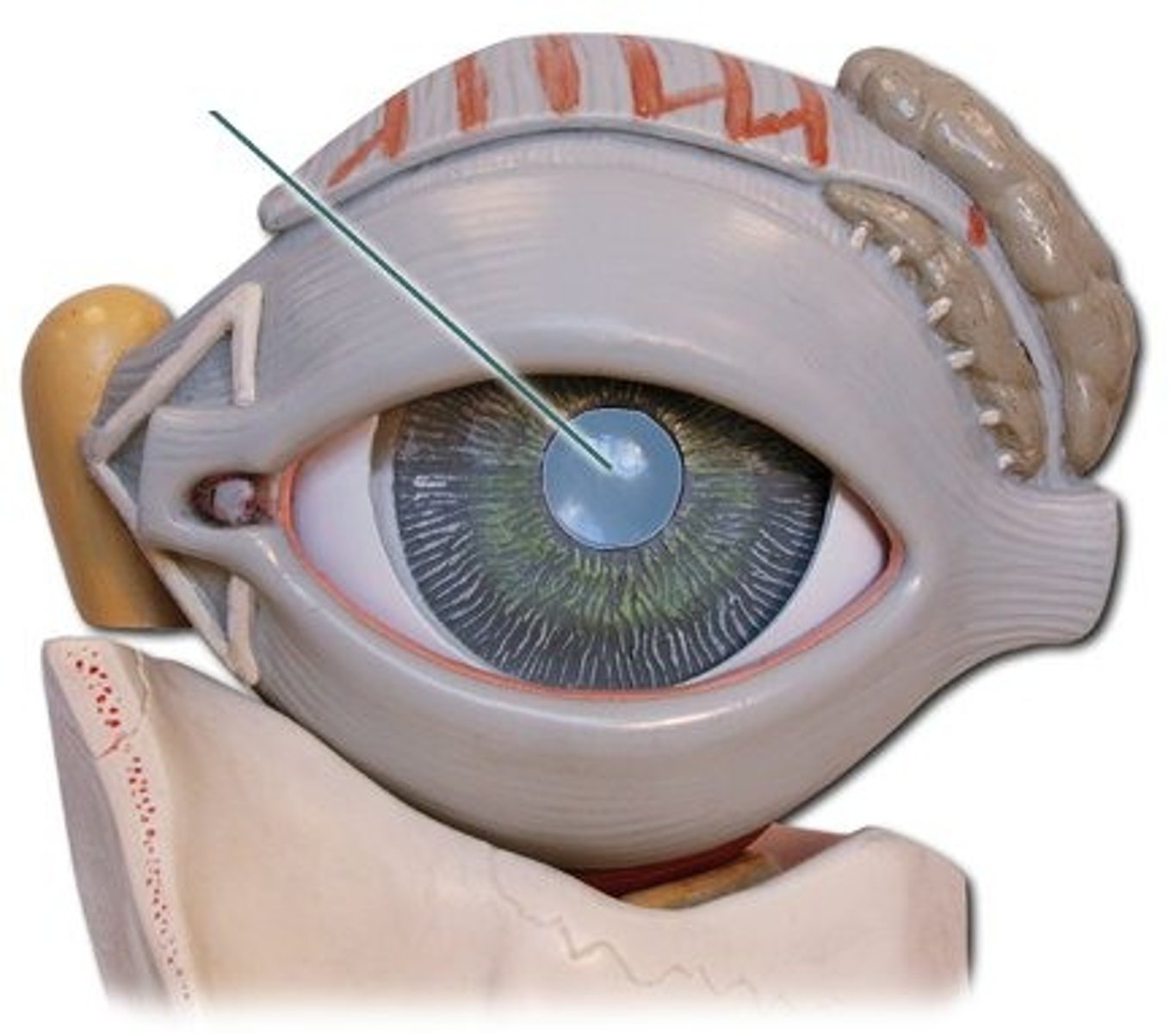
Pupil
The opening through which light enters the eye
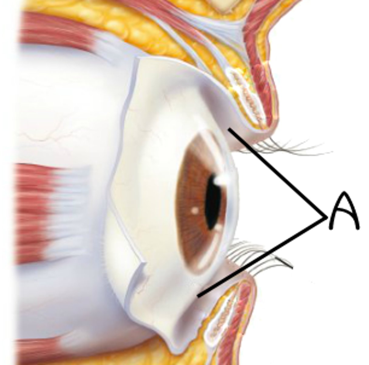
Conjunctiva
Mucous membrane that lines the eyelids and outer surface of the eyeball. non-keratinized, stratified columnar epithelium. contains goblet cells that secrete mucin, which generates the moisture that covers the surface of the eye and allows the eyelids to open and close easily.
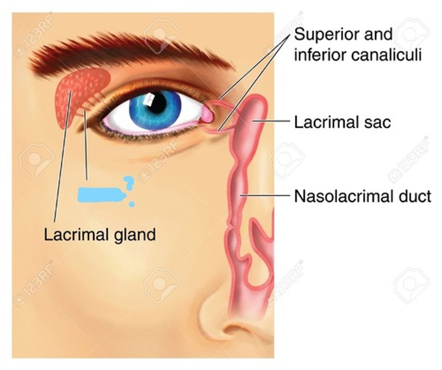
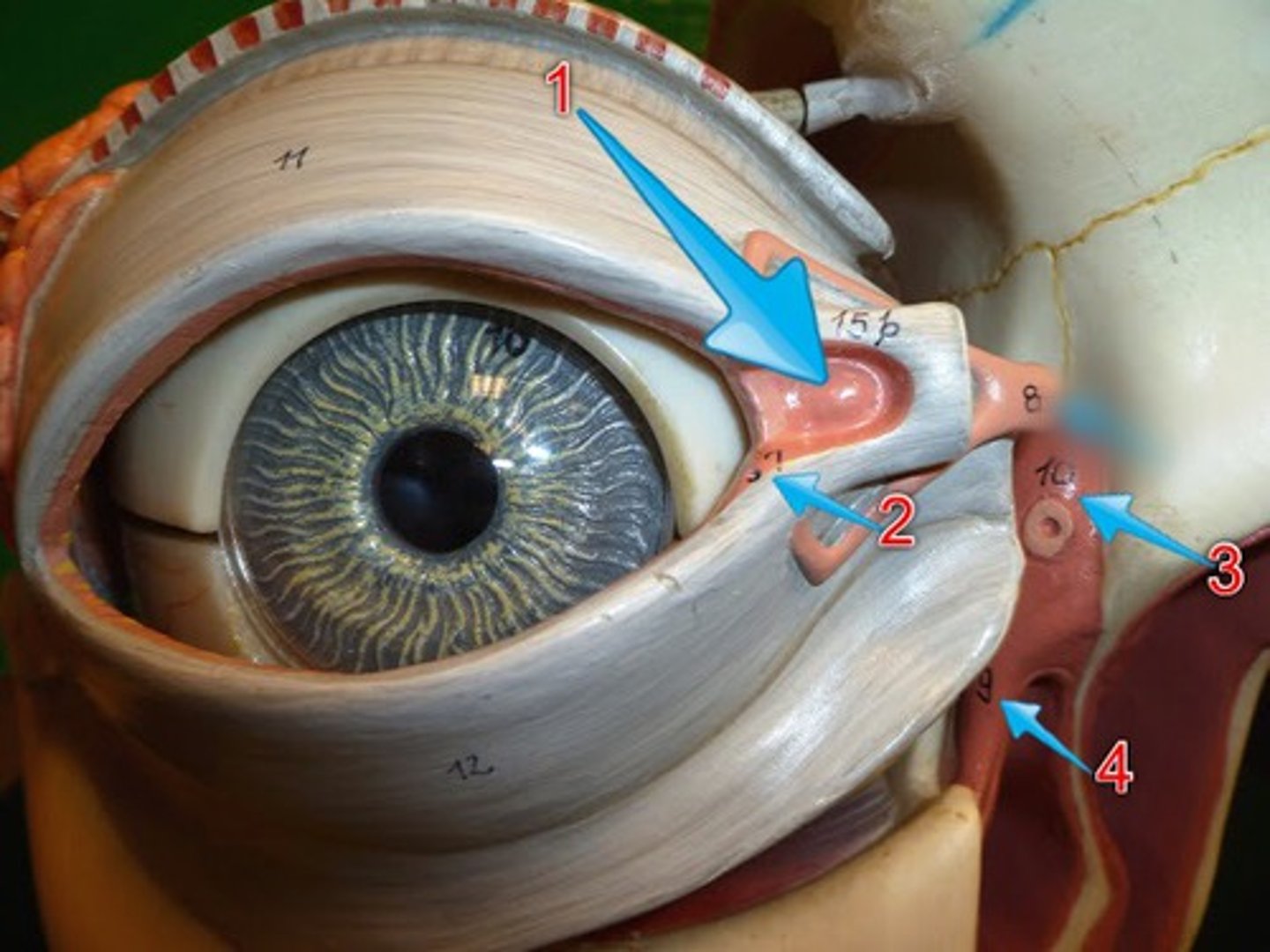
Lacrimal Apparatus
The structures that produce, store, and remove tears.
Made up of:
o lacrimal gland
o lacrimal duct
o lacrimal sac
o nasolacrimal duct
Lacrimal Gland
gland located in the upper outer region above the eyeball that secretes tears. produces tears which then flow into canals that connect to the lacrimal sac.
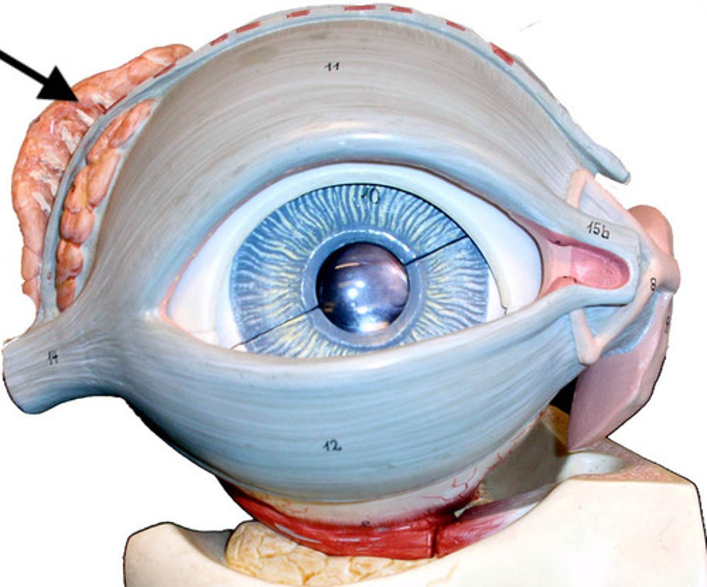
Lacrimal Sac
structure that collects tears before emptying into the nasolacrimal duct (white arrow)
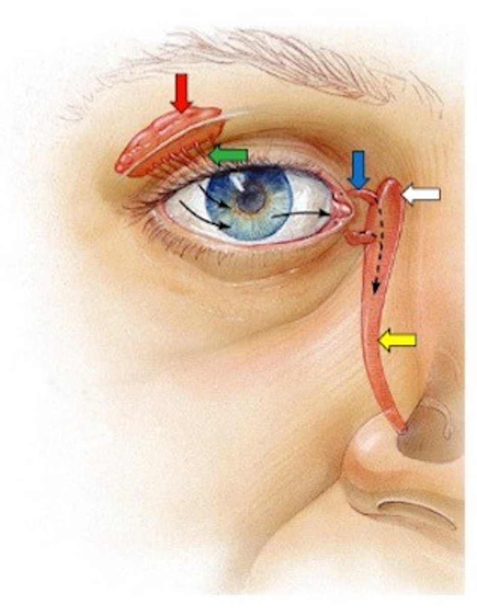
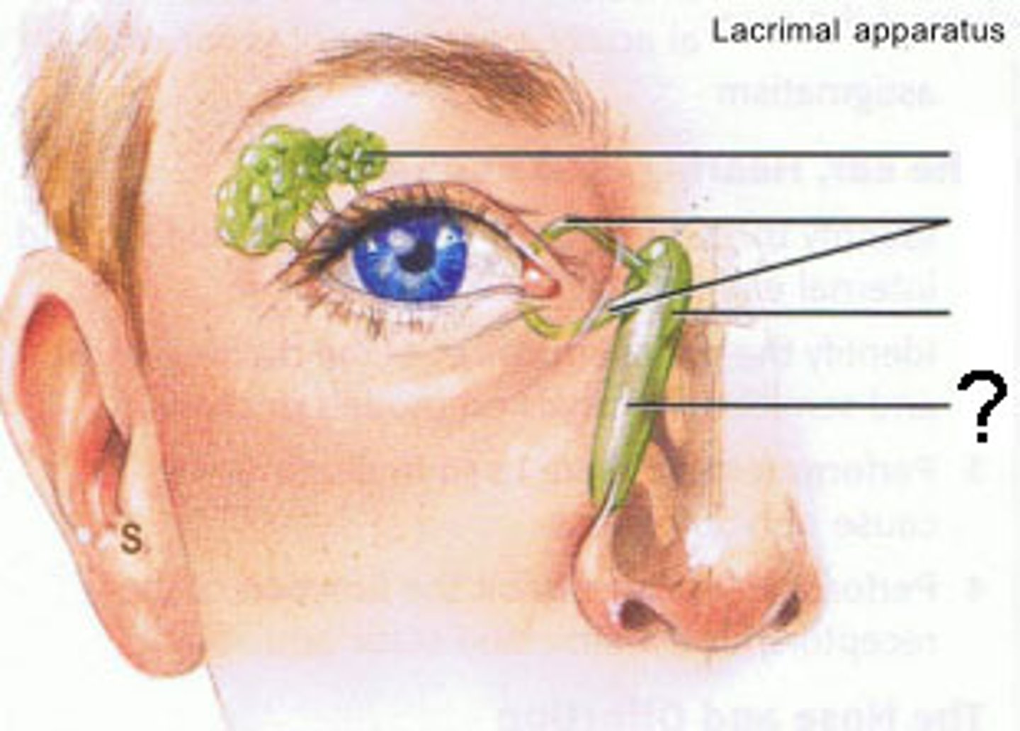
Nasolacrimal Duct
Passageway for tears from the lacrimal sac into the nose
Lateral Canthus
outer corner where the upper and lower eyelids meet
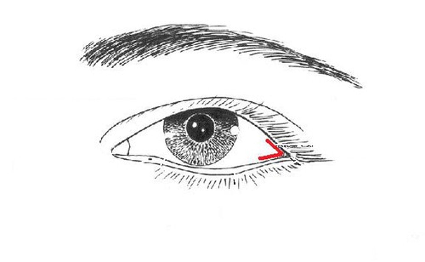
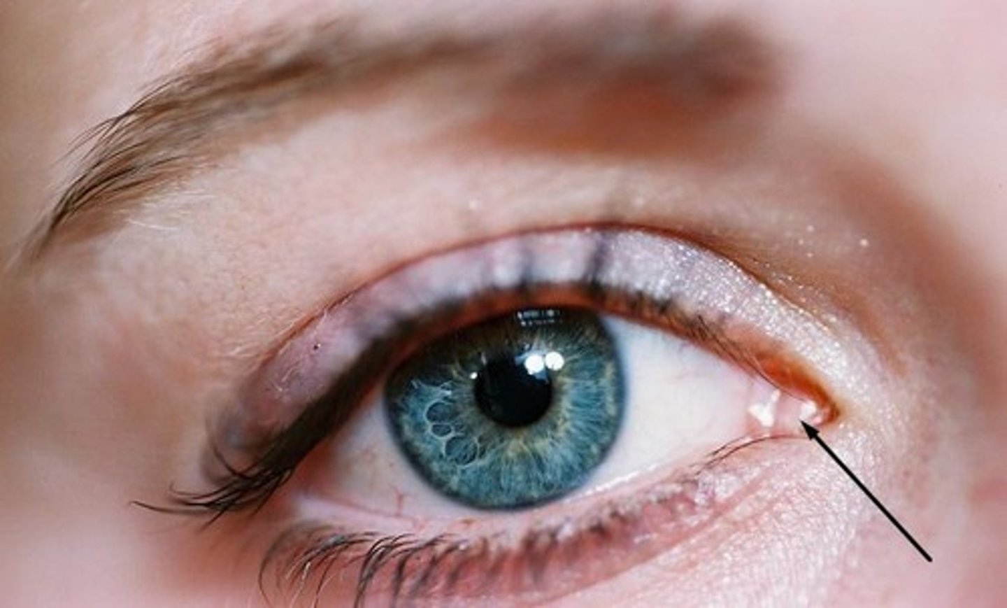
Medial Canthus
inner corner of the eye where the upper and lower eyelids meet
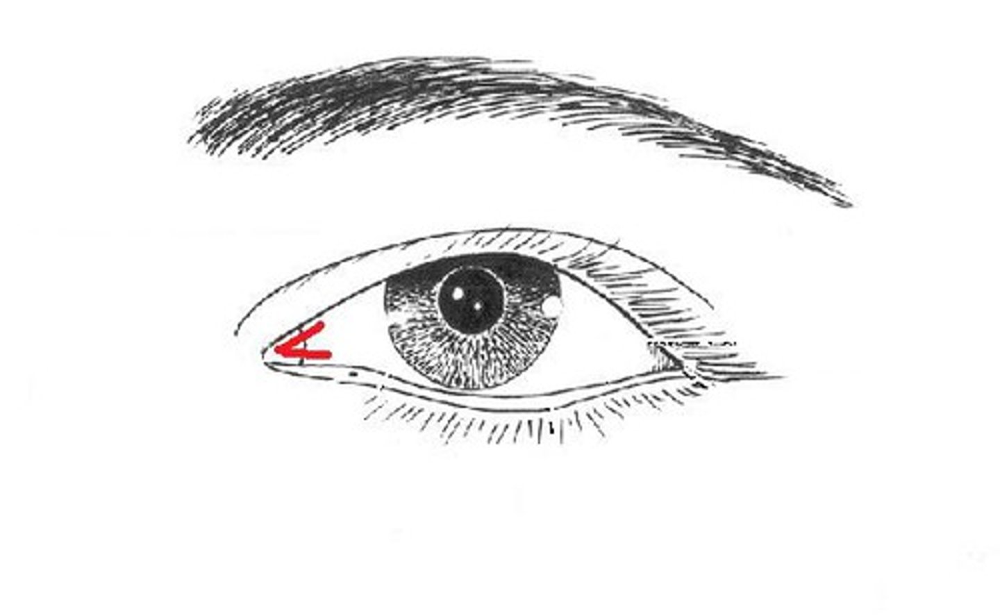
Caruncle
found in the medial canthus, it is a small fleshy mass containing sebacceous glands
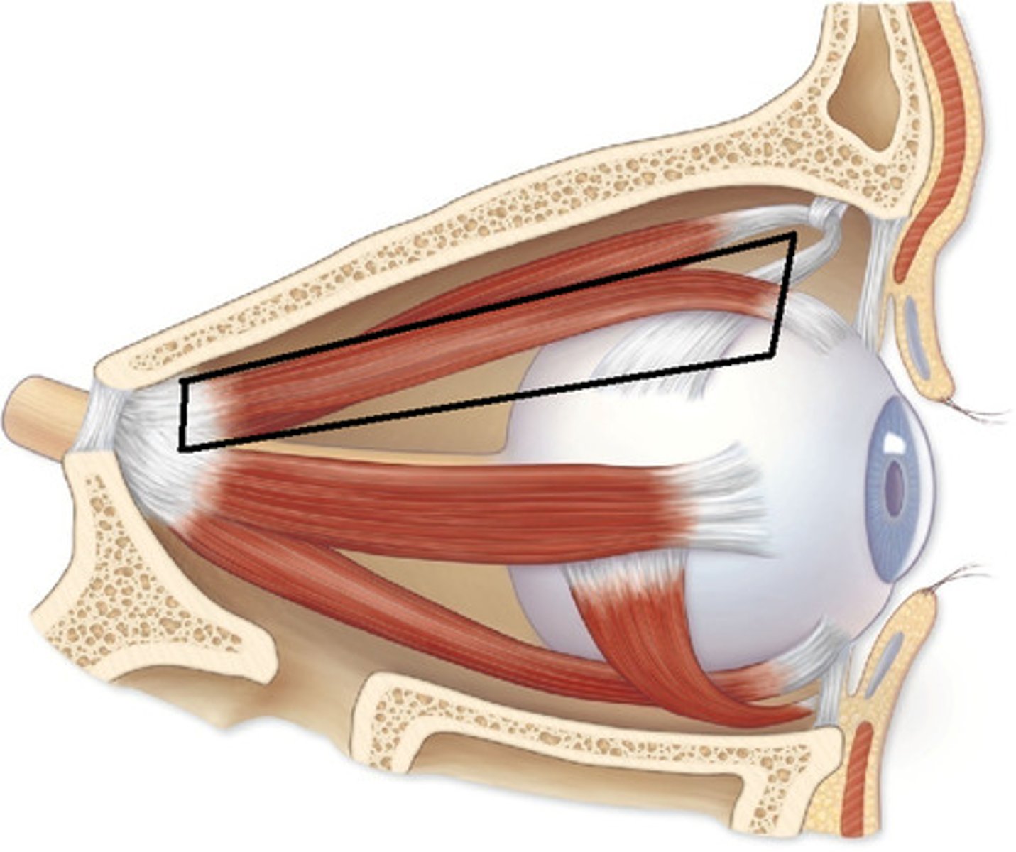
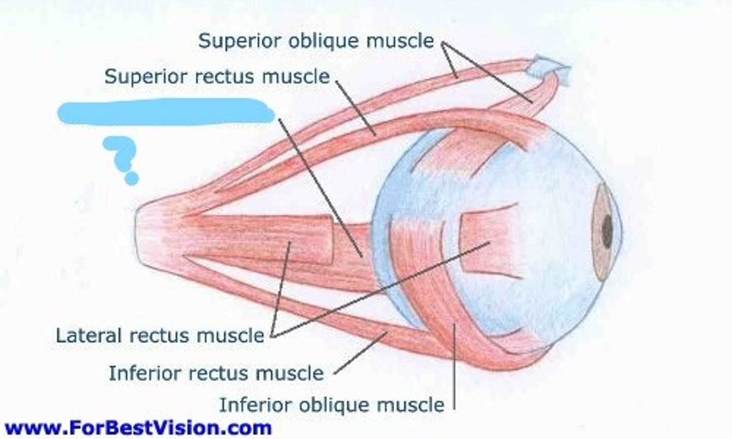
Superior Rectus of Eye
elevates eye and turns it medially, controlling cranial nerve: III occulomotor
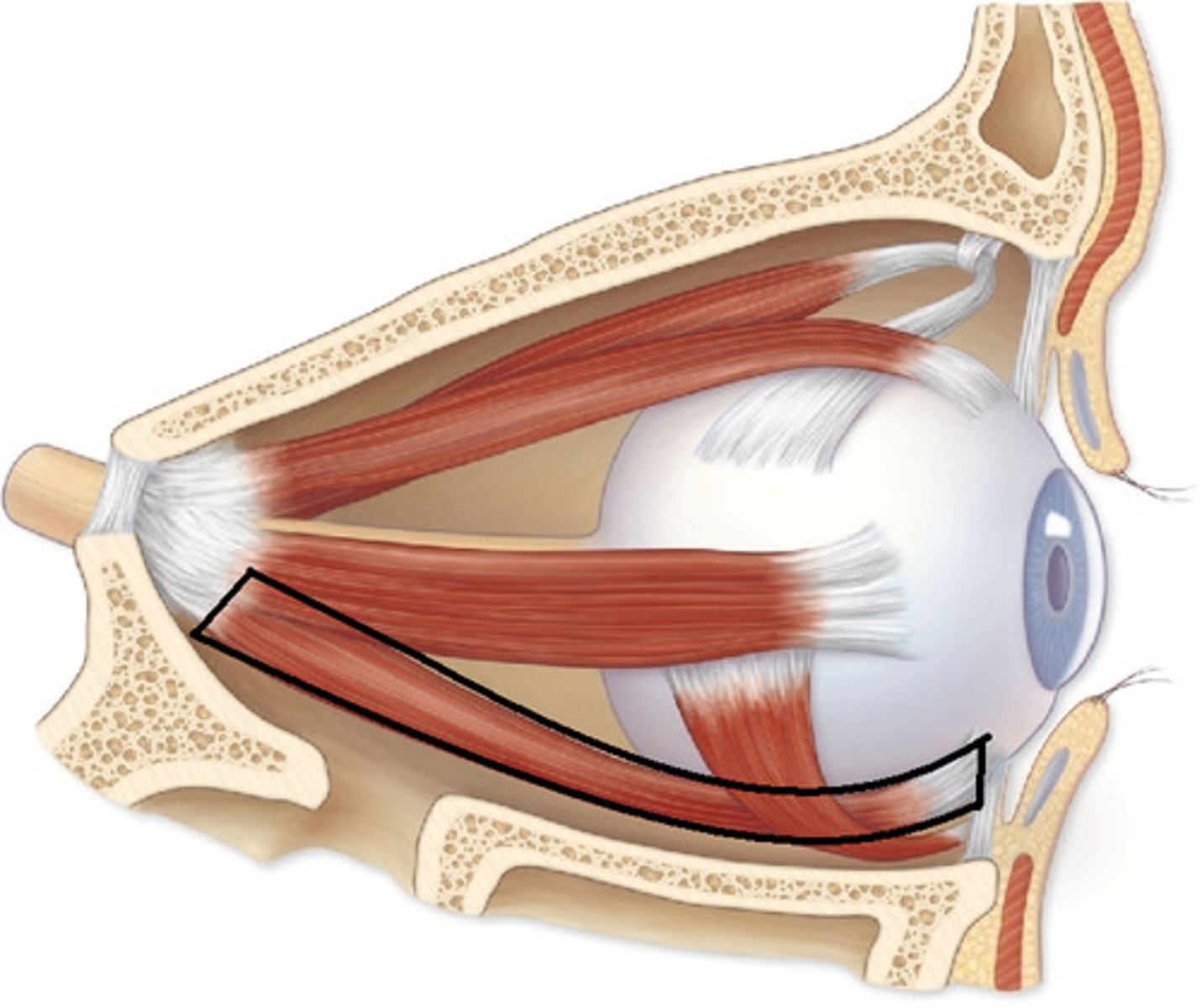
Inferior Rectus of Eye
depresses eye and turns it medially, controlling cranial nerve: III occulomotor
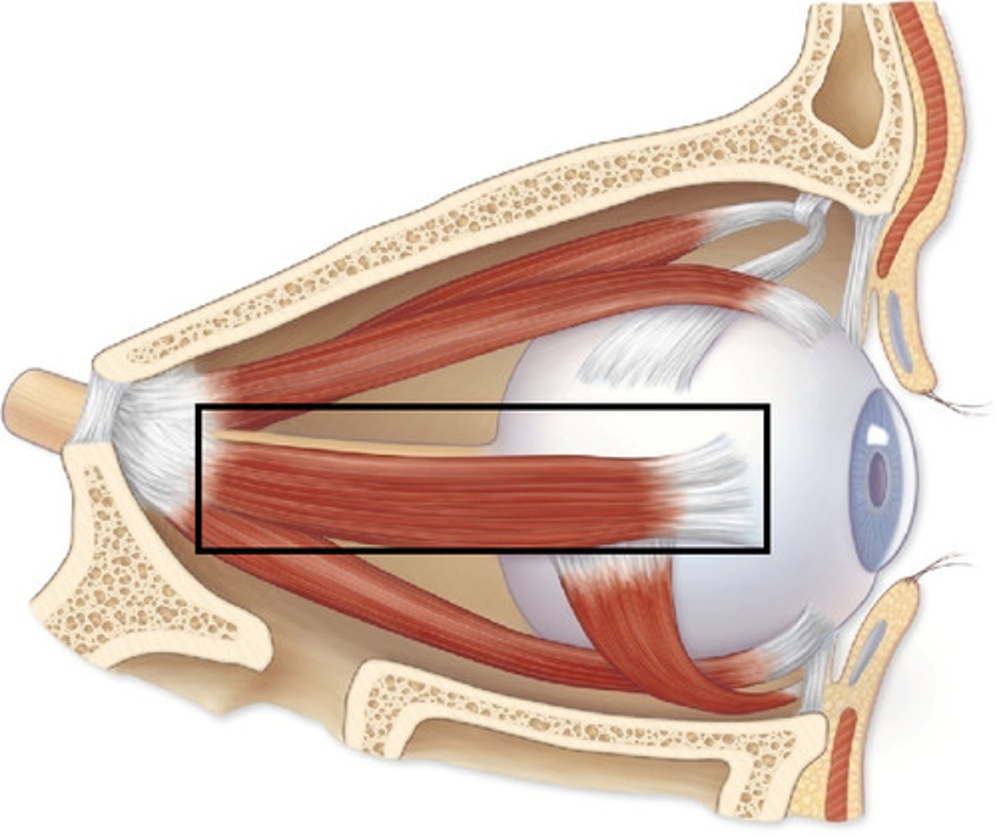
Lateral Rectus of Eye
moves eye laterally, controlling cranial nerve: VI abducens
Medial Rectus of Eye
moves eye medially, controlling cranial nerve: III occulomotor
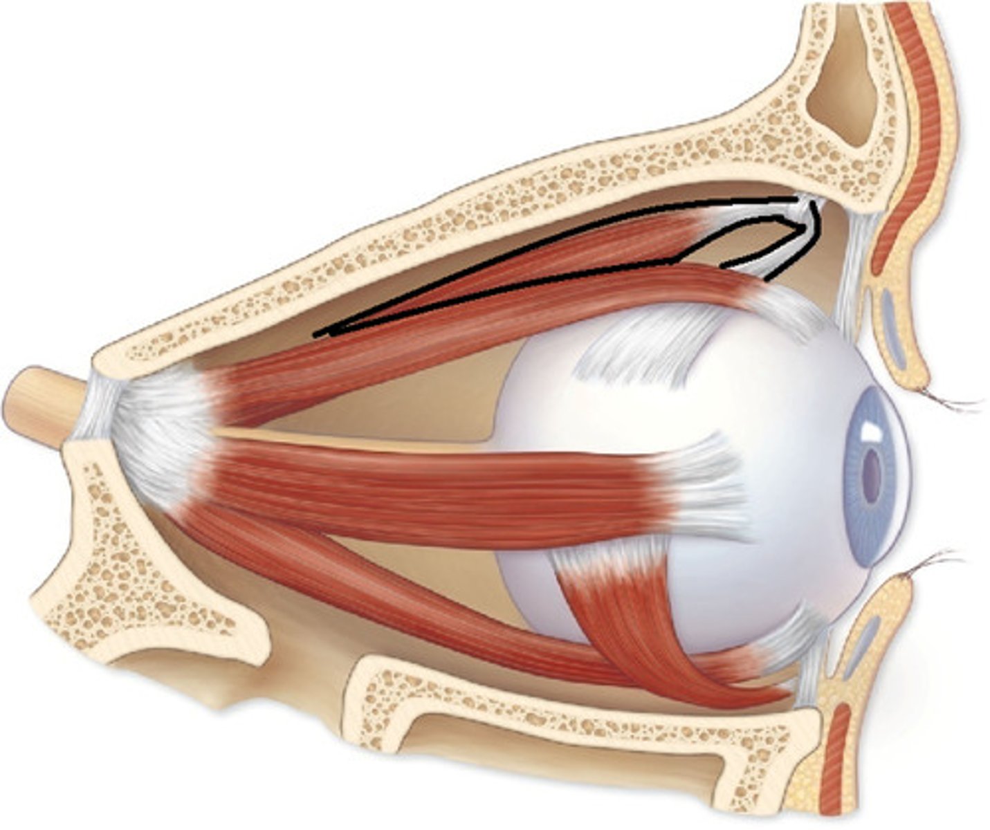
Superior Oblique of Eye
depresses eye and turns it laterally, controlling cranial nerve: IV trochlear nerve
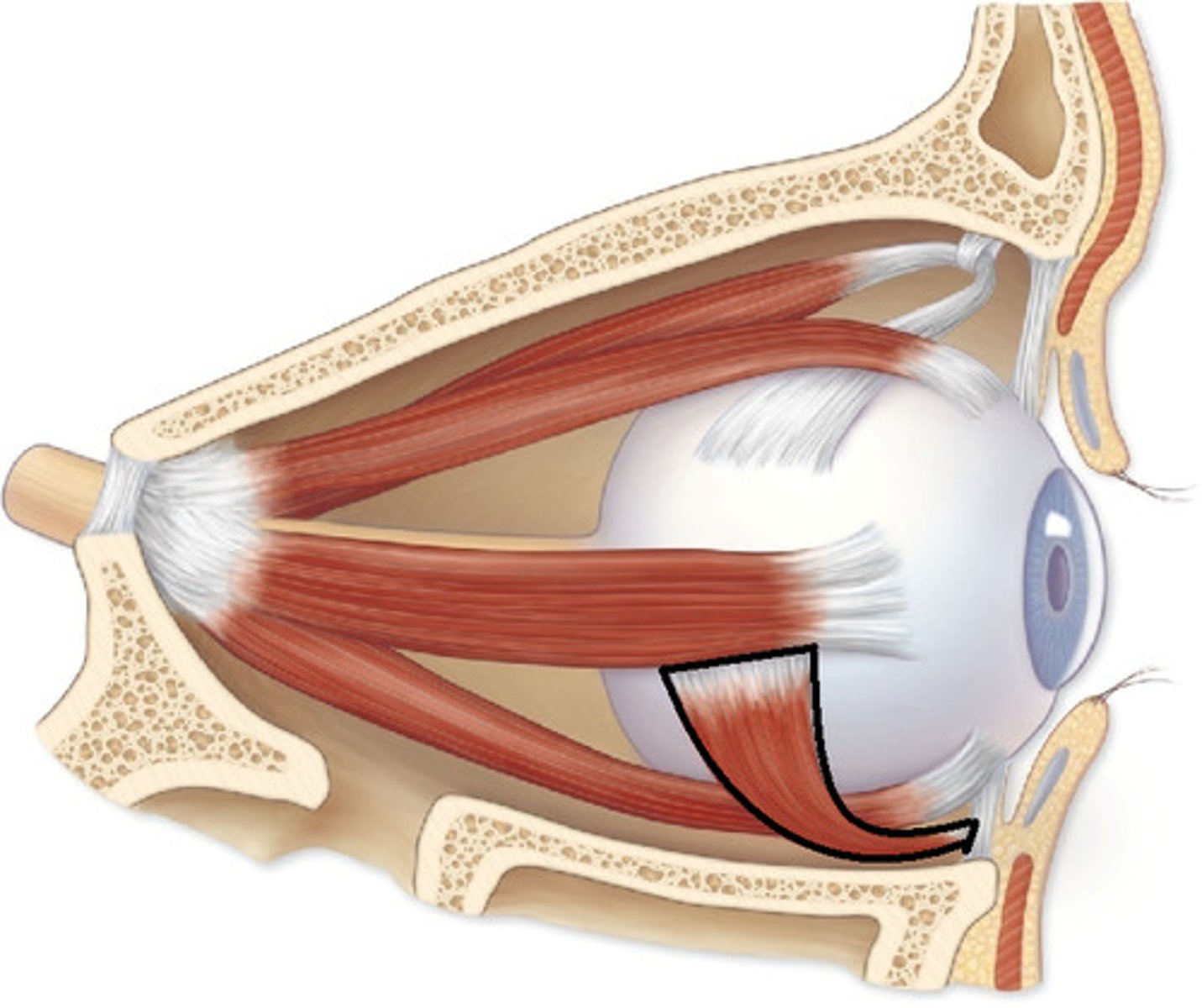
Inferior Oblique of Eye
elevates eye and turns it laterally, controlling cranial nerve: III occulomotor
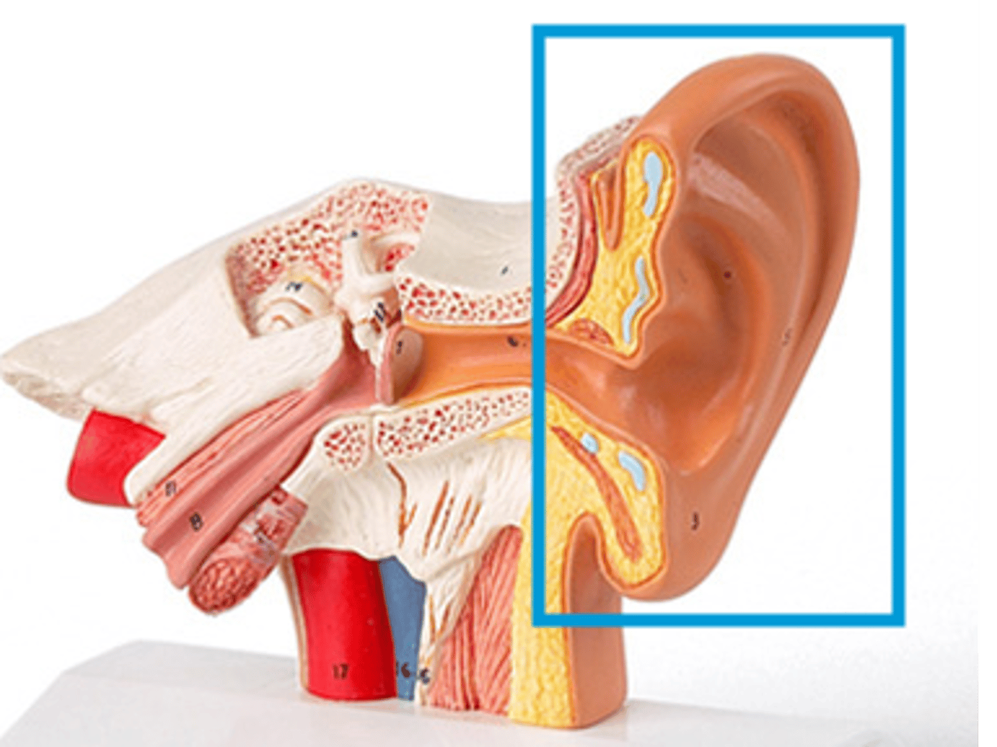
Auricle (Pinna)
external portion of the ear
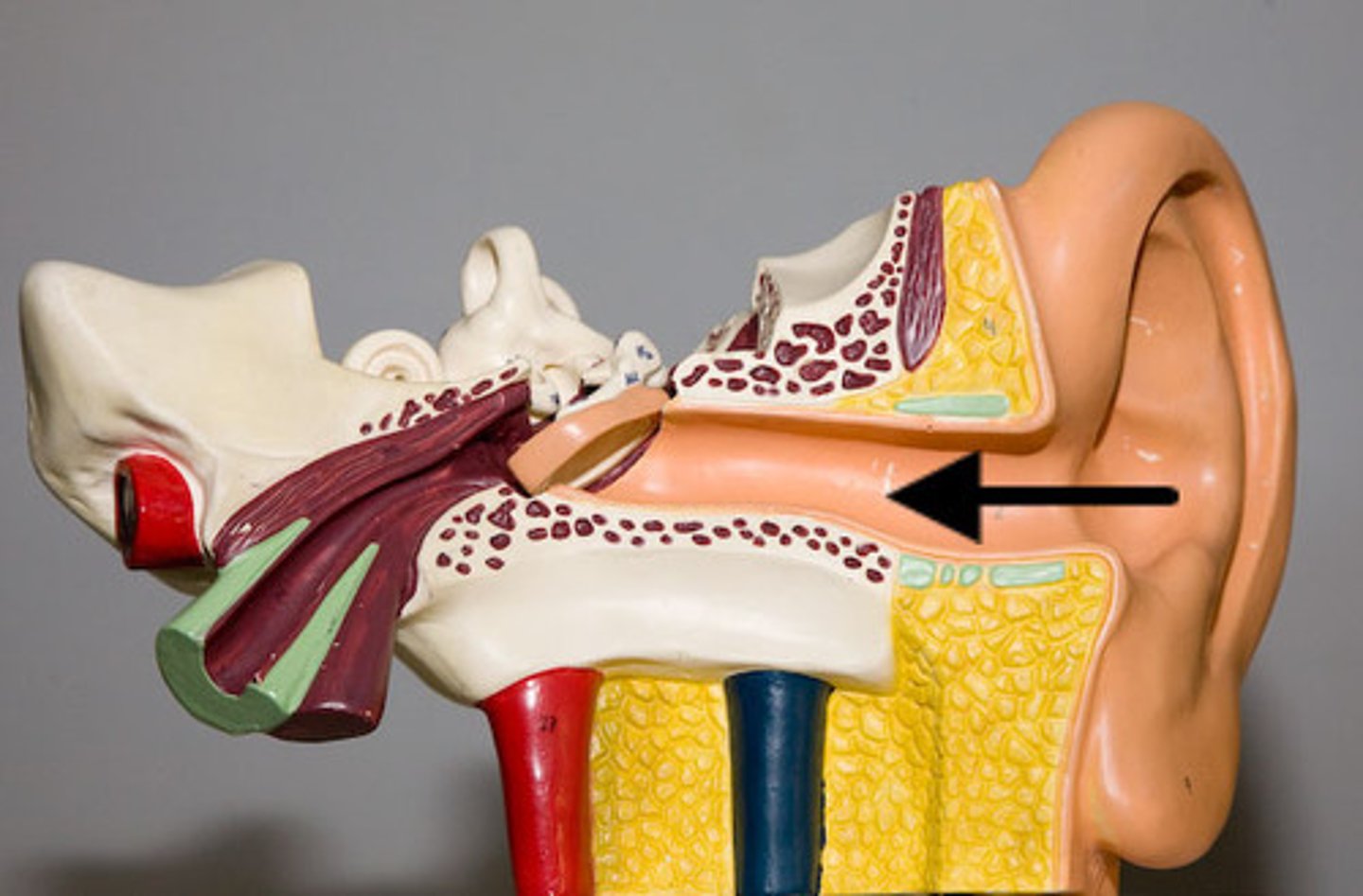
External auditory canal
transmits sound waves from the pinna to the tympanic membrane of the middle ear
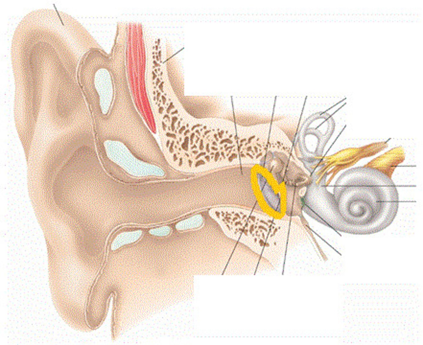
Tympanic Membrane
The eardrum. A structure that separates the outer ear from the middle ear and vibrates in response to sound waves.
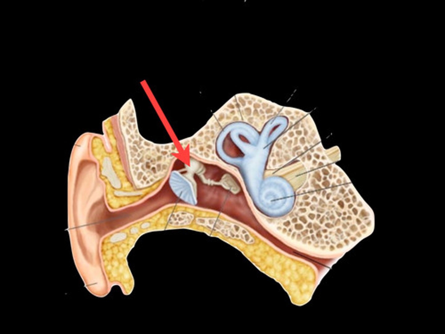
Malleus
a small bone in the middle ear that transmits vibrations of the eardrum to the incus.
Incus
middle of the three auditory ossicles of the middle ear
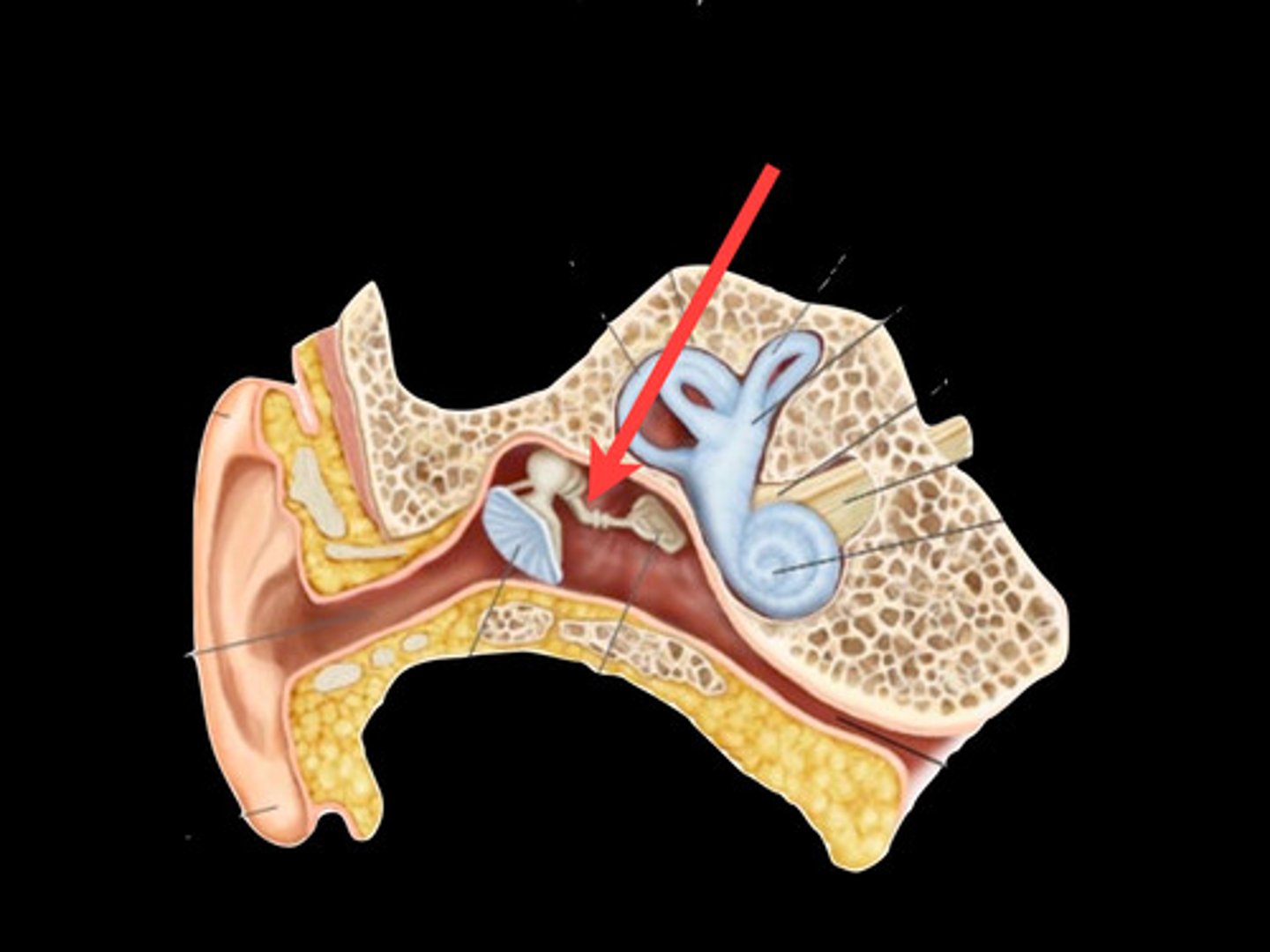
Stapes
stirrup; last of the three auditory ossicles of the middle ear
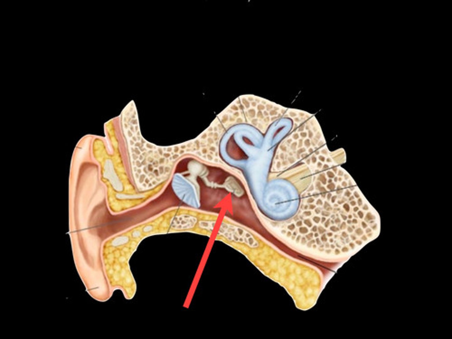
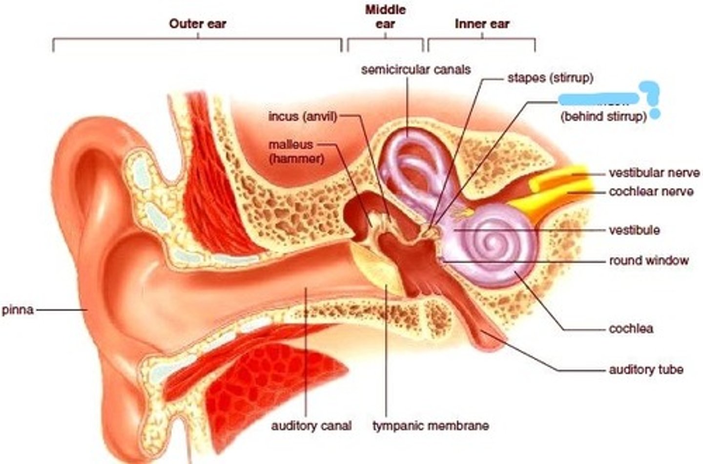
Oval (vestibular) window
covered by the base of the stapes; where mechanical vibrations enter the inner ear
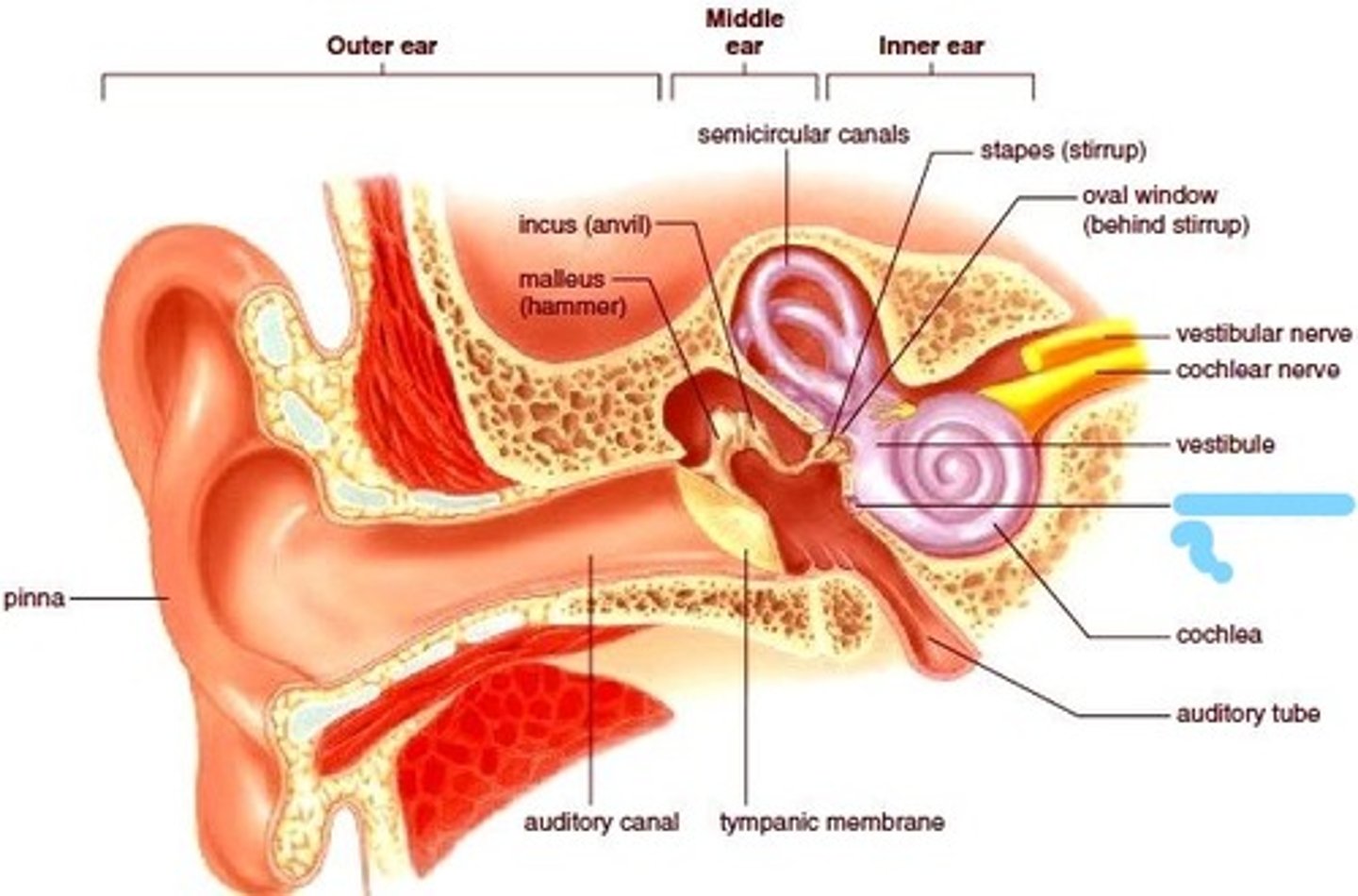
Round (cochlear) window
Covered by the secondary tympanic membrane. Acts as an outlet for pressure waves created in the fluid of the inner ear by movement of the stapes in response to sound.
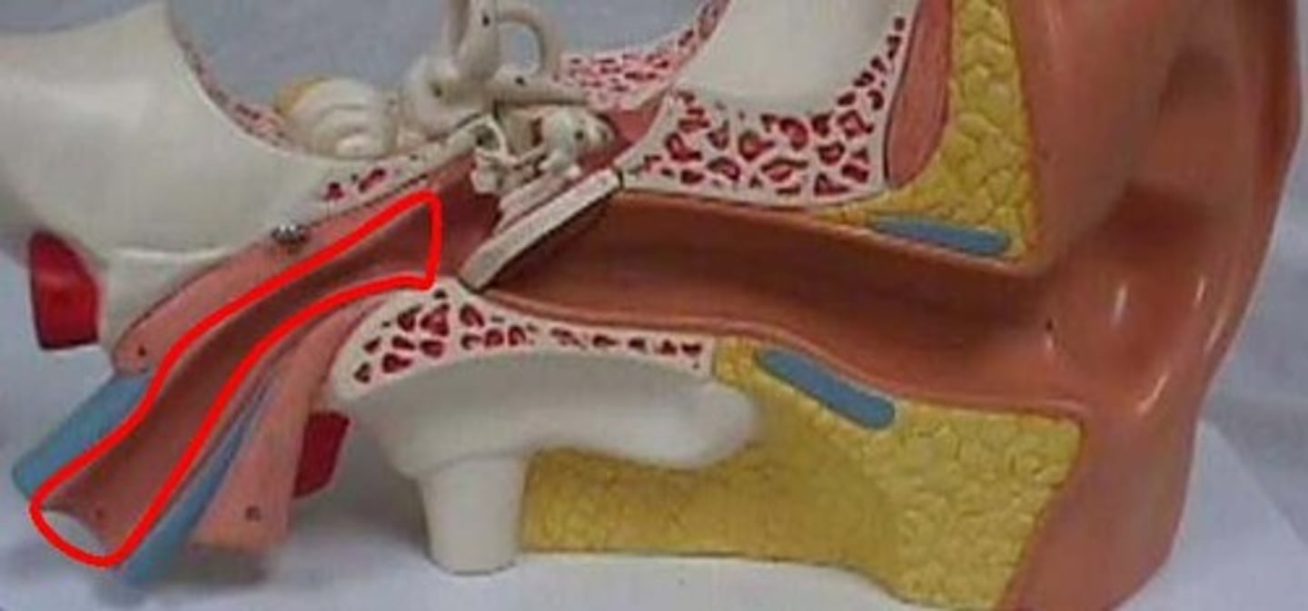
Eustachian tube
The primary function is to ventilate the middle ear space, ensuring that its pressure remains at near normal environmental air pressure. The secondary function is to drain any accumulated secretions, infection, or debris from the middle ear space.
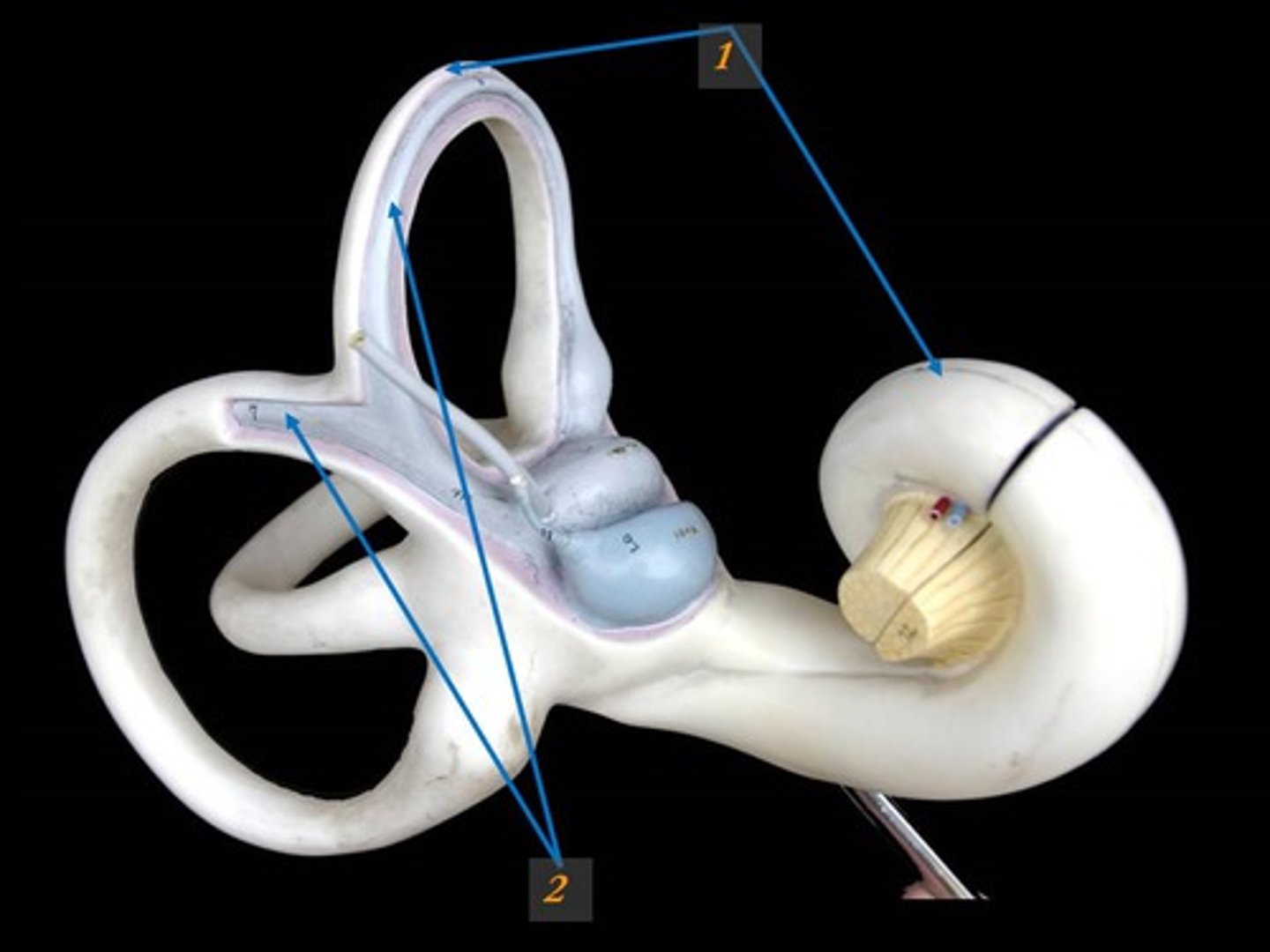
Osseous Labyrinth
Bony canal in temporal bone filled with perilymph
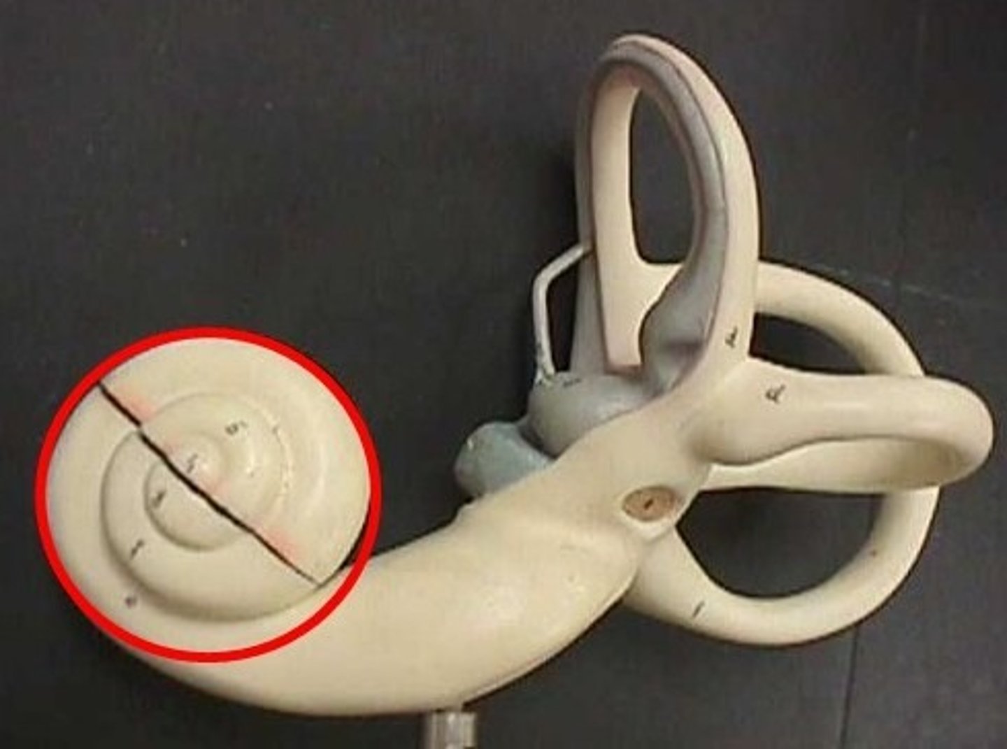
Cochlea
A coiled, bony, fluid-filled tube in the inner ear through which sound waves trigger nerve impulses.
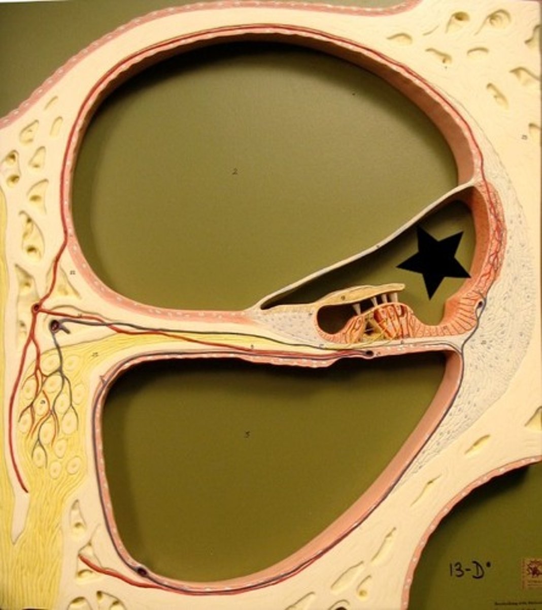
Cochlear Duct
A fluid filled cavity within the cochlea that vibrates when sound waves strike it.
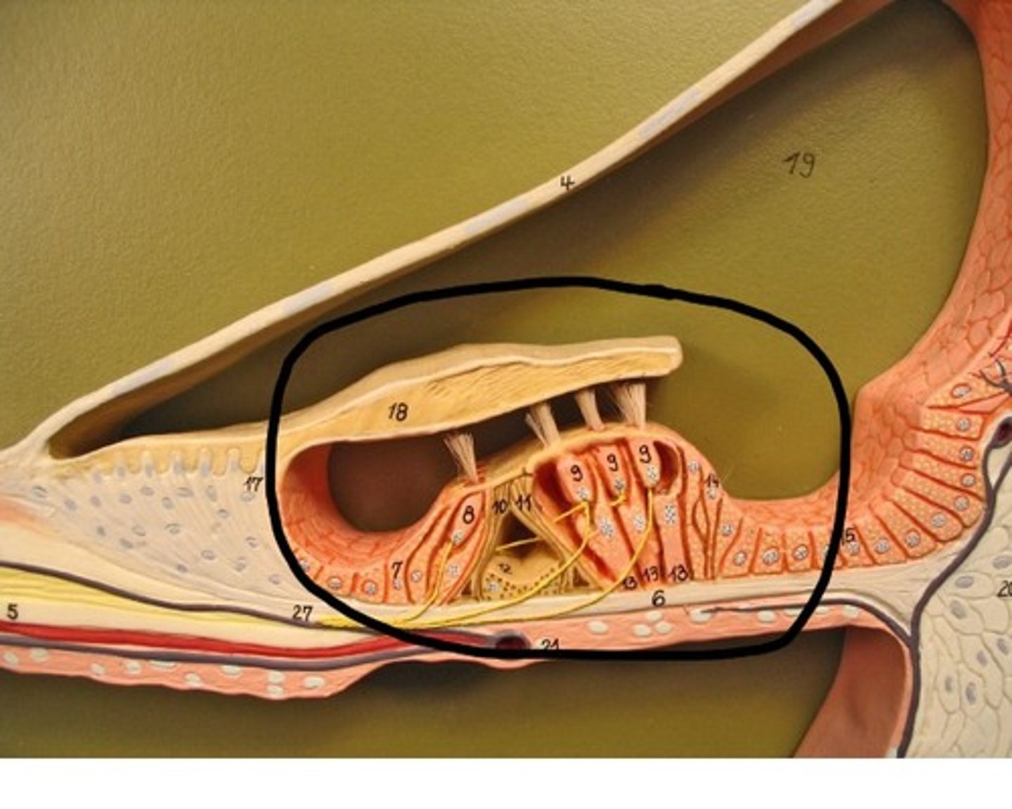
Organ of Corti
Center part of the cochlea, containing hair cells, canals, and membranes. Transduce mechanical sound vibrations into nerve impulses.
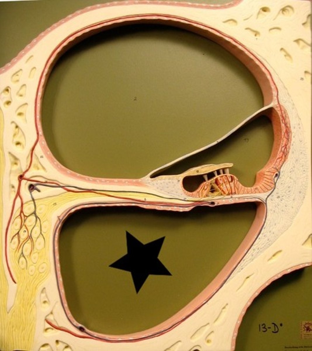
Scala Tympani
Terminates at round window; contains perilymph
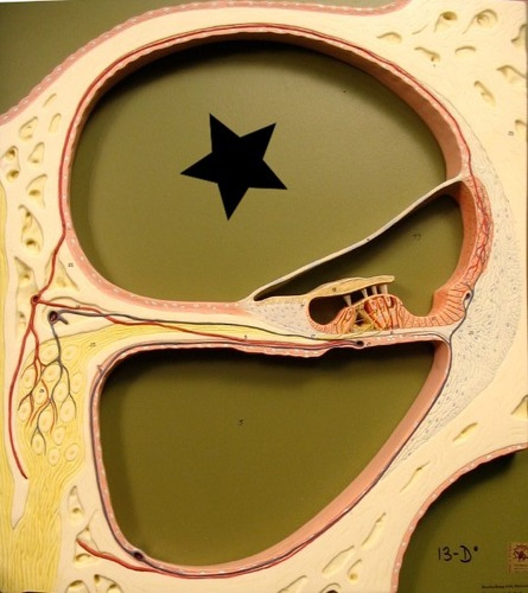
Scala Vestibuli
Leads from oval window to apex of cochlea
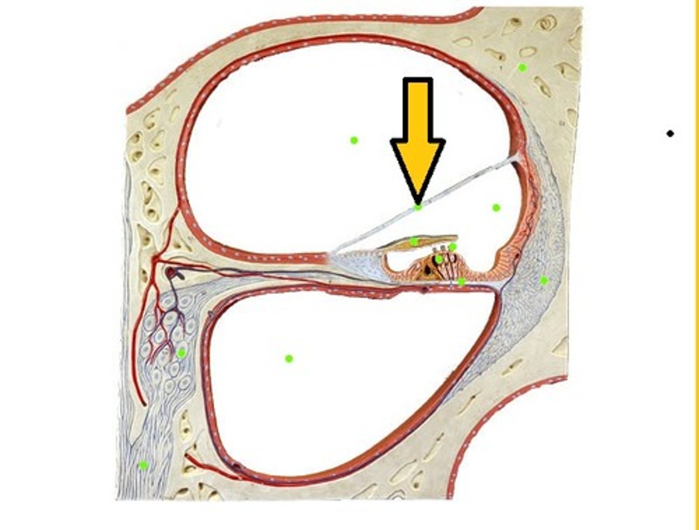
Vestibular Membrane
Separates the cochlear duct from the scala vestibuli. It primarily functions as a diffusion barrier, allowing nutrients to travel from the perilymph to the endolymph of the membranous labyrinth
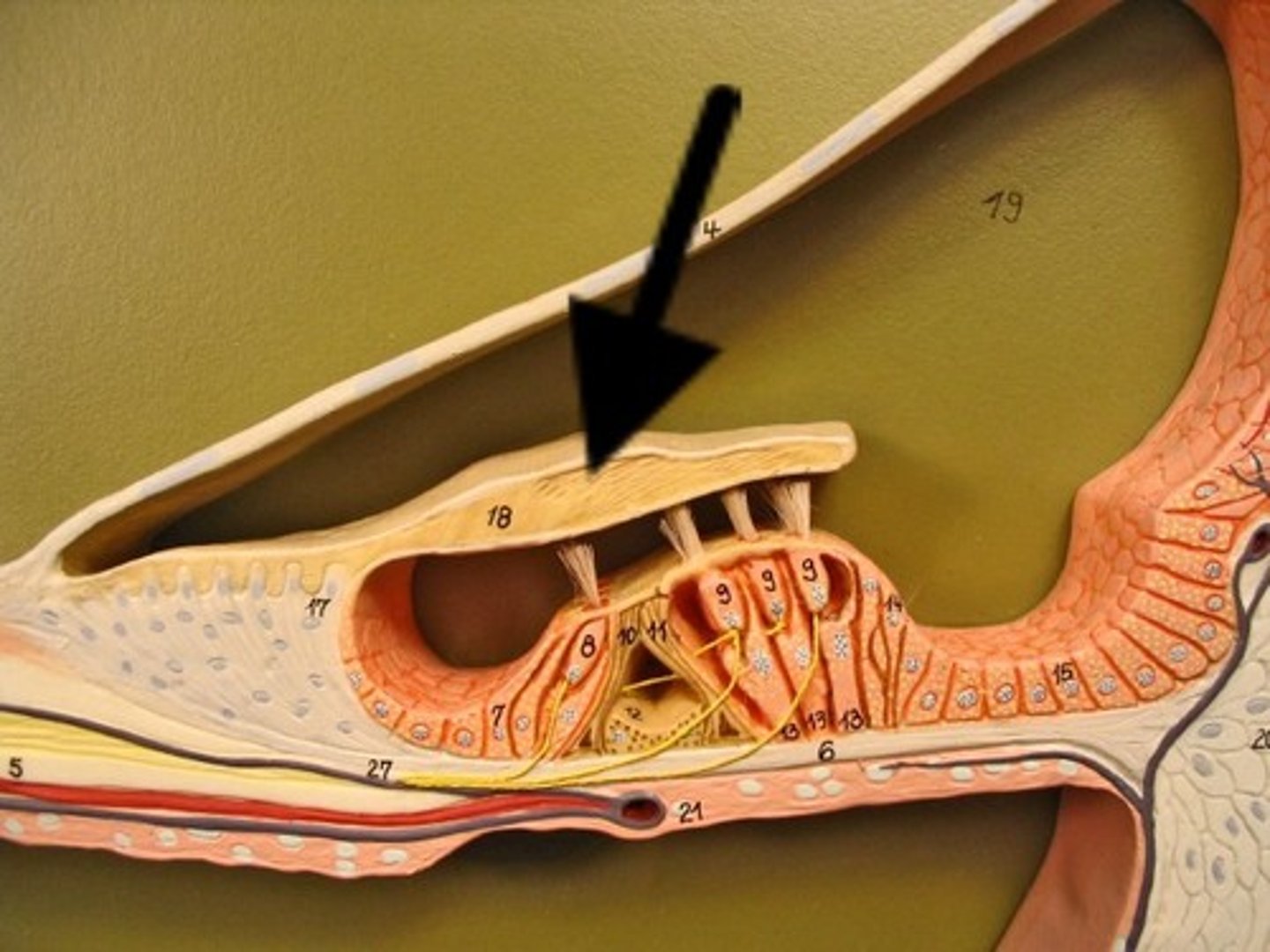
Tectorial Membrane
A membrane located above the basilar membrane; serves as a shelf against which the cilia of the auditory hair cells move
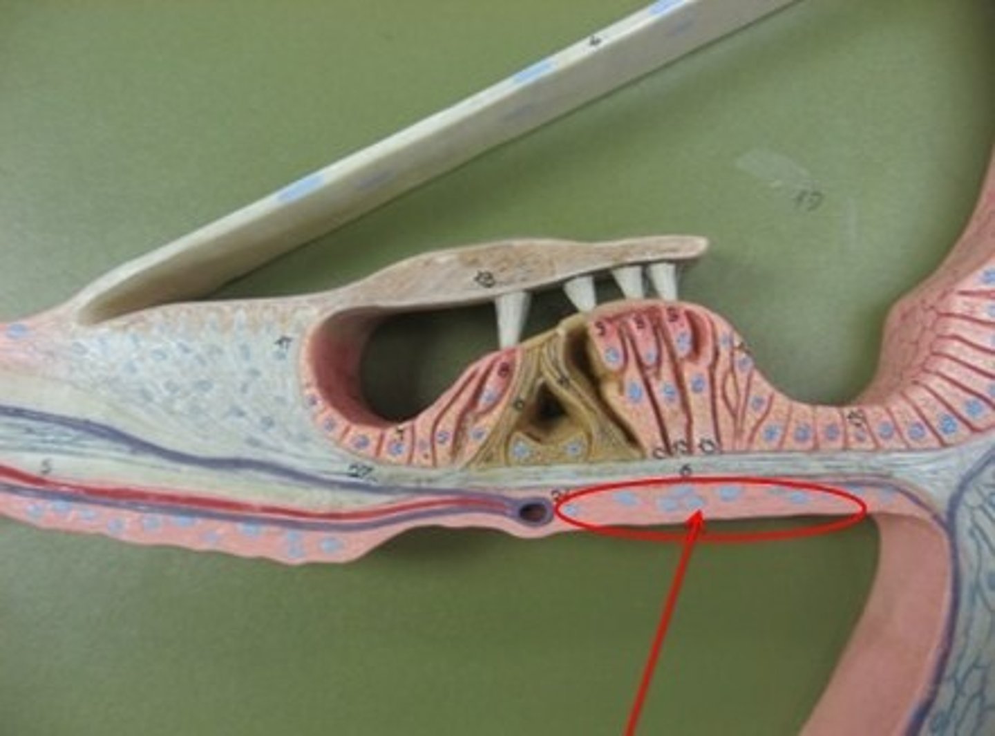
Basilar Membrane
A structure that runs the length of the cochlea in the inner ear and holds the auditory receptors, called hair cells.
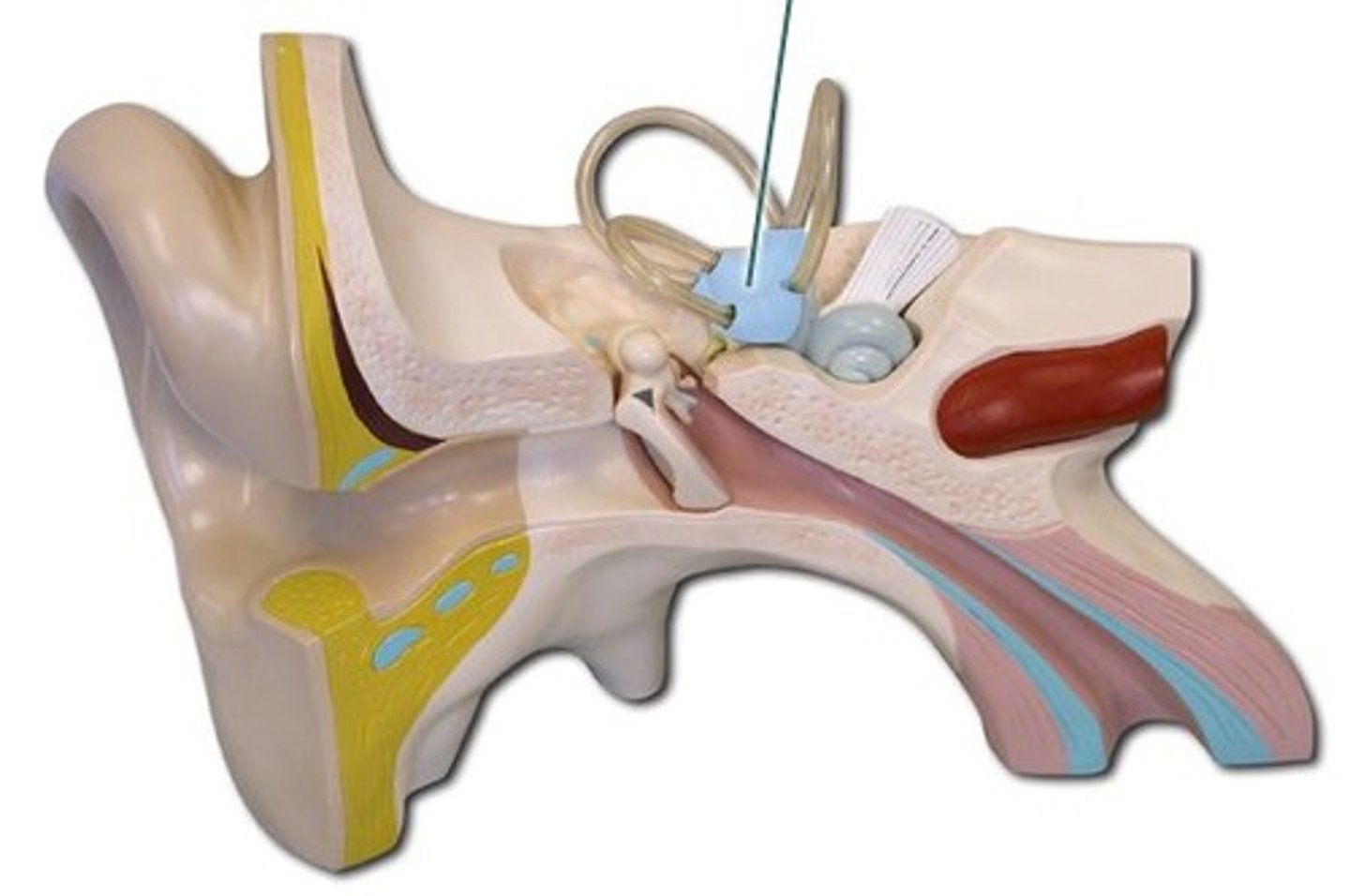
Vestibule
Main function is to detect changes in gravity and linear acceleration therefore, maintaining equilibrium.
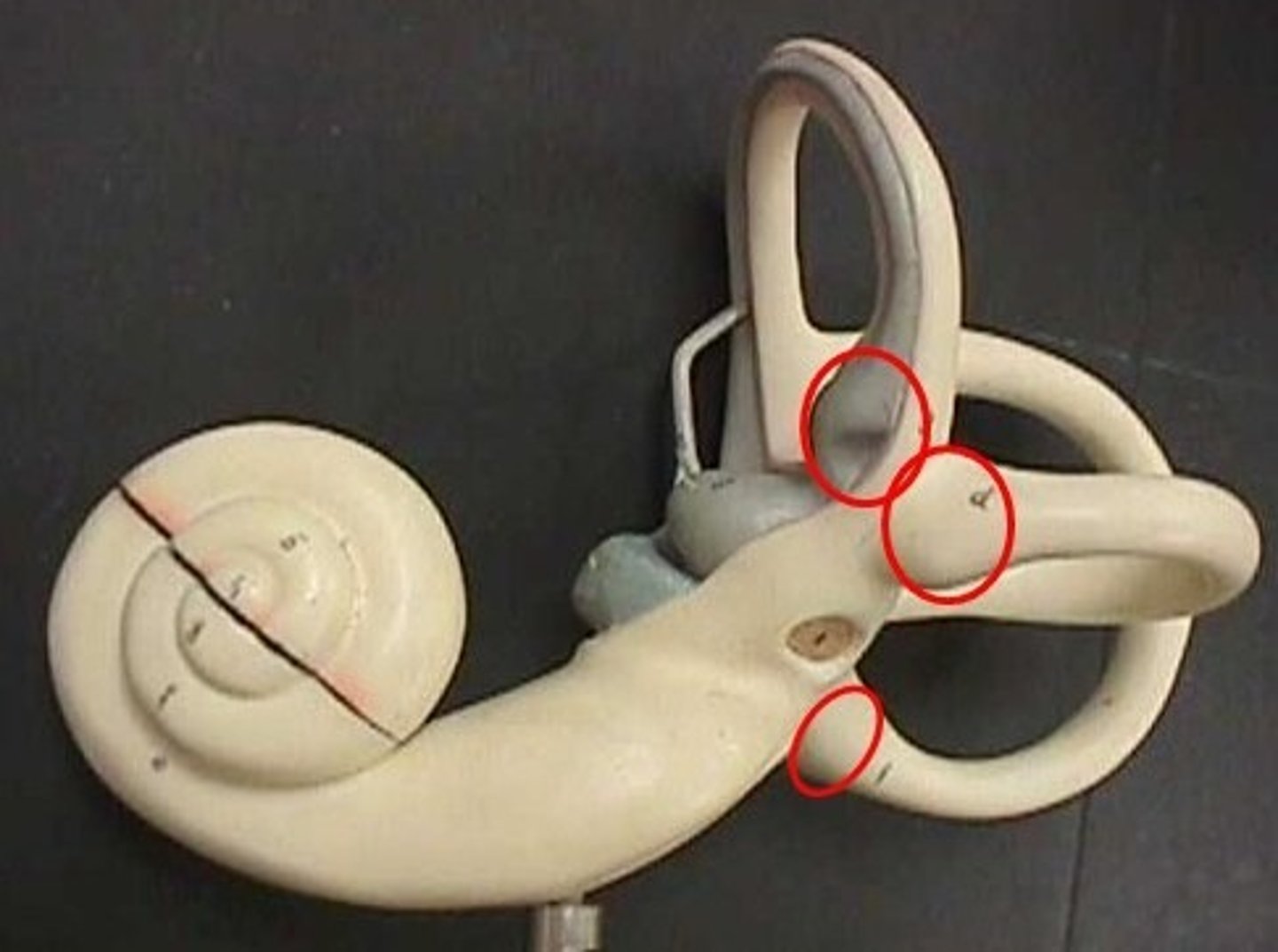
Ampullae
balance and sensory reception. ...canal has a swelling that contains the cupula, a cluster of sensitive hairs embedded in a jellylike mound.
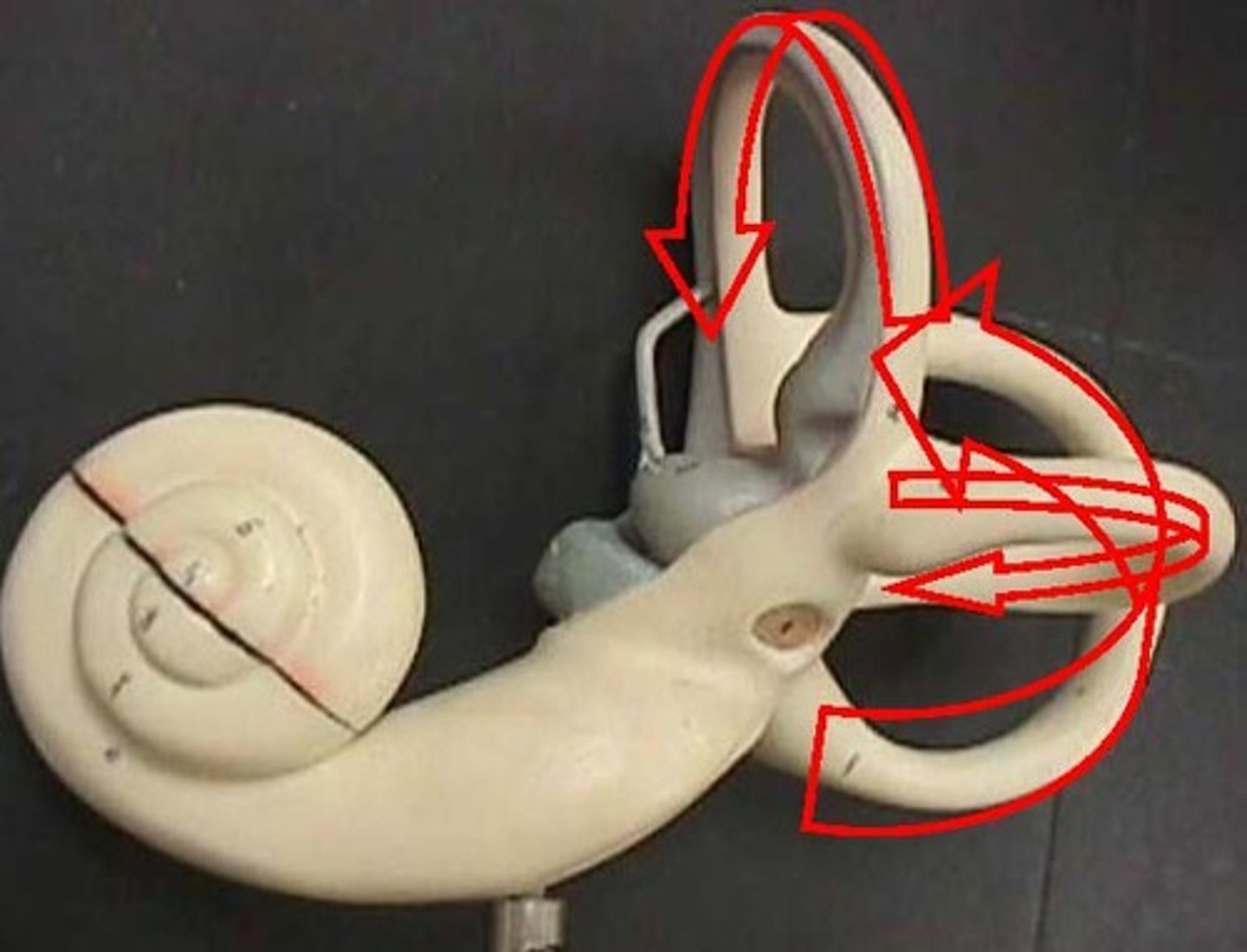
Semicircular Canals
passages in the inner ear associated with maintaining equilibrium
Lacrimal Duct
the passageway that drains excess tears into the nose