psyc 211 - synaptic communciation
1/37
There's no tags or description
Looks like no tags are added yet.
Name | Mastery | Learn | Test | Matching | Spaced | Call with Kai | Chat |
|---|
No analytics yet
Send a link to your students to track their progress
38 Terms
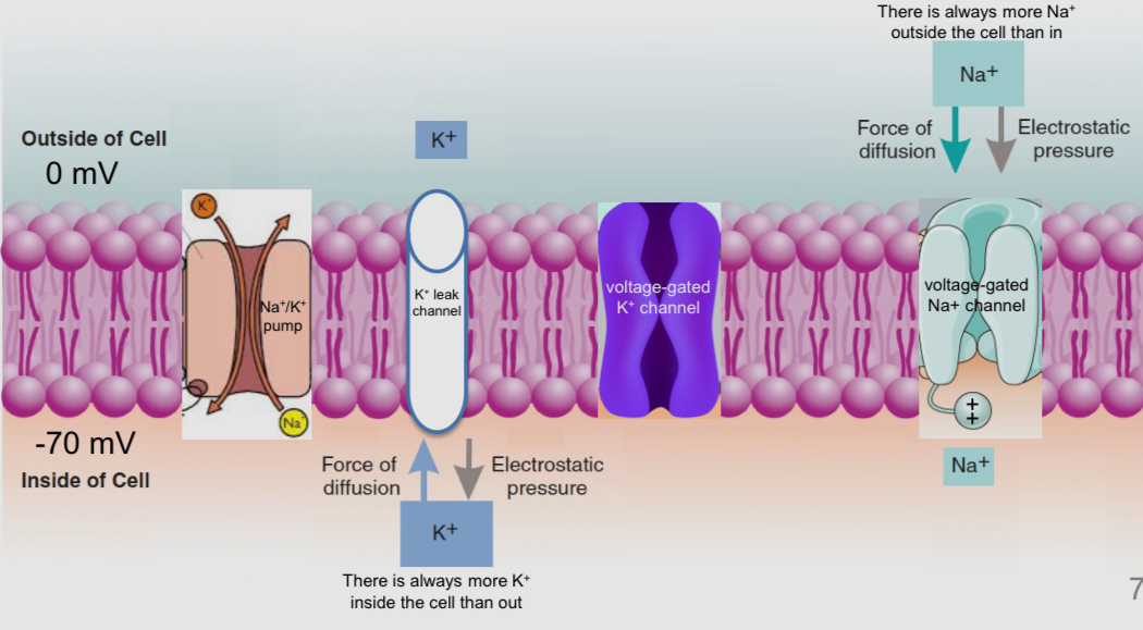
What are the 4 proteins embedded in the membrane of the axon
1) N’a+/ K+ pumps: always pumpking K+ in and Na+ out
2) K+ leak channels: always open
3) voltage gated Na+ channels: responsible for the upward swing of an action potential
4) voltage gated K+ channels: responsible for the downward swing of action potential
How can an ion channel be permeable to K+ but not Na+
K+ had more protons, neutrons, and electrons. It aaslo has the same charge as Na+ when dissolved in water (+1)
What do we know about K+ and Na+ ion channels
their DNA code that is used to make the proteins. So, we also know the string of amino acids that form these proteins
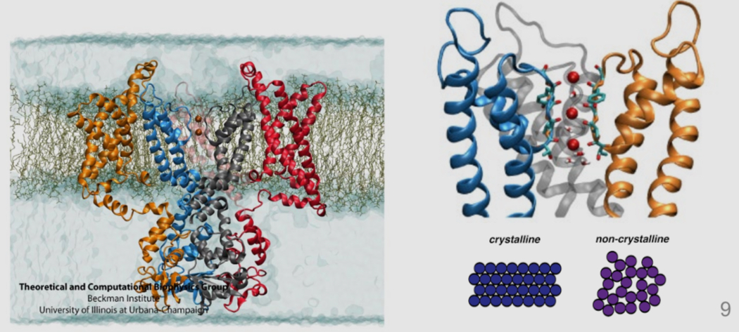
Their precise 3-dimensional shape: the position and arrangement of every atom (via x-ray calligraphy)
What is glycine and what do we know about is
it is one of the bumps that line the pore of the voltage gated potassium channel
We know the 3-nucleotide sequence that’s codes for this amino acid (GGC)
This code can be modified to make cells put a different amino acid in that part of the protein ( GGC → GCC; glycine → alanine
How do we study how ion channels work
Researchers create modified DNA in a lab, and they inject it into a cell, causing it to make alternative versions of the protein of interest
What is a gene promoter
It is the DNA that precedes the gene
It indicates where the gene starts
It indicates which cells should read and when
Ex: a gene promoter might say “if you are a herr cell and are not getting enough sugar, read this gene starting here”
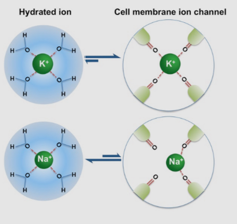
What is an hydrated ion
it is an ion surrounded by a shell of water molecules, when in water
Ion channel selectivity filters are design de to replace the hydration shell of a specific ion
K+ ions are comfortable when surrounded by water and when they pass through the por of a potassium ion channel
Unhydrated Na+ ions are to small to fit comfortably in the pore of a potassium ion channel and are too big to fit when they have their hydration shell
How many genes does the human genome contains for the voltage gated potassium channel
it contains 40 genes
The first voltage gated potassium channel evolved over a billion years ago and today, our genome contains many version of this gene
What are the two main categories of cells in our central nervous system
Neurons: they have an action potential and are responsible for communicating information about sensations and movement
Glial cells: they servo a variety of support functions for neurons
What is the function of astrocytes ( type of glial cell)
They provide a structural matrix
They physically surround blood vessels, regulating blood flow and nutrient distribution
They physically synapses, regulation the concentration of ions in then extracellular space and helping to clear away neurotransmitters
What is the function of Ependymal cells (glial cells)
they are primarily in the middle of the brain and spinal cord
They help circulate the fluid that surrounds neurons (i.e. extracellular fluid)
What is the function of microglia (glial cell)
they are the brain’s clean up crew and the smallest of the glial cells
They remove dead cells and other debris
They serve an immune function to protect the brain from invading micro-organisms
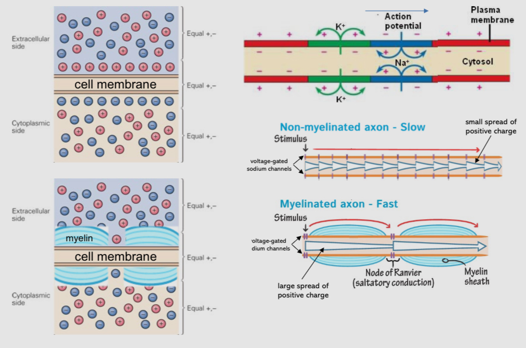
What is the function of oligodendrocytes
they produce the myelin sheaths, which speed up neuronal action potentials
How do myelin sheaths form
Oligodentrocytes produce large branches of cell membrane. Each branch wraps many times around a nearby axon, which forms the myelin sheath
Myelin is just a wrapping of fat which electrically insulates the bacon and speeds up the action potential
Most axons in the brain are heavily myelinated
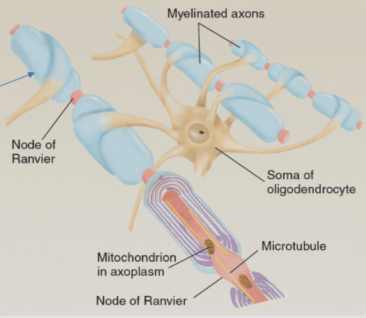
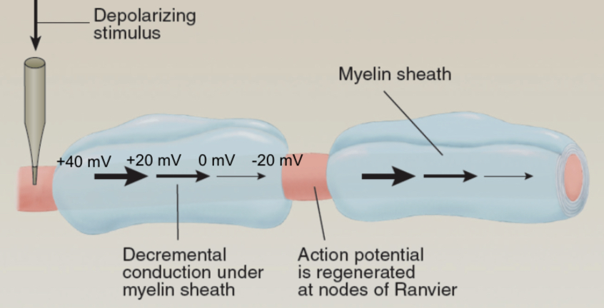
What are nodes of Ranvier
They are the exposed segments of an axon covered by myelin sheath
These are the only places where myelinated axons feel the charge differences between inside and out
They are perfectly space out sock the influx of positive current at one node is strong enough to reach the voltage gated channels at the next node
What is the distribution of ions within an axon
What is the impact of myelination
myelination speeds up conduction of the action potential 20x
Action potential in myelinated axons appears to jump from one node of Ranvier to the next (satisfactory conduction)
The amplitude of the action potential (+40 mV) is regenerate at each node of Ranvier because this is the only place where a myelinated axons feel had access to extracellular fluid
All the voltage gated ion channels in a myelinated axons appears are concentrated at the nodes of Ranvier
Action potential speed also depends on the thickness of the axon. Thick → fast and thin → slow
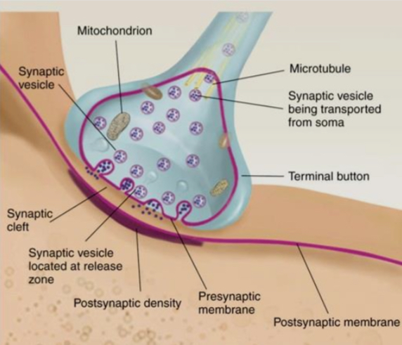
What is a synapse and how does it communicate
It is a junction between the axon terminal of the sending neuron and the cell membrane Of the receiving neuron
Communication of the synapse is mediated by the réalises of a signaling molecule (a neurotransmitter) from the axon terminal
When a neurotransmitter activates a receptor on the receding neuron, the consequences can be excitatory, inhibitory or modulatory (i.e. depolarizing, hyperpolarizing, or more complicated)
What are the parts of the synapse
synaptic vessel; contains neurotransmitters dock at the presynaptic membrane
Axon terminal: end of the axon
Presynaptic membrane: where the neurotransmitters are released
Synaptic cleft: the space between the pre and postsynaptic membranes where neurotransmitters are released
Postsynaptic membrane: the area where neurotransmitters diffuse across from the synapse and where they can actrice receptors in the postsynaptic density
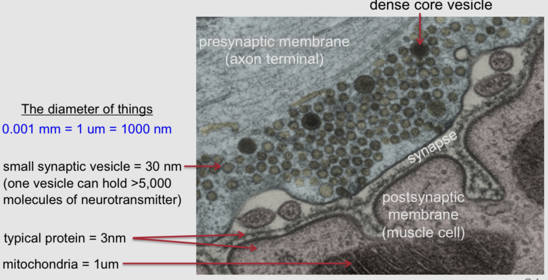
What is electron microscopy
it allows to see small anatomical structures like synaptic vessels.
It was used to create a photo of a synapse between a motor neuron and a muscle cell in the common frog
What is a ligand
it is a receptor activated by a signaling molecule
sign,aiding witching and between cells occurs through ligand-receptor interactions
What are the two categories of neurotransmitter receptor
ionotropic receptros: ion channels
metabotropic receptors: not ion channels; they mostly mediate their effects through intracellular G protein signaling cascades
Where are intracellular and surface receptors located
intracellular: inside the cell
surface: on the cell membrane
What are the types of surface receptors and where are they located
postsynaptic → on the synaptic membrane
Presynaptic→ on the presynaptic membrane
Extrasynapric → near but outside a synapse
What is a ligand
it is a general term for a signaling molecule that can bind to a receptor
Neurotransmitters are ligands
What is a binding site
It is the place on a receptor where a ligand binds
What is a postsynaptic receptor
it is a receptor located on the postsynaptic membrane
It can be ionotropic or metabotropic (most synapses contain both
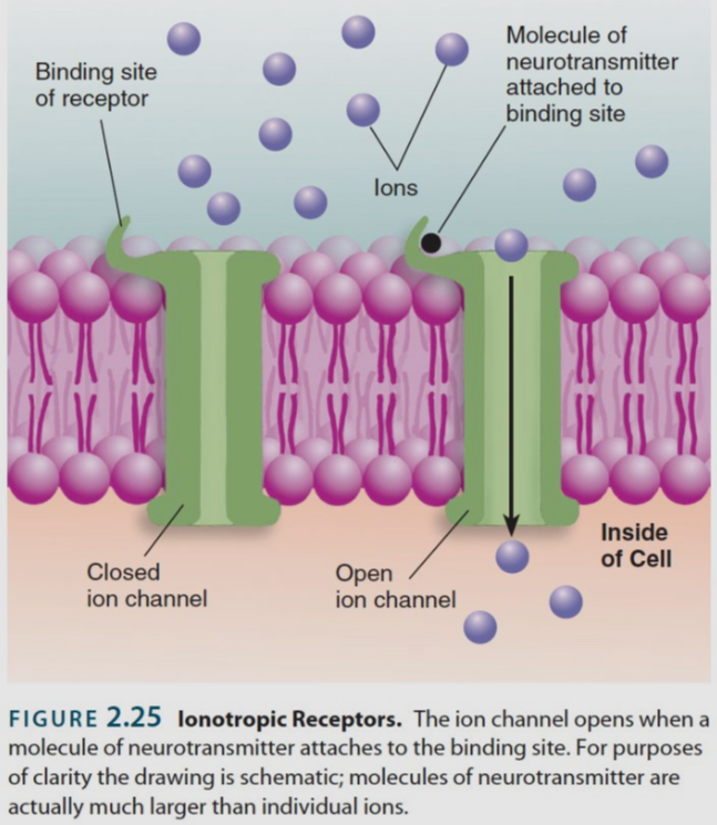
What is an ionotropic receptor
it is a ligand-gated ion channels
it is an ion channels that opens up in response to ligand binding
Its effects on the membrane potential are very brief and peak within a few milliseconds
What is a metabotropic receptor
it is a receptor that is not an ion channel
Ligand binding usually triggers an intracellular G protein signaling cascade, which can have diverse effects on cell function
These sign,sing cascades take time and effects are usually not evident for at least 100ms of not much longer
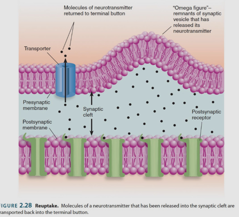
What after the three mechanisms involved in neurotransmitters signaling in the synapse
diffusion: passive movement from areas of high concentration to area of low concentration
Enzyme deactivation: destruction of a neurotransmitter by an enzyme
Reuptake: they recycle neurotransmitters by pulling them back into the cell that just released them (e.g. the serotonin reuptake transporter)
What is a postsynaptic potential
it is when a neurotransmitter binds to a postsynaptic receptor and changes the membrane potential of the synaptic cell. Ionotropic receptors → produce rapid postsynaptic potentials. Metabotropic receptors → do not always produce postsynaptic potentials, but when they do, they are relatively slow.
Excitatory postsynaptic potentials: the result of positive sodium ions entering the postsynaptic cell, causing membrane de polarization and perhaps an action potential
Inhibitory postsynaptic potentials: the result of negative chloride ions entering the cell, causing membrane hyper polarization and no action potentials
Ionotropic receptors are classified as inhibitory or excitatory based on whether they let in Na+ or Cl- ions
Metabotropic receptors are classified based on wether they cause Na+ or Cl-
What is depolarization in the membrane potential
it is when the membrane potential of a cell becomes less negative than it normally is at rest
The opening of Na+ ion channels will depolarize a neuron making it more likely to have an action potential
What is hyper polarization in the membrane potential
it is when the membrane potential of a cell becomes more negative than it normally is at rest
The opening of Cl- ion channels can hyperpolarize a neurons, making it less likely to have an action potential
What is neural integration
it is the interaction between excitatory and inhibitory synapses on a neuron
IPSPs decrease the likelihood that a neuron will fire
When EPSPs and IPSPs occurs at the same time, the influx of negatively charged chloride ions diminish the impact of the positively charged sodium ions

How is exhibition or inhibition of a neurotransmitter is determined
each type of neurotransmitter can activate multiple types of receptors (14 kinds of serotonin receptors → 1 kind of serotonin molecule
Some receptors are inhibitory, while some are excitatory
It is the receptor that is expressed by the postsynaptic cell that determines wether a neurotransmitter will be excitatory or inhibitory, not the neurotransmitter itself
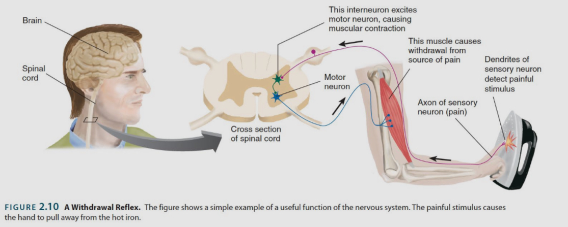
what are the steps of the reflex neural circuits
A sensory neuron spikes and the message travels down its axon to the spinal cord
it releases a neurotransmitter onto an interneuron, causing the cell to depolarize and spike
The inter neuron relaxes neurotransmitter into a motor neuron, causing it to depolarize and spike
The motor neuron releases neurotransmitter onto a muscle finer, causing it to contract (withdrawal reflex)
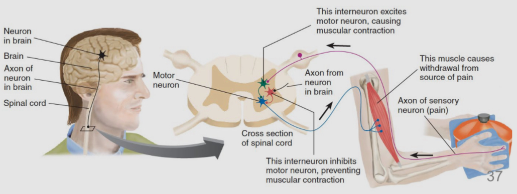
What happens when a neuron in the cerebral cortex can anticipate pain
it will send an action potential down the spinal cord to override this chain of event
The cortical neuron can trigger an action potential in a different interneuron in the spinal cord, one that had an inhibitory influence on motor neurons
When the interneuron spikes and releases neurotransmitter onto a motor neuron, it causes the motor neuron to hyperpolarize and not spike, thus counteracting the withdrawal reflex
This circuit depicts a context between two competing drives ( drop the pan or keep holding it)
Neural excitation and behavioural excitation
inhibition of inhibitory neurons generates motor behaviour
The firing of excitatory neurons in the brain does not necessarily cause movement and the firing of inhibitory neurons does not necessarily inhibit movement