Upper Extremity Venous Anatomy and Evaluation
1/16
There's no tags or description
Looks like no tags are added yet.
Name | Mastery | Learn | Test | Matching | Spaced |
|---|
No study sessions yet.
17 Terms
What are radial veins?
Paired vessels that originate from dorsal metacarpal veins and terminate at antecubital fossa
What are ulnar veins?
Paired vessels that originate from palmar arch and terminate at antecubital fossa
What are brachial vein(s)?
Vessel(s) that joins basilic vein to become axillary vein
What is the axillary vein?
Continuation of brachial vein(s) that joins cephalic vein to become subclavian vein
What is the subclavian vein?
Vessel formed by axillary and cephalic junction that joins IJV to become innominate or brachiocephalic vein
What is the internal jugular vein (IJV)?
Vessel formed by confluence of venous sinuses in brain that joins subclavian vein to become innominate or brachiocephalic vein
What is the brachiocephalic or innominate vein?
Vessel formed by confluence of subclavian vein and IJV
What is the SVC?
Vessel formed by confluence of right and left brachiocephalic or innominate veins
What is the basilic vein?
Superficial vessel in forearm and deep vessel in proximal arm that originates at dorsal venous arch and courses along medial arm to join brachial vein
What is the cephalic vein?
Superficial vessel that originates at dorsal venous arch and courses along lateral arm to join axillary vein
What is the antecubital vein or medial cubital vein?
Superficial vessel that connects cephalic and basilic veins in antecubital fossa
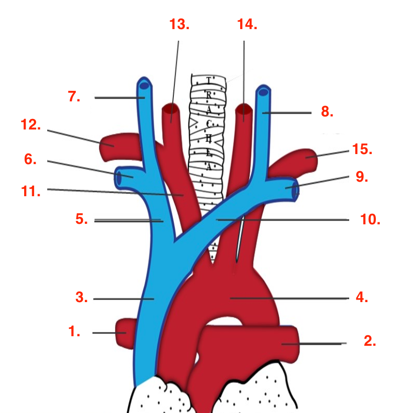
Identify this image.
Right pulmonary artery
Left pulmonary artery
SVC
Aortic arch
Right innominate or brachiocephalic artery
Right subclavian vein
Right IJV
Left IJV
Left subclavian vein
Left innominate or brachiocephalic artery
Innominate or brachiocephalic artery
Right subclavian artery
Right CCA
Left CCA
Left subclavian
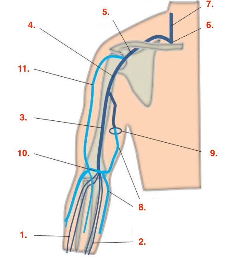
Identify this image.
Radial veins
Ulnar veins
Brachial vein
Axillary vein
Subclavian vein
Brachiocephalic or innominate vein
IJV
Basilic vein
Superficial basilic vein becomes deep basilic vein
Antecubital vein
Cephalic vein
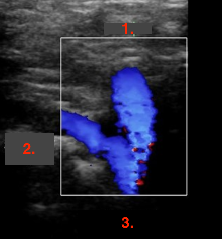
Identify this image.
IJV
Subclavian vein
Brachiocephalic or innominate vein
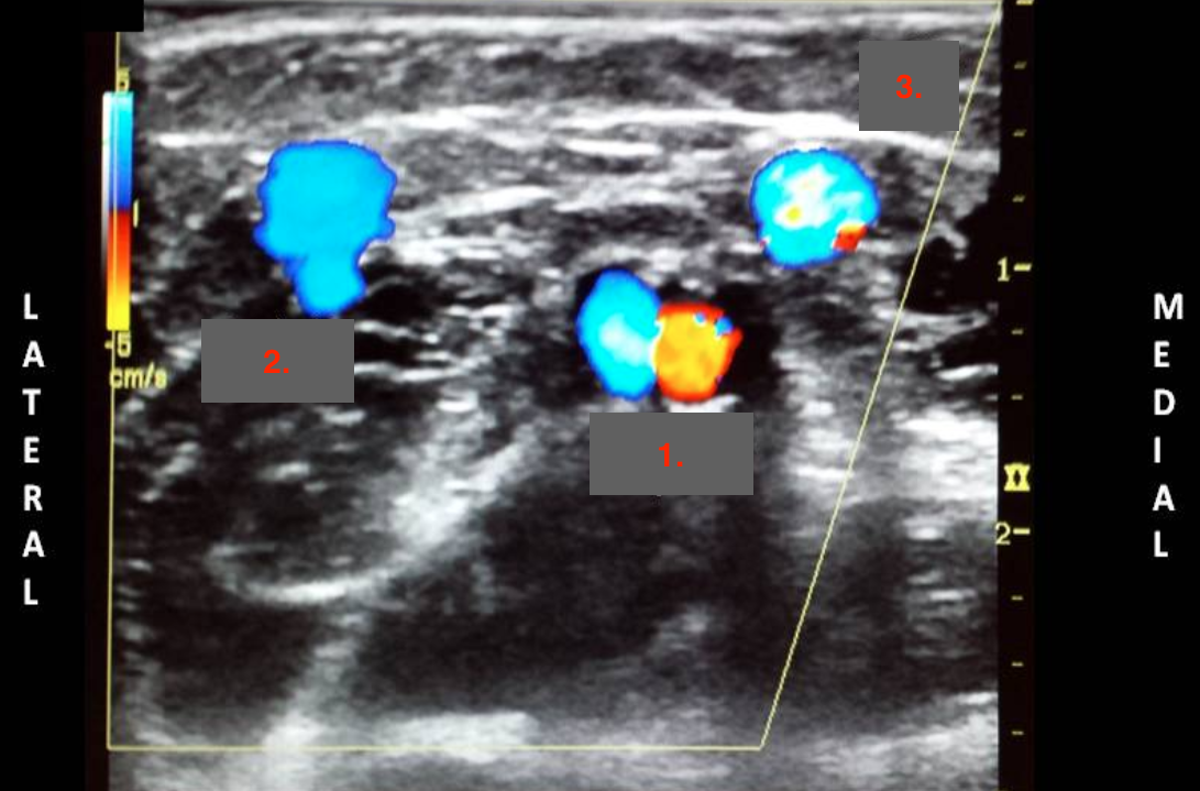
Identify this image.
Brachial vein paired with brachial artery
Cephalic vein
Basilic vein
How is an upper extremity venous exam performed?
Evaluate IJV and subclavian veins in supine
Ask patient to sniff to visualize IJV and subclavian compressibility
Evaluate remainder of upper veins with head of bed elevated
What is the normal sonographic appearance of an upper extremity venous exam?
Veins of shoulder and upper arms demonstrate spontaneous and phasic flow WITH cardiac pulsatility
Forearm veins DO NOT demonstrate spontaneous flow
Superficial veins DO NOT demonstrate spontaneous flow