Lecture 10- Edema, congestion, hyperemia
 Call Kai
 Learn
Learn Practice Test
 Spaced Repetition
 Match
Match Flashcards
 Knowt Play
1/77

There's no tags or description
Looks like no tags are added yet.
Name | Mastery | Learn | Test | Matching | Spaced | Call with Kai | Chat |
|---|
No analytics yet
Send a link to your students to track their progress
78 Terms
what percent of the lean body weight is water
60%
what is the distribution of body water
66% intracellular
25% extracellular
8% of total body water in blood plasma
tissue well being also requires a normal
fluid balance
abnormalities in vascular permeability or hemostasis can result in
injury even with an intact blood supply
normal fluid homeostasis encompasses maintenance of
vessel wall integrity as well as intravascular pressure and osmolarity within certain physiologic ranges
what all affects the net movement of water across the vascular wall
changes in vascular volume, pressure, or protein content
Alterations in any of the factors which regulate
normal fluid distribution resulting in fluid
accumulation in the interstitium results in
edema
edema
increased fluid extravasation into interstitial/ extracellular spaces
what is edema easily recognized by
clear to yellow gelatinous material
where is edema and effusion found
subcutiss
brain
lungs
any tissue
how does edema manifest microscopically
as clearing and seperation of the intracellular matrix elements
what does effusion appear as
clear to yellow fluid
starling’s forces describes
movement of fluid between blood and tissues with hydrostatic and oncotic pressures being the major forces involved
push pressure
hydrostatic
pull pressure
oncotic
swollen
distended, tends to gravitate ventrally (sagging tissues)
tissue pits
indetations remain after pressure is removed
what temperature is edema usually to touch
cool
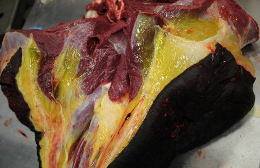
edema
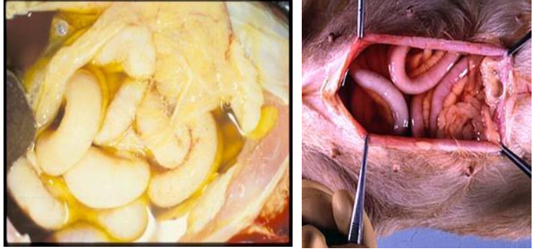
effusion
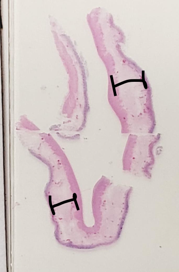
edema
with edema what will be seen with lymphatic vessels of histology
will be dilated as they are the natural channel for removal of excess fluid
what prefix is added to indicated effusion
hydro
hydrothorax
fluid in pleural cavity
hydropericardium
fluid in pericardial sac
hydrosalpinx
fluid in uterine tube
hydrocephalus
accumulation of fluid in the brain
hydrocoele
fluid filled cyst anywhere in body
hydroperitoneum (ascites)
fluid in the peritoneal cavity
anasarca
severe and generalized edema, with profound SQ tissue swelling
non inflammatory effusion
transudate
protein levels of transudate
low
transudate
fluid accumulation due to hydrostatic imbalances between intravascular and extravascular compartments DESPITE normal vascular permeability
clear, colorless, or slightly yellow
modified transudate
• In-between where protein content is high but not as
high as with exudate
• Most common in FIP
inflammatory effusion is also known as
exudate
eudate
related to increased endothelial permeability
high protein levels
caused by leakage of plasma proteins (albumin) and leukocytes
usually opaque
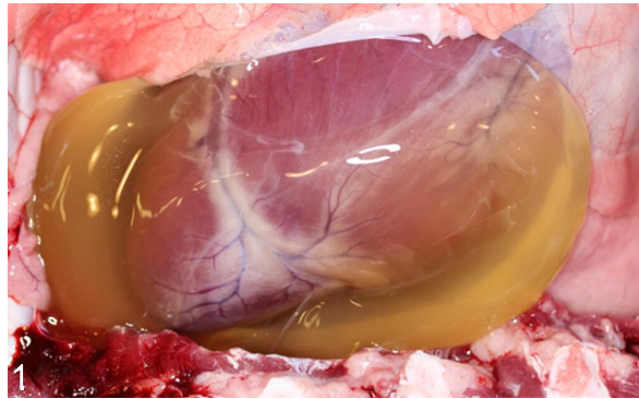
transudate
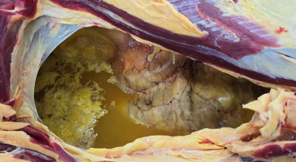
exudate
intracellular edema
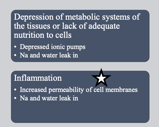
extra cellular edema
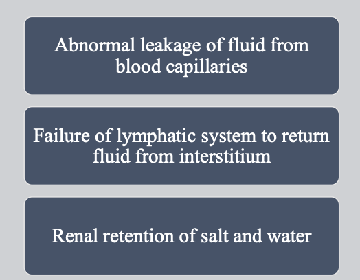
mechanisms of edema ***
increased microvascular permeability
increased intravascular hydrostatic pressure
decreased oncotic pressure
decreased lymphatic drainage
in increased microvascular permeability what does inflammation or endothelial damage result in
increased capillary permeability to fluids, salts, and colloids
during increased microvascular permeability inflammation results in release of
mediators to cause vasodilation
vasodilation results in
widening of the interendothelial gaps
water moves from ______ into _________
intravascular space into the interstitium
increased microvascular permeability manifest grossly as
edema/ effusion
venous obstruction or impaired venous outflow
• Results in increased hydrostatic pressure as blood
backs up in the venous system
• Causes leakage of sodium and fluid into the
interstitial tissue
• No significant leakage of colloids
main thing that occurs with increased intravascular hydrostatic pressure
venous obstruction or impaired venous outflow
mechanisms of increased intravascular hydrostatic pressure
• Increased blood flow = hyperemia
• Passive accumulation of blood = congestion
• Increased blood volume
increased intravascular hydrostatic pressure causes
congestive heart failure
cirrhosis of the liver
obstruction or narrowing of the veins
capillary blood contains a decreaed quantity of colloids due to
• Decreased hepatic synthesis of proteins
• Increased protein loss through the kidney or GI tract
what happens as a result of hypoproteinemia
fluid and sodium are not reabsorbed at the venous end of the capillary
fluid accumulates in the interstitium as edema
decreased oncotic pressure causes
• Kidney disease
• Cirrhosis of the liver/liver failure
(hypoalbuminemia)
• Malnutrition/starvation
• Protein-losing gastroenteropathies or nephropathies
• Gastrointestinal parasitism
• Blood loss
decreased lymphatic drainage causes
Normally, small quantities of fluids, salts, and
colloids escape from capillaries and are drained
from the interstitium by lymphatics
why do lymphatic obstructions occur with decreased lymphatic drainage
these materials
accumulate because of the reduced ability of the
lymphatic system to remove excess fluid
decreased lymphatic drainage causes
• Inflammation
• Lymphatic vessel compression
(neoplasia or inflammation)
• Lymph vessel constriction (fibrosis)
• Lymph vessel blockage (thrombus or embolus)
• Post-surgical
• Post-irradiation
hyperemia
an active process resulting in increased tissue blood flow due to arteriolar dilation
excess blood of arterial origin
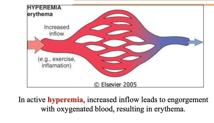
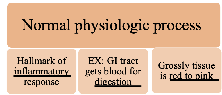
examples of hyperemia
skeletal muscles during exercise
at sites of inflammation
what color is hyperemia
red because of engorgement with oxygenated blood
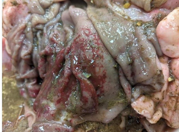
hyperemia
congestion
a passive process resulting from impaired outflow from a tissue
interference with venous drainage
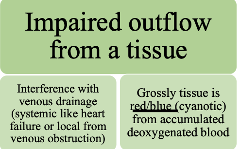
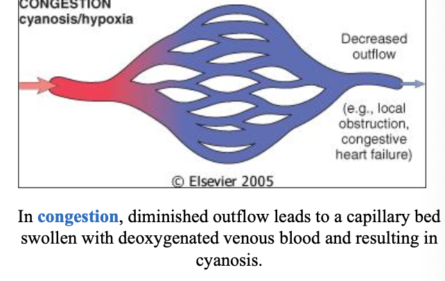
what might congestion interference with venous drainage be
systemic as in heart failure
local due to isolated venous obstruction
what color is congestion
red-blue (cyanosis) due to the accumulation of deoxygenated blood
congestion causes
• Heart failure (generalized slowing of blood everywhere)
• Left heart – lung most affected (chronic pulmonary congestion)
• Right heart – liver (chronic passive congestion)
• Venous occlusion (localized) – thrombus, pressure, twisting
• Hypostasis – hypostatic congestion
blood flow
active hyperemia
passive congestion
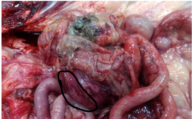
active hyperemia
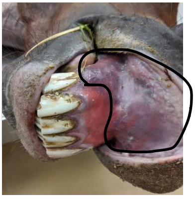
passive congestion
hypostatic congestion
Blood pooling in organs and tissues on the lower side of a recumbent animal (contrast to livor mortis)
chronic passive congestion
Caused by disease that
results in right heart failure
chronic pulmonary congestion
Caused by any disease that
results in left heart failure
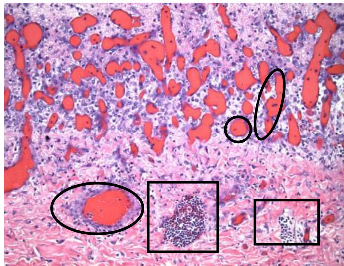
hyperemia
what occurs in the histology of hyperemia
• Arterioles and capillaries are dilated and filled with
blood (may appear more numerous)
• If the cause is inflammatory (usually is), other morphologic features of inflammation may also be present
• Inflammatory cells
• Necrotic cells
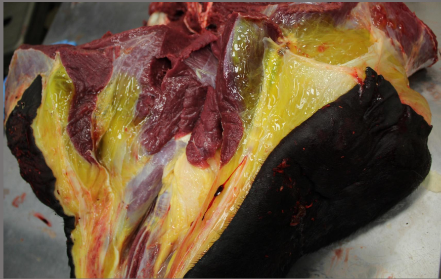
SQ tissue edema
transudate
locally extensive
severe
acute
notice that it is gelatin like
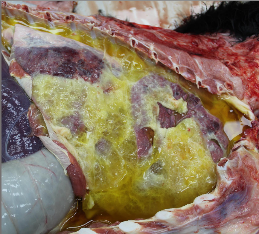
lungs: pleural edema, multifocal to locally extensive, severe, acute
thoracic cavity: thoracic effusion (or hydrotorax), diffuse, moderate, acute
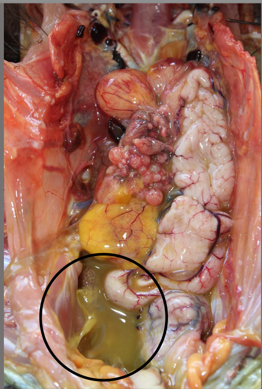
exudative effusion
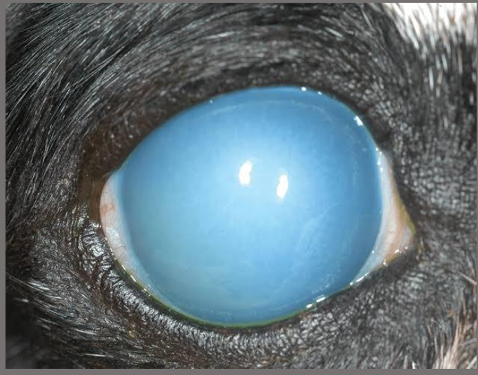
corneal edema
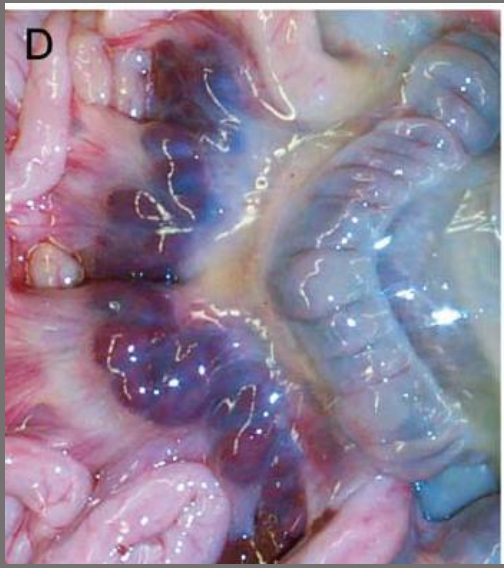
small intestine: mesenteric edema, locally extensive, mild, acute
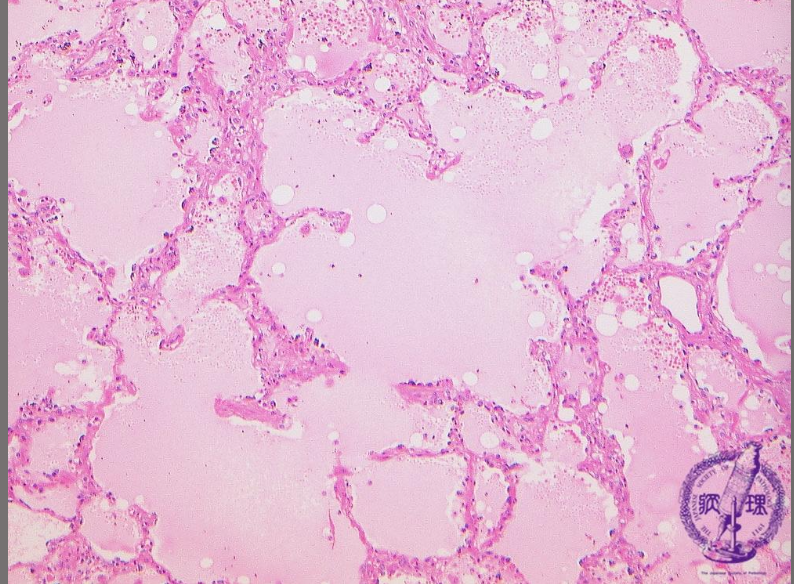
lung: pulmonary edema, diffuse, severe, acute