Chapter 2 Microbiology Practice (Light and Microscopes)
1/75
Earn XP
Description and Tags
DSfiohfdsfaklfhjdaahdskmotherkjdhfkhadflkjahslkdjfhdksahkjsadhfdsaf
Name | Mastery | Learn | Test | Matching | Spaced | Call with Kai | Chat |
|---|
No analytics yet
Send a link to your students to track their progress
76 Terms
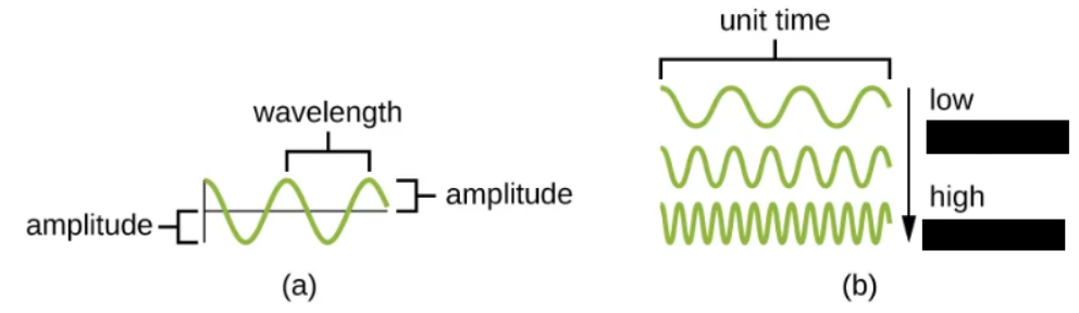
Wavelength
The distance between one peak of a wave and the next peak
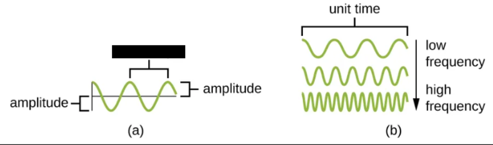
Amplitude
The height of each peak or depth of each trough
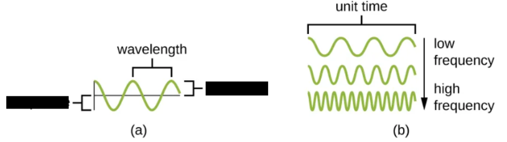
Frequency
The rate of vibration of a wave, or the number of wavelengths within a specified time period.
Reflection
Occurs when a wave bounces off another material
Absorbance
Occurs when a material captures the energy of a light wave
Transmittance
The process of transmission of light, occurring when a wave travels through a material
Transparency
an object's permeability to light
Opacity
An object's ability to block the passage of light
Interference
Creating complex patterns of motion (two light waves can interact with each other in this way)
Diffraction
When light waves interact with small objects or openings by bending and scattering
Refraction
Occurs when light waves change direction as they enter a new medium.
Refractive Index
The extent to which a material slows transmission speed relative to empty space
Image Point
Also called the focus. It is where all the light that is refracted by the lens comes to hit.
Focal Point
The image point where light entering the lens is parallel
Focal Length
The distance to the focal point for convex/concave lenses
more
Higher frequency waves contain ____ energy than lower-frequency waves. The energy is delivered as elementary particles called photons.
red
The lowest frequency of visible light appears as the color ___
violet
The highest frequency of visible light appears as the color ______
Dispersion
Separation of colors by means of refraction, such as white light through a prism. Occurs because the refractive index is different for different frequencies of light through a given material
Fluorescent
Having the ability to absorb ultraviolet or blue light, and then use the energy to emit photons of a different color, giving off light instead of vibrating.
Phosphorescence
The process of photons being emitted after absorption with a delay. Ex. glow-in-the-dark plastic
Magnification
The ability of a lens to enlarge the image of an object when compared to the real object
Resolution
The ability to tell that 2 separate points or objects are separate. Low-________ appears fuzzy, and high-________ appears sharp
Numerical Aperture
A measure of a lens’s ability to gather light. The higher, the better the resolution
Compound Microscope
A microscope that utilizes many lenses to multiply the magnification of an image
Simple Microscope
A microscope that utilizes a single lens
Light Microscopes
Microscopes that use light to visualize images
Brightfield Microscopes
Compound microscopes with 2+ lenses that produce a dark image on a bright background. Some are monocular and some are binocular.
Monocular
Utilizes one eyepiece
Binocular
Utilizes two eyepieces
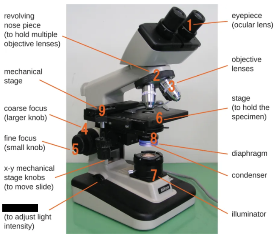
Ocular lens
Lens that typically magnifies images 10 times, usually located on the eyepiece
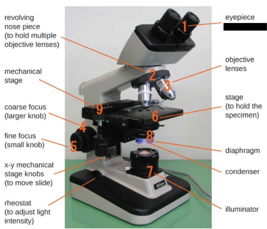
Objective Lens
A lens on a rotating nosepiece, ranging from 4-100 times magnification
Total Magnification
The product of ocular magnification times objective magnification.
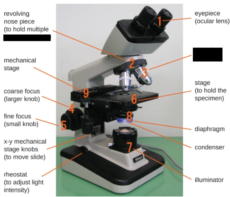
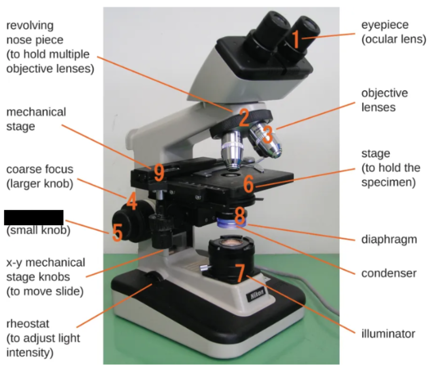
Stage
Platform of the microscope
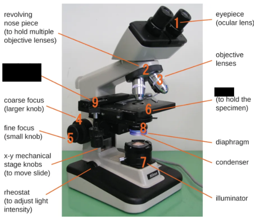
X-Y Mechanical Stage Knobs
Knobs that move the slide on the surface of the stage, but do not raise or lower the stage
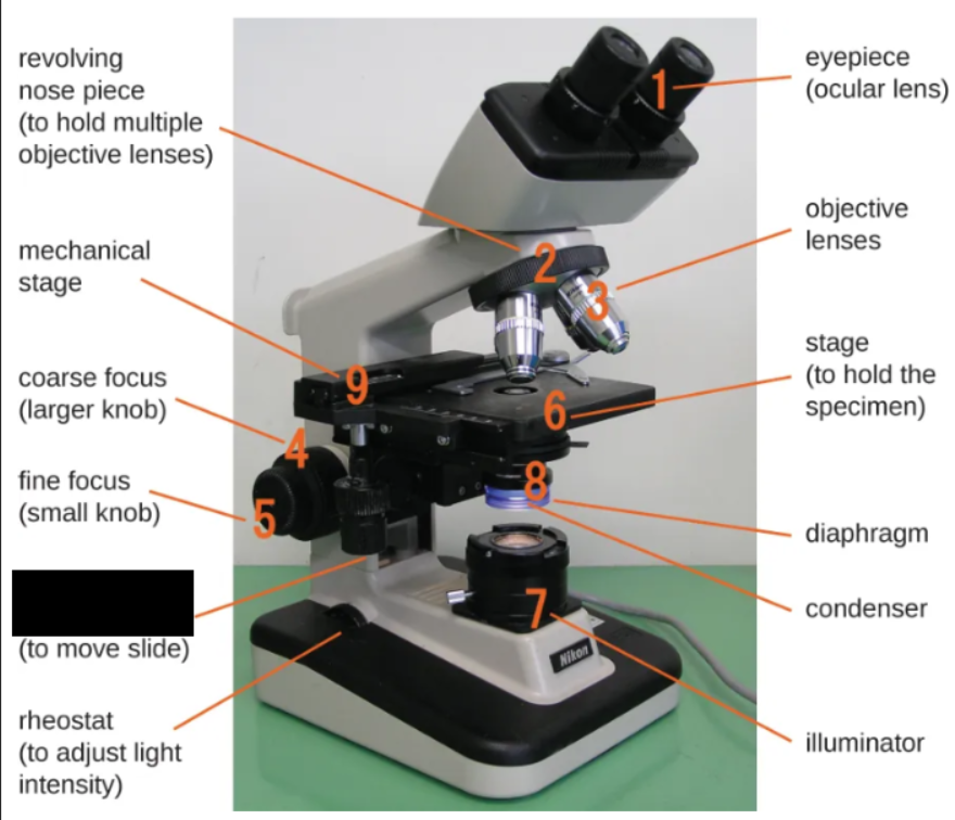
Coarse Focusing Knob
Knob used for large-scale movements with 4-10x objective lenses
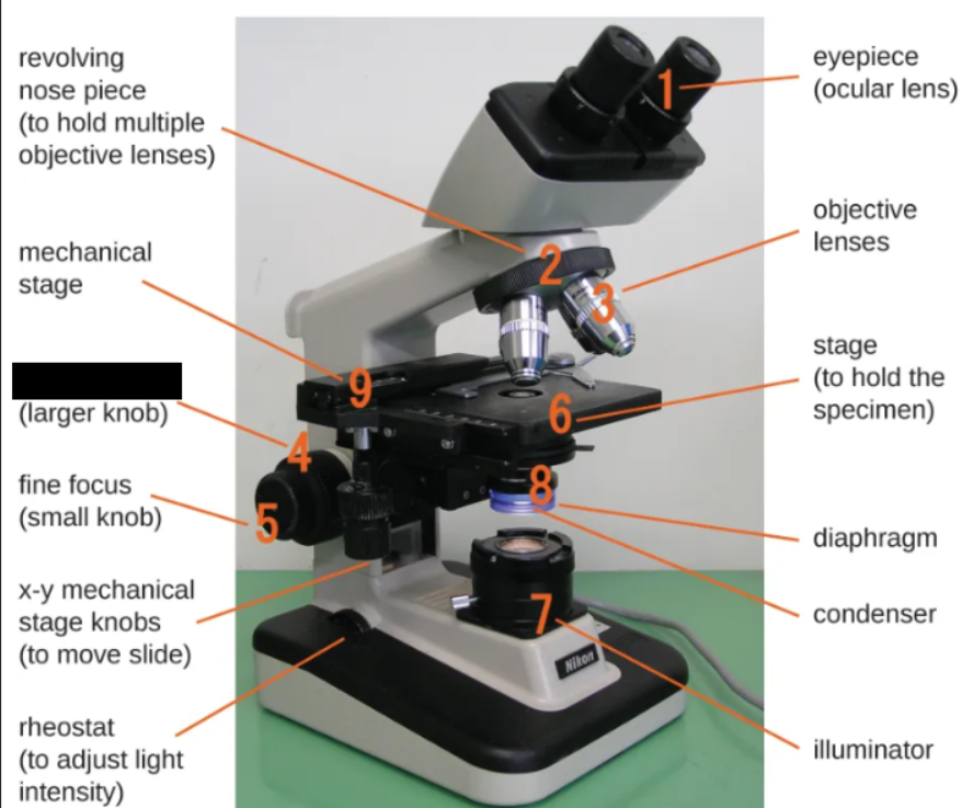
Fine Focusing Knob
Knob used for small-scale movements, typically with 40-100x objective lenses
dimmer
When images are magnified, they become ________ because there is less light per unit area of image.
Illuminator
A high-intensity bulb below the stage
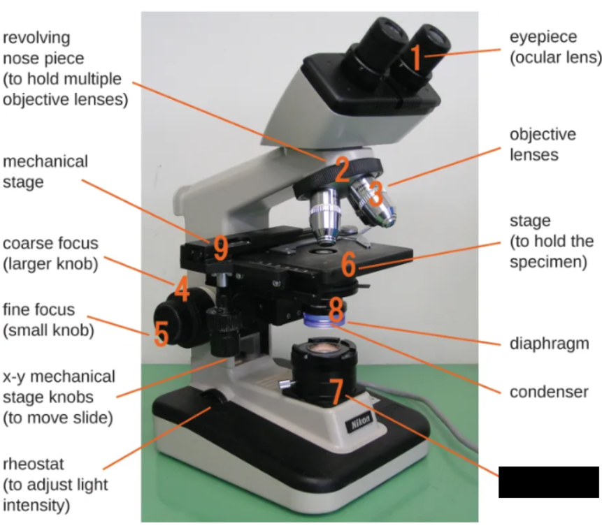
Condenser Lens
Lens below the stage that focuses light on the specimen
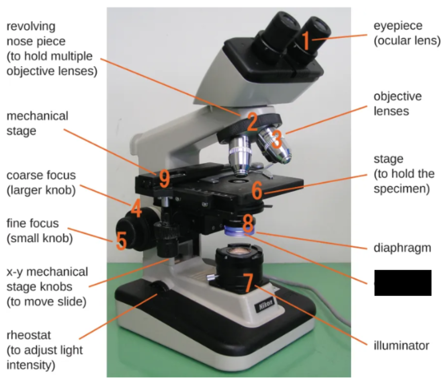
Diaphragm
Can adjust the light striking the specimen by opening and closing
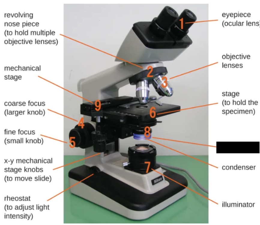
Rheostat
A dimmer switch that controls the intensity of the illuminator
Chromophores
Pigments that absorb and reflect particular wavelengths of light. They can be added to the specimen using stains
Oil Immersion Lens
Lens that utilizes a drop of immersion oil to fill the space between itself and the specimen. Used to heighten resolution at high magnifications
Darkfield Microscope
A brightfield microscope that has a small but significant modification to the condenser. A small opaque disc (1cm diameter) is placed between the illuminator and the condenser lens, called an opaque light stop. It blocks most light from the illuminator and the resulting image shows bright objects on a dark background.
Doesn't require the use of stains, which is useful for viewing live specimens that the stains could kill or compromise
Phase-Contrast Microscopes
Microscopes that use refraction and interference caused by structures in a specimen to create high-contrast, high-resolution images without staining. The oldest and simplest type of microscope.
DIC Microscopes
Also known as Differential Interference Contrast Microscopes, use two beams of light created in which the direction of wave movement differs. The beams pass through the space (with or without specimens) and are recombined, resulting in high-contrast images of living organisms with a 3d appearance.
Fluorescence Microscope
Microscope that uses fluorescent chromophores called flurochromes, which are capable of absorbing energy from a light source and then emitting the energy as visible light
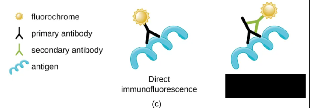
Immunofluorescence
A technique used in fluorescence microscopy used to identify certain disease-causing microbes by observing whether antibodies bind to them. There are two approaches.
DFA
Immunofluorescence technique where specific antibodies are stained with a fluorochrome. If the specimen contains the targeted pathogen, one can observe the antibodies binding to the pathogen under the fluorescent microscope
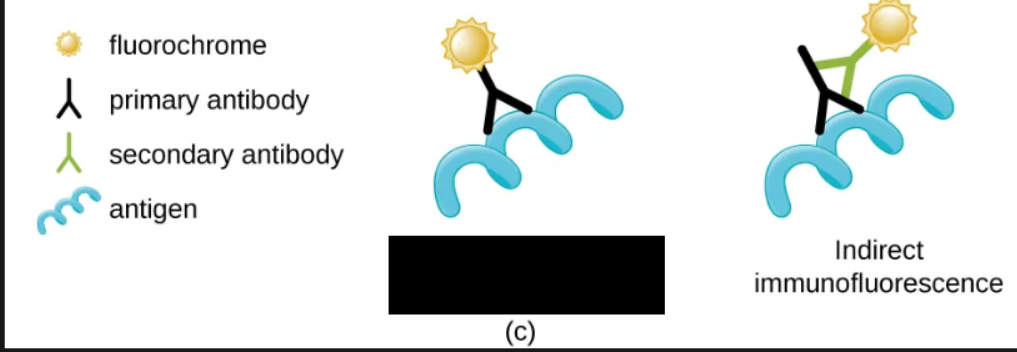
IFA
Immunofluorescence technique where secondary antibodies are stained with fluorochrome rather than primary antibodies. Secondary antibodies do not attach directly to the pathogen, but they bind to primary antibodies. When the primary antibodies bind to the pathogen, the secondary antibodies can be observed. Makes it easier to visualize features in the specimen.
Confocal Microscope
A microscope that uses a laser to scan multiple z-planes (depth) successively. Produces numerous 2d high-resolution images at various depths, which can be constructed into a 3d image by a computer. Stains are generally used to increase contrast and resolution
Two-Photon Microscope
A microscope that uses a scanning technique, fluorochromes, and long-wavelength light (like infrared) to visualize specimens. Useful for examining living cells with intact tissues, brain slices, embryos, whole organs, and entire animals, since it’s low energy light is less damaging to cells. The long-wavelengths make it easy to penetrate through the specimen
Electron Microscope
A microscope that uses short-wavelength electron beams rather than light to increase magnification and resolution. Can produce sharp images magnified up to 100,000x. There are two types: TEM and SEM
TEM
Also known as the transmission electron microscope, uses an electron beam from above the specimen that is focused using a magnetic lens (rather than a glass lens) and projected through the specimen onto a detector.
SEM
Also known as a Scanning electron microscope, forms images of surfaces of specimens, using electrons that are knocked off of specimens by a beam of electrons, creating highly detailed images that look 3d. These images are displayed on a monitor.
STM
Also called the scanning tunneling microscope, is a microscope that uses a probe that is passed above the specimen, as constant voltage bias creates an electric current between the probe and the specimen. This maps the structure of surfaces at a resolution at which individual atoms can be detected
AFM
Also called the atomic force microscope, is a microscope that uses a probe above the specimen like an STM, however it uses forces between the atoms to move up and down, constructing images of the surface of the specimen
Wet Mount
Preparation where the specimen is placed on the slide in a drop of liquid
Fixation
Preparation where the cells are attached on the slide, either by heating or chemically treating the specimen. It kills microorganisms in the specimen
Smear
A thin layer of the specimen spread on the slide in heat-fixing. It is briefly heated over a heat source.
Basic Dye
A dye that contains a colored positively charged ion
Acidic Dye
A dye that contains a colored negatively charged ion
Positive Stain
A dye that will be absorbed by the cells or organisms being observed
Negative Stain
A dye that will be absorbed by the background, but not by the cells or organisms in the specimen
Simple Staining
A single dye is used to emphasize particular structures in the specimen
Differential Staining
Staining that distinguishes organisms based on their interactions with multiple stains.
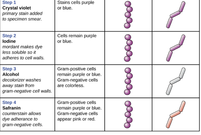
Gram Stain Procedure
A staining procedure with multiple steps developed by Danish microbiologist Hans Christian Gram in 1884 as a method to distinguish between bacteria with different types of cell walls.
A primary stain called crystal violet is applied to a head-fixed smear, giving cells a purple color
A mordant (substances used to set or stabilize stains or dyes) called Gram’s iodine is added to make the stain clump and stay contained in thick layers of peptidoglycan in the cell walls
A decolorizing agent is added to wash the dye out of cells with thinner peptidoglycan layers, making them colorless
A counterstain, usually safranin, is added, staining the decolorized cells pink and is less noticeable in cells that contain crystal violet dye.
Acid-Fast Stain
A staining technique able to differentiate two types of gram-positive cells.
Ziehl-Neelsen Technique
Acid-fast staining technique using heat to infuse carbolfuchsin into acid-fast cells
Kinyoun Technique
Acid-fast staining technique that does not utilize heat
Capsule Staining
A negative staining technique that stains the background but does not penetrate capsules of certain bacteria and yeast
Endospore Staining
A staining method that uses two stains to differentiate endospores (structures produced within certain bacteria cells that allow them to survive harsh conditions) from the rest of the cell.
Flagella Staining
Staining technique that stains flagella, which typically cannot be seen under a light microscope without it
Giemsa Stain
A Romanowsky stain used in the microbiology laboratory for the staining of malaria, blood parasites, chlamydia, borrelia, yersinia, histoplasma, and pneumocystis
Cytoplasmic Inclusion Stains
Staining that identifies intracellular deposits of starch, glycogen, polyphosphates, hydroxybutyrate, and other substances