gross descriptions
 Call Kai
 Learn
Learn Practice Test
 Spaced Repetition
 Match
Match Flashcards
 Knowt Play
1/83

There's no tags or description
Looks like no tags are added yet.
Name | Mastery | Learn | Test | Matching | Spaced | Call with Kai | Chat |
|---|
No analytics yet
Send a link to your students to track their progress
84 Terms
parenchyma
generalized term that describes the functional tissue/cells forming an organ
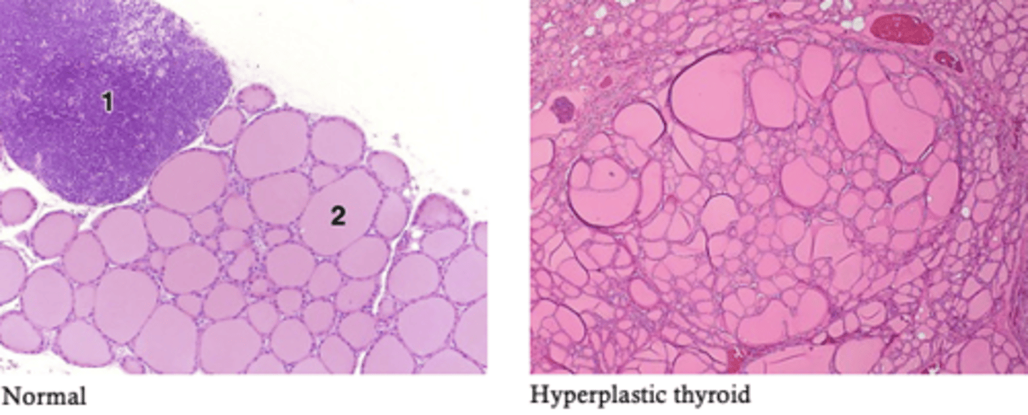
Hyperplasia
increase in number of cells
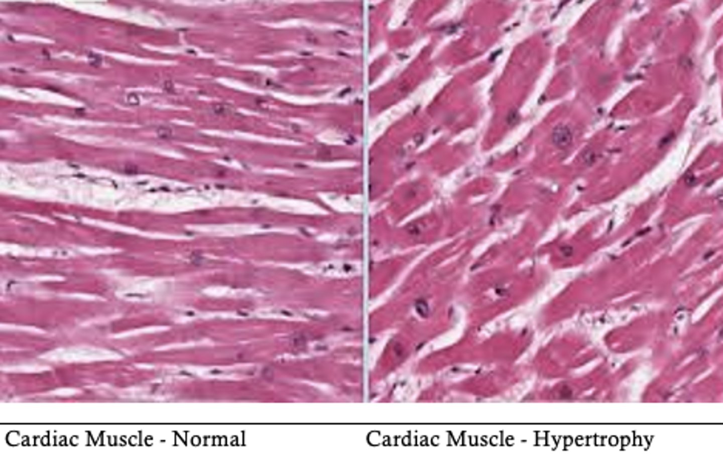
Hypertrophy
increase in cell size
metaplasia
Mature cell type is replaced by a different mature cell type
hyperplastic
an increase in size due to increase in cell number. Can be due to physiologic or pathologic instances
hypertrophic
increase in organ size due to an increase in size of the cells
serosa
describes the outer surfaces of an organ
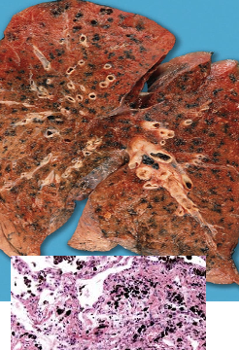
anthracosis
black pigmentation referring to lung tissue and occurs with inhalation of particulate material that is stored in the lungs. Appears as patchy or streaks of black discoloration
asymmetrical
not symmetrical
atrophic
showing signs of tissue degradation or breakdown
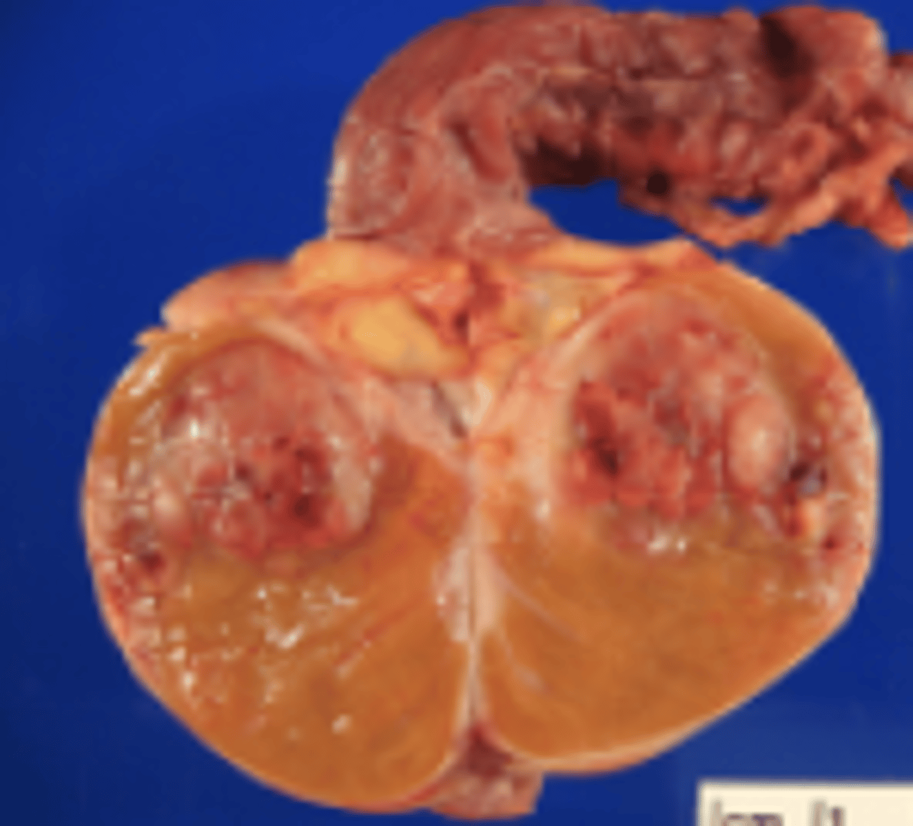
bivalve
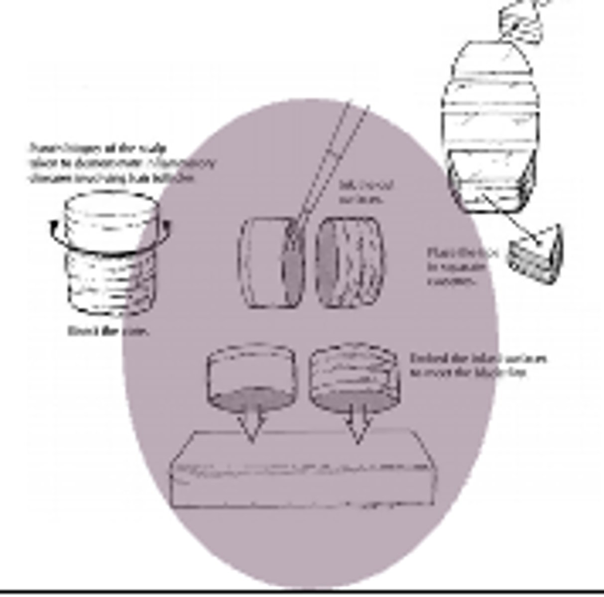
to split a specimen exposing the inner contents like a clam (testicles, kidney, uteri)
bisect
describes how something is cut refers to cutting something into roughly two equal halves (biopsies)
bosselated
knobby covered in small rounded bumps like a raspberry
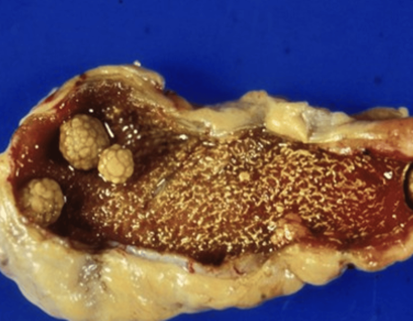
calculus
calcified material
congested
an accumulation of a (any) fluid resulting in distention or fullness. May indicate increased flow to the area or decreased output.
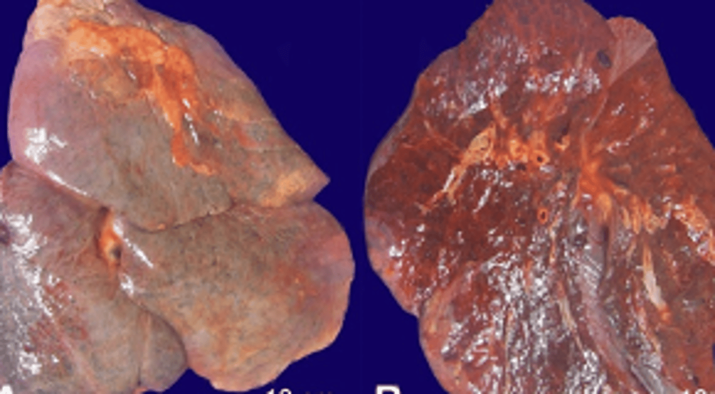
cystic
forming large open spaces filled with fluid (lesion)
chalky
resembling chalk in texture
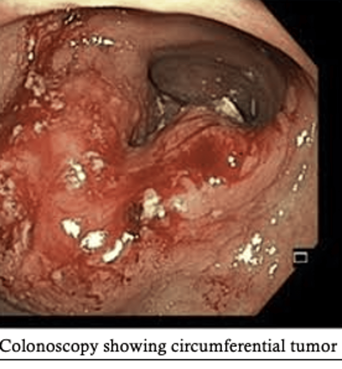
circumferential
involving all surfaces of a circular structure like the bowel lumen. used most often when referring to bowel tumors and the growth pattern tumors within a lumen
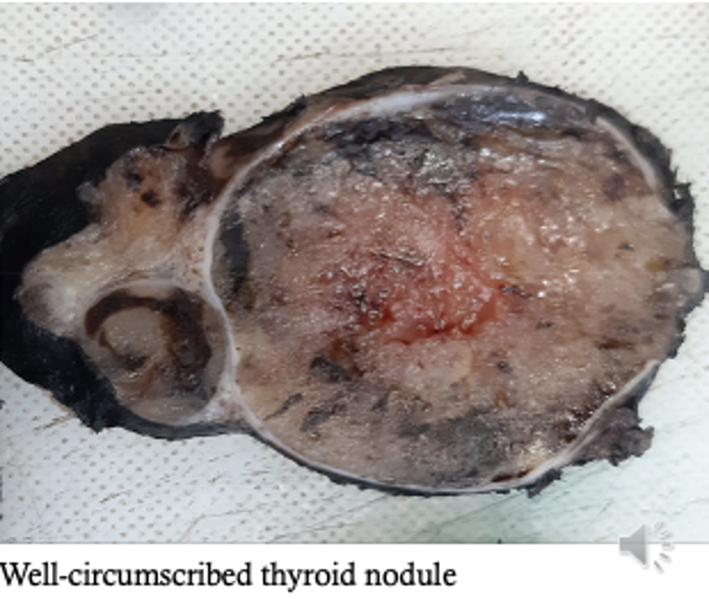
circumscribed
a circular shape often bordered or contained by a capsule. used to describe circular or spherical structures
defect
fault
demarcated
defined with boundaries
diffuse
spread throughout not confined or localized
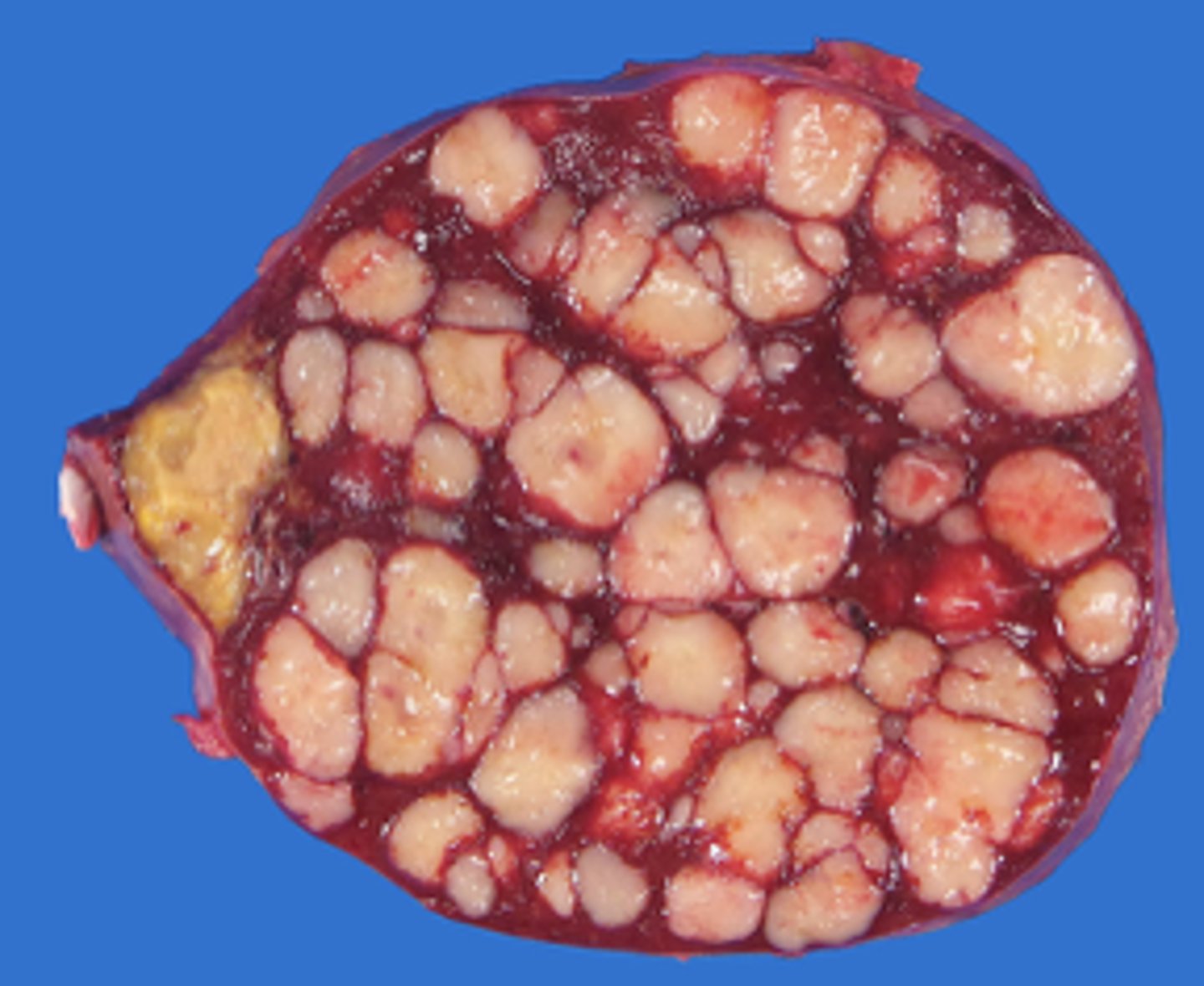
eccentric
off center within tissue or organ
ellipse
oval shaped
eroded
worn away
erythematous (erythema)
having red coloration typically referring to increased blood flow, inflammation
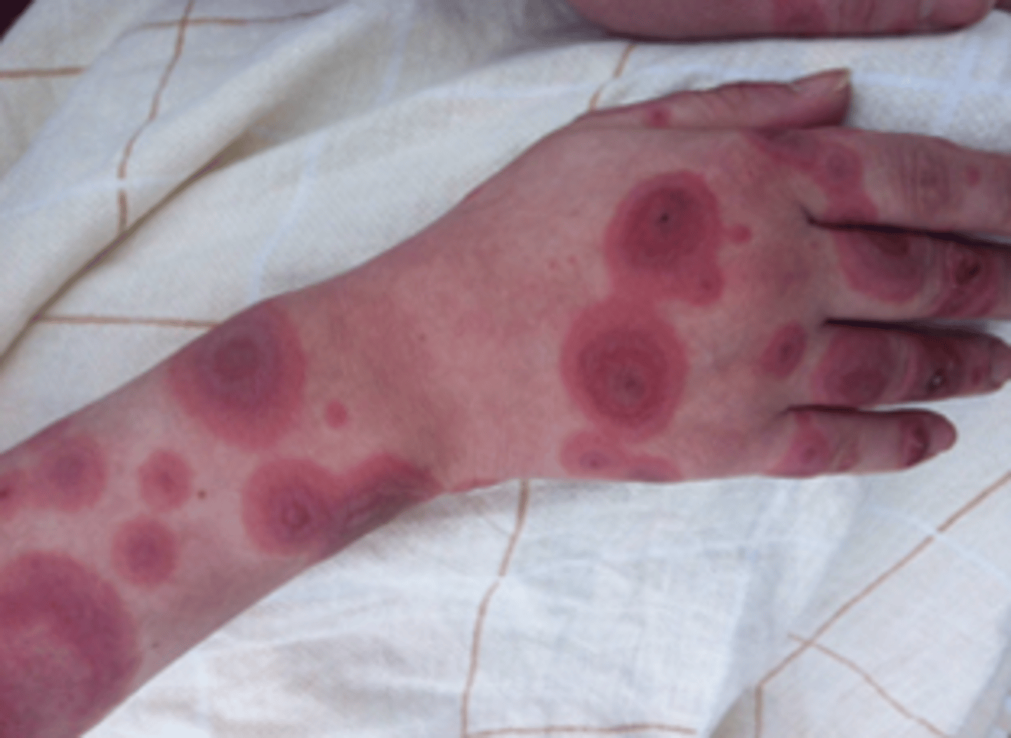
excoriated
scraped, abraded, or missing its top layers
excrescence
abnormal growths arising in tissue
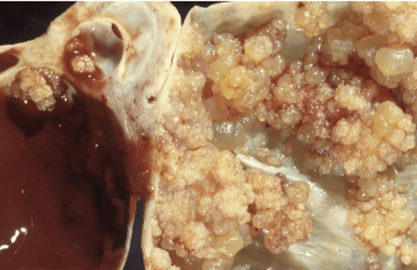
exophytic
projects outward from the surface of an organ or tissue
exudative
a fluid, cells, or other solid substances (like proteins) that has seeped out of blood vessels and into surrounding tissues or onto body surfaces
fibrous
appears tough, dense, white or gray, and stringy
filiform
thread-like
firm
dense, rubbery, or resistant to pressure
fleshy
the tissue is solid, dense, and resembles normal muscle or organ tissue
focal
isolated or localized in a single specific location
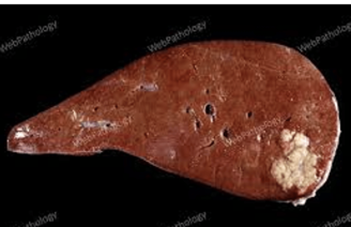
friable
delicate, easily falls apart or crumbles
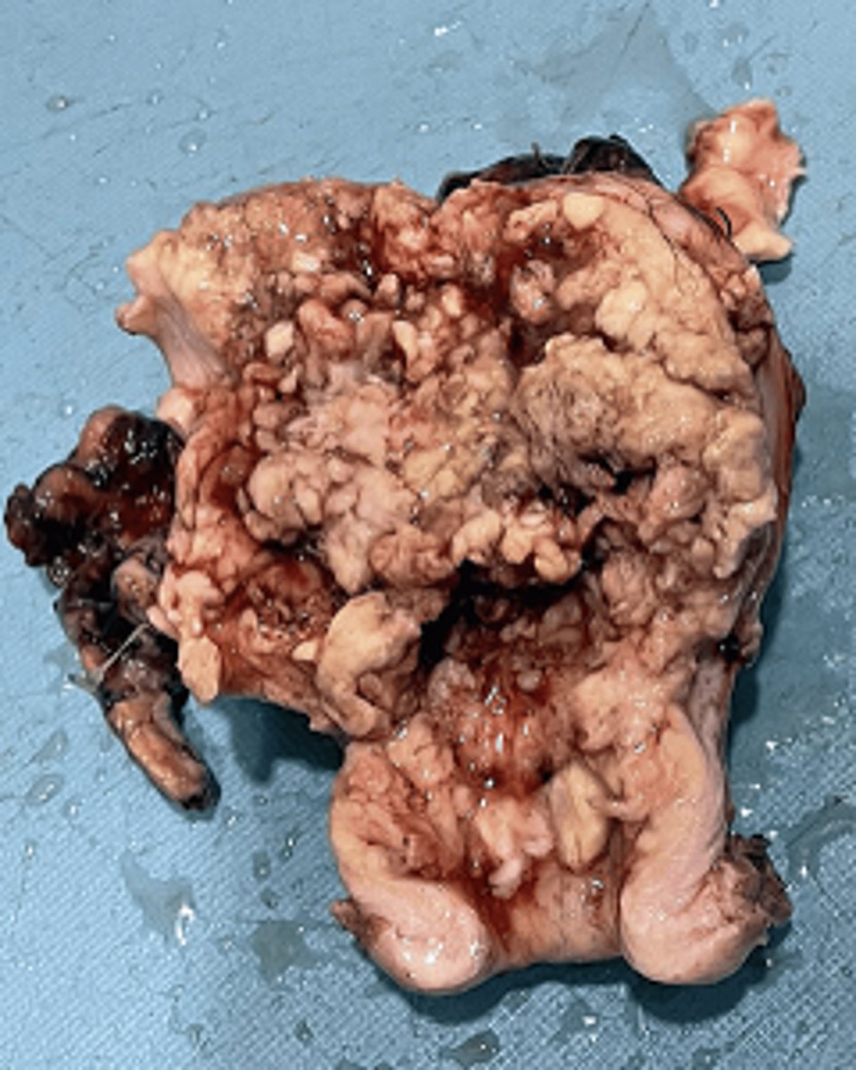
fungating
an outward growth pattern similar to a mushroom
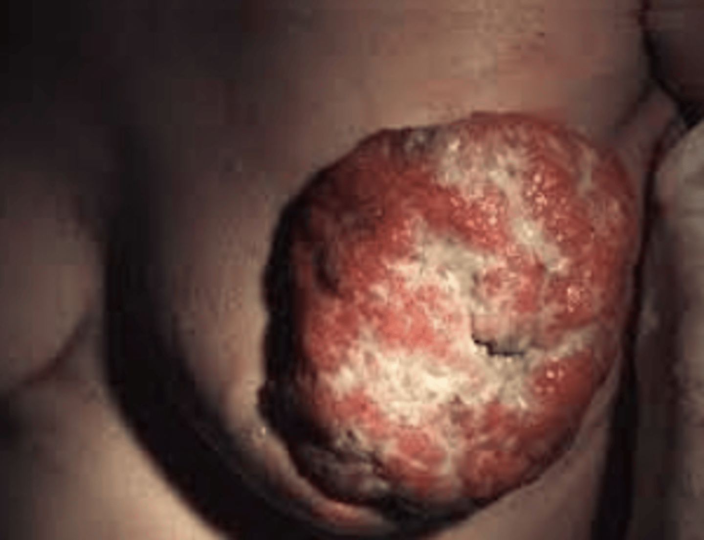
homogenous
uniform appearance comprised of one or similar components
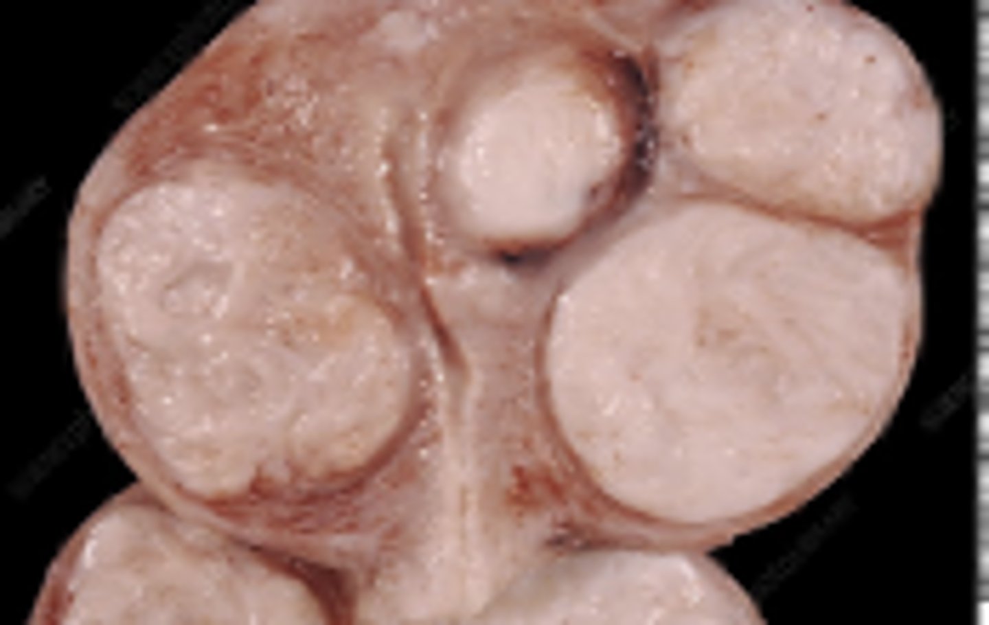
heterogenous
a varied appearance comprised of multiple components
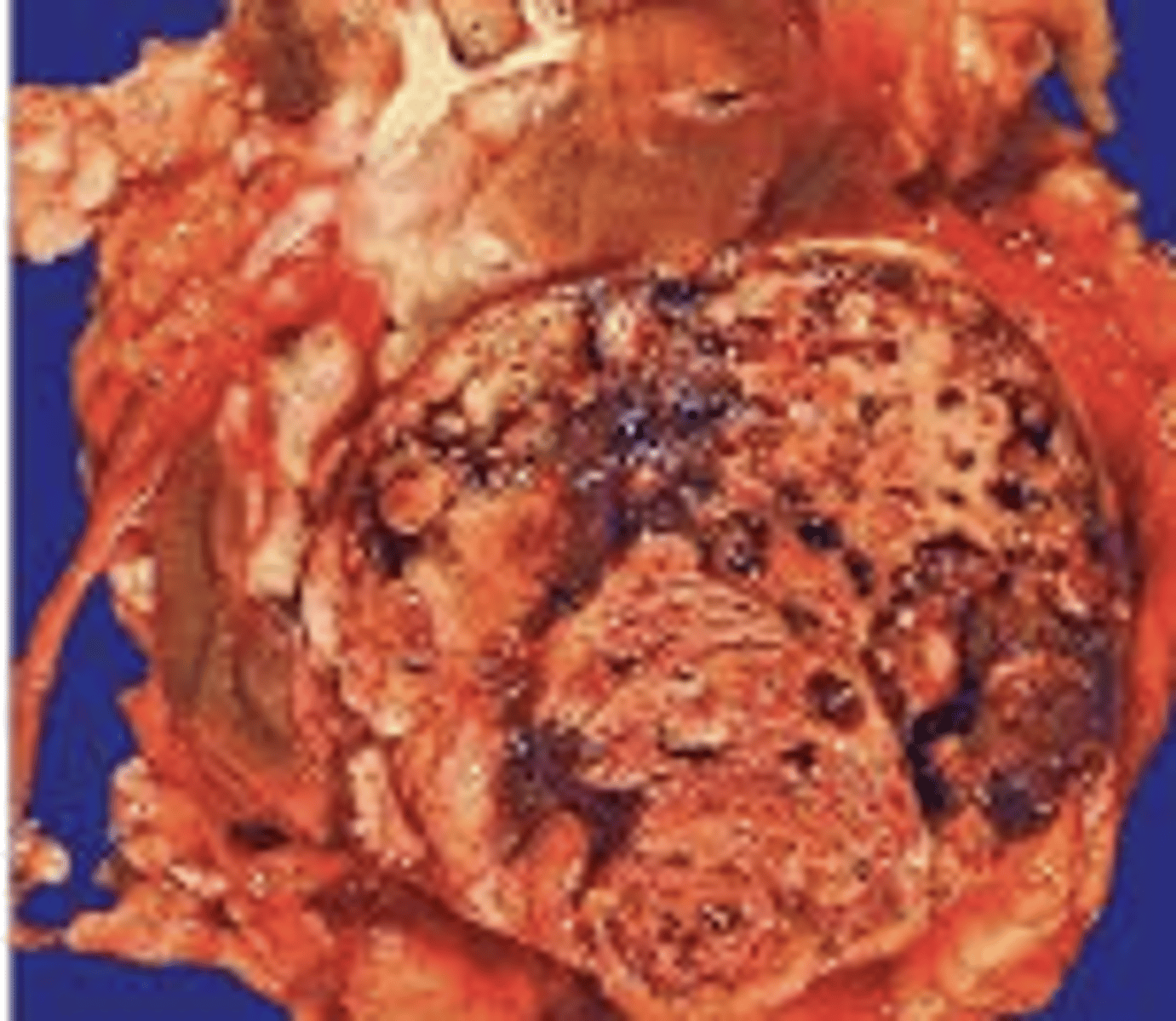
hyperemic
refers to an excessive amount of blood in a part or area
ill-defined
no clear borders or irregular shape
ischemic changes
a tissue or organ is suffering from a lack of adequate blood flow
lobulated
tissue that has taken on a bumpy, lumpy, or segmented appearance
intact
complete/whole unit without being broken fragemnted or cut
irregular
a generalized term for "other than normal" used to describe abnormla things
linear
lesion or feature is straight, strip-like, or thread-like in appearance
loculated
the tissue or fluid is divided into multiple small, separate compartments or pockets rather than being free-flowing
macule
a flat non raised skin or mucosal lesion (less than 1cm in size)
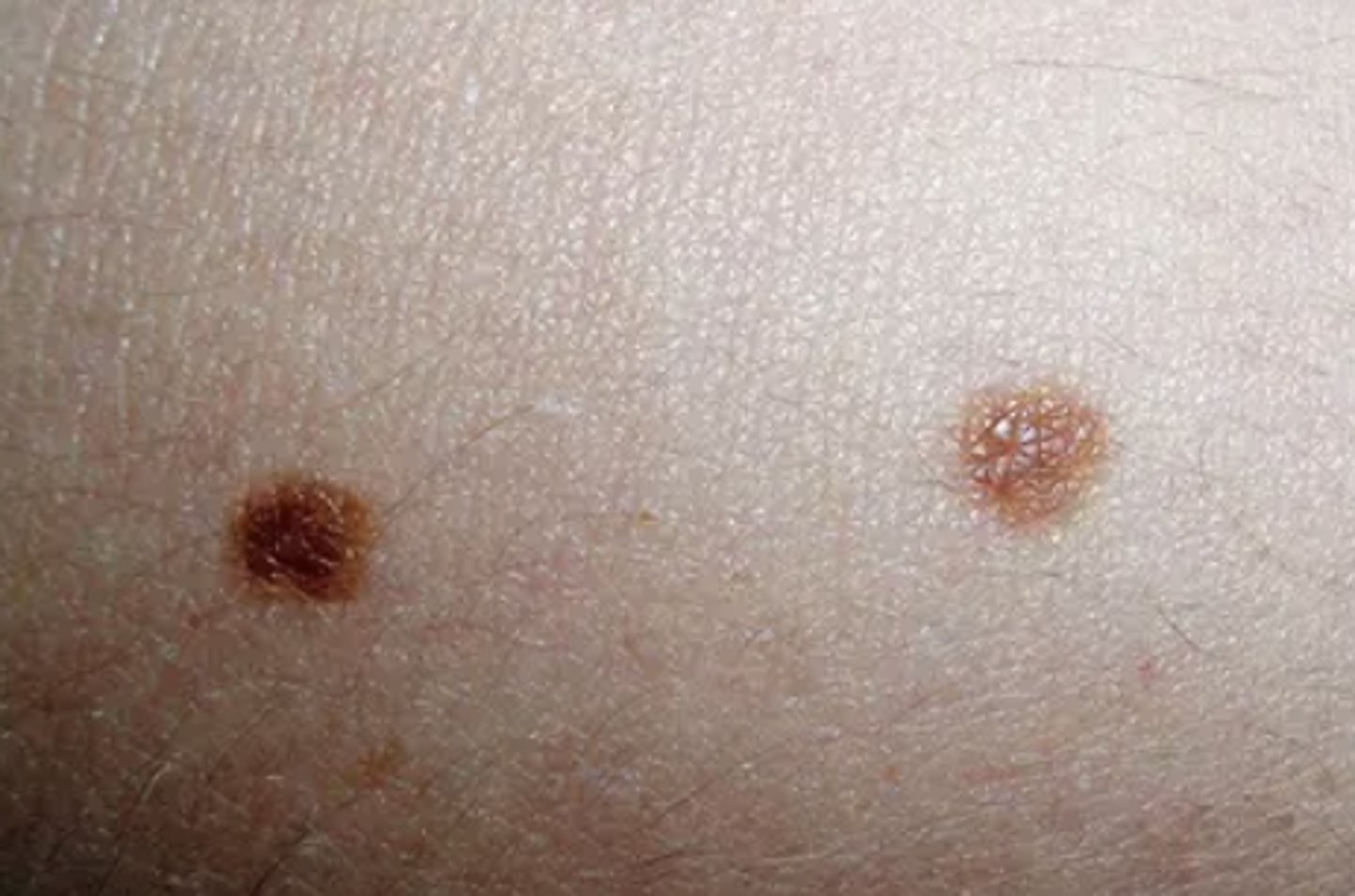
patch
a flat non raised skin or mucosal lesion (greater than 1cm in size)
mottled
patchy or blotchy discoloration. describes post mortem changes or ischemic changes
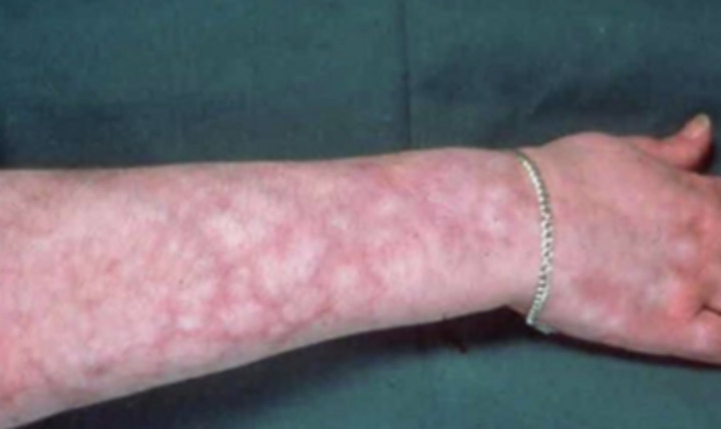
mucinous
a tumor or tissue produces abundant mucin (a thick, jelly-like, sugar-protein fluid, the main component of mucus)
multicystic
mutiple cysts
multifaceted
multiple flat sides or faces
multifocal
occurring in more than one district spot or location within
mummified
a tissue sample is completely dried out, shriveled, and dark brown or black
necrotic
decaying dead tissue
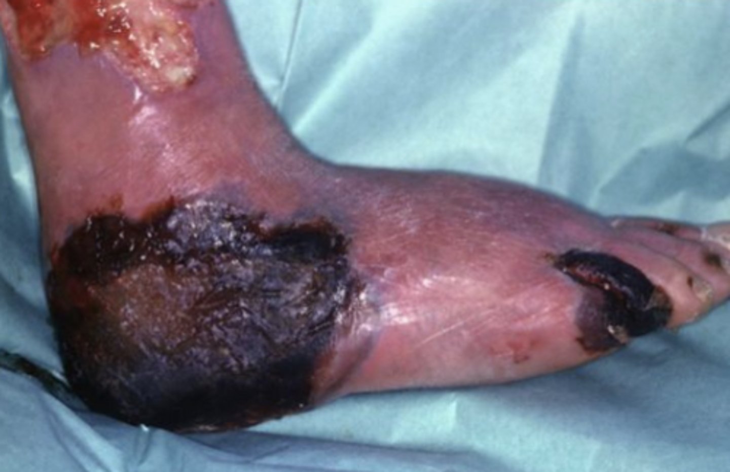
ovoid
egg-shaped
papillary
shaped like tiny, finger-like or nipple-like projections.
papule
a small raised skin or mucosal lesion refers to lesions less than 1cm
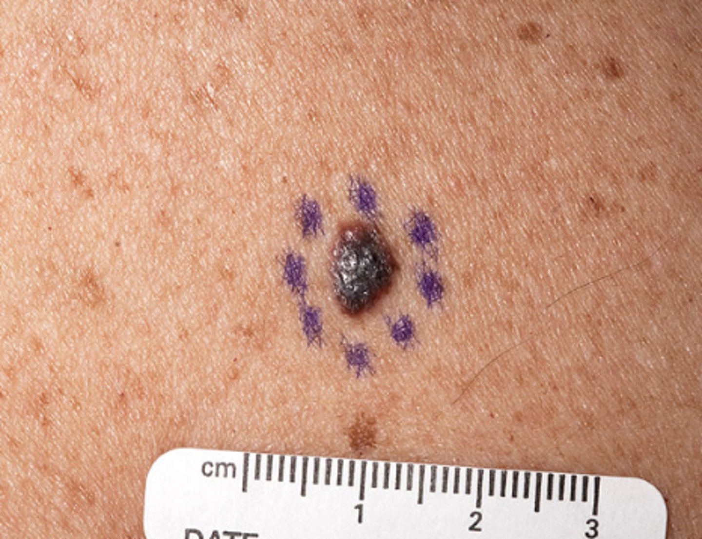
nodule
a small raised skin or mucosal lesion refers to lesions greater than 1cm
patent
open or unobstructed lumen such as ducts vessels or bowel
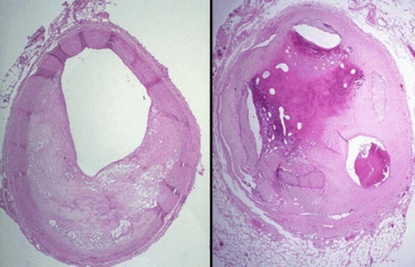
Pendunculated
stalk or stem
hyperpigmented
darker than surroundings
hypopigmented
lighter than surroundings
plaque
a flat, raised patch or growth on a surface that is wider than it is thick
purulent
containing, consisting of, or forming pus
raised
slightly elevated
roughened
surface bumpy course or uneven
ruptured
burst, torn, broken open
scaly
flaky, rough, and covered in peeling layers of dead cells
septate
divided into separate chambers or sections by internal walls
serosal
relating to the serosa
sessile
flat and attached directly by the base, without a stalk.
sloughing
dead tissue is peeling, shedding, or separating away from the living tissue underneath
stellate
star-shaped, featuring a central point with lines or points radiating outward
smooth
even, level, and free from roughness, bumps, or ridges
stenotic
narrowed or constricted passage of a body opening; vessels, ducts, bowel, esophagus, larynx
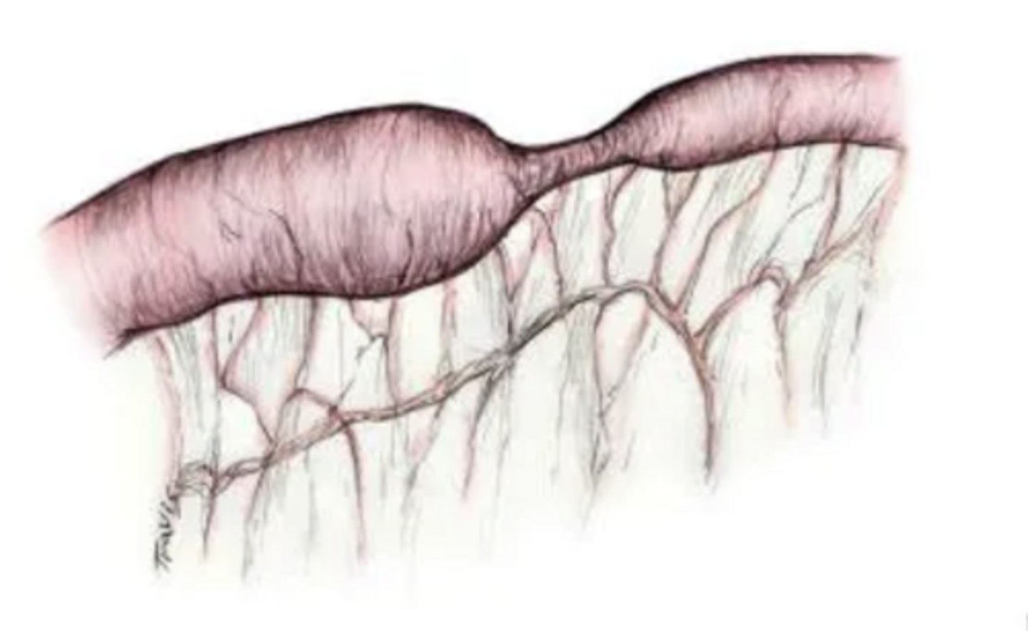
submucosal
relating to submucosa
subserosal
related to subserosa
tortuous
twisted, winding, or full of sharp turns and bends
trabecular
a network of tiny beams, bars, or interlocking struts
variegated
streaked, spotted, or patched with different colors or textures
verrucoid
wart-like
Whorled
swirling, circular, or concentric pattern