BFA - Exam 1 (Ch. 1-4)
 Call Kai
 Learn
Learn Practice Test
 Spaced Repetition
 Match
Match Flashcards
 Knowt Play
1/202

There's no tags or description
Looks like no tags are added yet.
Name | Mastery | Learn | Test | Matching | Spaced | Call with Kai | Chat |
|---|
No analytics yet
Send a link to your students to track their progress
203 Terms
Quality Assessment (QA)
A programs of checks and balances to ensure the quality of a laboratory’s services
Must involve a mechanism for error detection and provide an opportunity to improve services
Encompasses all aspects of the urinalysis laboratory: Preanalytical, analytical, postanalytical
Preanalytical components
all procedures occurring before testing:
ensuring appropriate test ordering processes
specimen collection, transport, storage, and handling
patient education, preparation, and instructions
Procedures for handling inappropriate or unacceptable specimens
documentation of any problems or unusual situations
accurate, up-to-date, available procedure manuals
adequate personnel training and supervision
Analytical components
Components directly involved in lab testing
Equipment/instrumentation
Reagents and supplies
Procedure manuals w/ standardized procedures
Analytical methods
Monitoring of analytical methods/ quality control
Lab personnel technical skills, training, monitoring, technical competence, and continuing education
Analytical components - Equipment/instrumentation
requires calibration, preventive maintenance, and monitoring and documentation of these actions
Analytical components - reagents
use of reagent-grade or analytical-grade reagents
distilled or deionized water and quality monitoring
verification of newly prepared reagents b4 use
Analytical components - procedure manuals
should be current, complete, readily available, comprehensive, and comply w/ Clinical Laboratory Standards Institute (CLSI) standards
Analytical components - Quality Control (QC)
QC materials used to monitor and assess analytical error
internal QC materials mimic patient samples in physical and chemical characteristics
Commercially obtained or laboratory prepared
Results recorded, and tolerance limits as well as troubleshooting and corrective action procedures established
Monitor personnel’s analysis of same specimen
Analytical components - Proficiency testing
Measures to monitor and evaluate a laboratory’s performance compared w/ that of other facilities
testing surveys or programs in which many labs use the same lot of QC materials
Reports useful for detecting quantitative small changes in systematic error in quantitative methods
Accredited laboratories must participate in proficiency testing
what can cause proficiency testing to fail?
all examples of analytical components, such as improper training, expired reagents, ill-maintained instrumentation, etc.
Postanalytical components
results reporting and interpretation
efficient and effective communication of results
standardized reporting format w/ reference ranges and informative comments listed
immediate reporting critical values to health care provider, according to laboratory policy
Appropriate documentation and evidence of review
Critical values
What’s the goal of effective quality assessment program
to obtain consistently accurate and reproducible results
test results should reflect patient’s condition, rather than being modified due to procedural or personnel variations
in the spirit of QA, an action has not been performed if it has not been documented
UA Quality Assurance Examples (not inclusive)
Procedural Factors
Volume of urine examined (10, 12, 15 mL)
Speed of centrifugation
Length of centrifugation
Conc. of sediment
Volume of sediment dispensed
Reporting Factors
Each lab should publish its own normal values (based on system used and patient population)
All personnel must use the same terminology
All personnel must report results in standard format
All abnormal results should be flagged for easy reference
True/False: Procedure manuals are part of the pre-analytical, analytical, and post-analytical components of a QA program
True
Safety in the Urinalysis Lab
practices to prevent spread of infection and ensure patient/employee safety
lab employees exposed to numerous biological, chemical, electrical hazards
Occupational Health and Safety Act of 1970
Established formal regulation of health and safety for all employees
Administered by Occupational Safety and Health Administration (OSHA)
Occupational Health and Safety Act of 1970
Established formal regulation of health and safety for all employees
Administered by Occupational Safety and Health Administration (OSHA)
Biological Hazards - Universal precautions
all body fluids, secretions, and excretions except sweat considered potentially infectious and capable of disease transmission
Biological Hazards - Transmission-based precautions
apply to specific patients with known or suspected infections
Three categories of transmission based precautions
contact precautions
droplet precautions
airborne precautions
Three routes of infection or disease transmission:
inhalation
ingested
direct inoculation or skin contact
What are the three categories of transmission based precautions
contact
droplet
airborne
what are the three routes of infection or disease transmission
inhalation
ingested
direct inoculation or skin contact
Biological hazards - What is required/ common procedures when dealing w/ specimens in the lab?
Appropriate PPE when handling body fluids
All specimens must be handled as if they are infectious and disposed of properly
Contaminated sharps must be disposed of in puncture-resistant containers and incinerated or autoclaved
Lab surfaces and equipment must be decontaminated daily w/ 10% bleach or a phenolic disinfectant
Chemical Hazards - OSHA rule (of Jan. 1990)
Requires each facility to have a Chemical Hygiene Plan defining safety policies and procedures for all hazardous chemicals
requires educating personnel annually about chemical safety policies and procedures
Components include:
Proper labeling of chemicals
Lab safety manuals
Use of PPE
Safety equipment such as an eyewash and shower
Safety Data Sheets (SDS)
ensures all employees are aware of potential chemical hazards in the workplace
Employee “Right to Know”
Requires chemical manufacturers and supplier to provide SDS for each chemical, and employers to make them available to employees
What general procedures should take place in the case of a chemical spill
consult SDS to determine appropriate action
make chemical spill kits accessible in lab
post-emergency treatment procedures for splashes and spill injuries
dispose of all chemical properly, according to the lab’s safety policies and applicable local, state, and federal laws
Procedure for flammable substances and fire
store in appropriately vented flammable storage cabinets
procedure for compressed gas tanks
tanks must be secured at all times, in use and in transit
valve caps must be in place at all times, except when in use
procedure for electrical hazards
monitor maintenance of equipment
report frayed cords or other needed maintenance
In case of emergency, generally, all personnel should be aware of:
location and operation of all safety equipment (fire extinguishers, alarm stations, emergency showers, eyewash stations)
exits and evacuations routes and procedures to follow during emergencies
What are multiple reasons to study urine?
fluid biopsy of the kidney
non-invasive → evaluate kidney
urine is the ultrafiltrate of plasma
Can be used to evaluate and monitor body homeostasis and many metabolic diseases
can monitor therapy effectiveness
specimen usually readily available
urine is the _______ of plasma
ultrafiltrate
Urinalysis is a ____ _____ of the kidney
fluid biopsy
non-invasive means to evaluate the kidney, body homeostasis, metabolic diseases, and therapy effectiveness
what collection container is used for pediatrics?
adhesive bags
what containers are used for 24hr specimens
large plastic containers
what kind of container is required for cultures?
sterile containers
Specimen collection containers
disposable, wide-mouthed, flat bottomed containers w/ caps are recommended
clear containers
at least 50 mL capacity
adhesive bags for pediatrics
large plastic containers for 24 hr specimens
sterile containers required for cultures
Specimen labeling - information on label
Patient’s name and unique identification number
Date and time of specimen collection
Additional info: DOB and ordering physician
LABEL ON CONTAINER, not lid
Specimen labeling - Requisition form
must accompany specimen
information must match label
time of receipt is stamped on requisition
type of specimen
interfering medications if applicable
what must accompany labeled specimen when its sent out for testing?
requisition form
Specimen types
First morning
empty bladder at night before going to bed
collect specimen first thing in the morning
most concentrated; often preferred specimen
Random
for routine screening
can be affected by excess fluid intake or exercise
Timed
collections for a predetermined length of time
collections at a specified time of day
timed specimen
collections for a predetermined length of time
collections at a specified time of day
Random specimen
for routine screening
can be affected by excess fluid intake or exercise
first morning specimen
empty bladder at night before going to bed
collect specimen first thing in the morning
most concentrated; often preferred specimen
What are some collection techniques for urinalysis?
routine void
Mid-stream “clean catch”
Catheterized specimen
suprapubic aspiration
pediatric collection
timed collections
drug specimen collection
collection technique - routine void
requires no patient preparation
collected by having patient void into appropriate container
patient normally needs no assistance other than clear directions
can be random or first morning specimen
collection technique - Mid-stream “clean catch”
For bacterial cultures or to prevent vaginal contamination and/or routine urinalysis
requires cleaning supplies, addition instructions to patient, and sterile container
requires thorough cleaning and rinsing of genital areas before collection
begin urination into toilet, collect midstream portion in container, finish voiding in toilet
collection technique - catheterized specimen
requires collection by medical personnel
sterile catheter inserted thru urethra into bladder
urine flows directly into collection bag
specimen obtained anytime from collection bag
good for culture and sensitivity
collection technique - suprapubic aspiration
requires collection by medical personnel
involves puncturing of abdominal wall and distended bladder by using needle and syringe
sample aspirated directly from bladder
bladder urine is normally sterile
used principally for cultures or in infants and usually when catheterization isn’t possible
pediatric collection techniques
often commercially available plastic urine collection bags that attach w/ a hypoallergenic skin adhesive are used
patient’s perineal area cleaned prior to bag attachment
specimen removed as soon as possible after collection
specimen appropriate for routine testing
Procedure for timed collections - 12 or 24 hr
may require addition of a chemical preservative to maintain integrity of analyte to be tested
begin and end w/ empty bladder, first void is discarded
keep on ice or refrigerate during collection period
check collection manual to identify necessary preservative
deliver specimen to lab immediately after completion of collection period
more accurate than random urine for urine chemistry tests
Drug specimen collection general procedure
proper collection, labeling and handling must be documented
if legal:
chain of custody → documentation from the time of specimen collection until the time of receipt of lab results → standardized form always accompanies specimen
specimen must withstand legal scrutiny → no tampering
points to consider for drug specimen collection
photo ID of urine donor or ID by employer
No unauthorized access to specimen
No adulteration, substitution or dilution of specimen
drug specimen collection: witnessed vs. unwitnessed collection
determined by individual ordering the test
both collection methods require that the specimen is handed immediately to the collector
drug specimen collection - adulteration tests
temp taken w/i 4 min - must be 32.5-37.7ºC
report temps outside of range immediately
collect another specimen asap
inspect urine color for anything unusual
-follow lab instruction for labeling, packaging and transport
What can determine whether a substance is urine or not
often need confirmation in drug screen collection or specimens collected by needle aspiration
urine creatinine conc. 50x higher than plasma
urea, sodium, and chloride higher in urine than in other body fluids
physiologic range is 1.002 to 1.035 for urine specific gravity and 4.0-8.0 for pH
urine from healthy people contains no protein or glucose , whereas many other body fluids do
urine from healthy people contains no ______ or ______, whereas many other body fluids do
protein, glucose
what is the physiologic range for urine sp. grav and pH
1.002-1.035 sp. grav
4-8 for pH
_____, _____, and _____ higher in urine than in other body fluids
urea, sodium, chloride
urine ______ conc. 50x higher than plasma
creatinine
what might cause specimen rejection
unlabeled containers
non-matching labels and requisitions
contaminated specimens (ex. stool, toilet paper)
Contamination on exterior of container
insufficient quantity
delayed or improper transport
lack of refrigeration
Note: labs have written policies for rejection
Changes in unpreserved urine: analyte color change to modified/darkened caused by
oxidation or reduction of metabolites
Changes in unpreserved urine: analyte clarity decrease caused by
bacterial growth and precipitation of amorphous material
Changes in unpreserved urine: analyte odor increase caused by
multiplication of bacteria or bacterial breakdown of urea to ammonia
Changes in unpreserved urine: analyte pH increase caused by
breakdown of urea to ammonia by urease producing bacteria/ loss of CO2
Changes in unpreserved urine: analyte glucose decrease caused by
glycolysis and bacterial utlization
Changes in unpreserved urine: analyte ketones decrease caused by
volatilization and bacterial metabolism
Changes in unpreserved urine: analyte bilirubin decrease caused by
exposure to light/photo oxidation to biliverdin
Changes in unpreserved urine: analyte urobilinogen decrease caused by
oxidation to urobilin
Changes in unpreserved urine: analyte nitrite increase caused by
multiplication of nitrate-reducing bacteria
Changes in unpreserved urine: analyte blood cells and casts decrease caused by
disintegration in dilute alkaline urine
Changes in unpreserved urine: analyte bacteria increase caused by
multiplication
How does color, clarity, odor, pH, glucose, ketones, bilirubin, urobilinogen, nitrite, blood cells and casts, and bacteria change in unpreserved urine?
Color: modified/darkened
clarity: decrease
odor: increase
ph: increase
glucose: decrease
ketones: decrease
bilirubin: decrease
urobilinogen: decrease
nitrite: increase
blood cells and cats: decreased
bacteria: increased
how to preserve specimen integrity?
tests w/i 2 hrs
for delayed testing: refrigerate, chemical preservatives
most problems are caused by bacterial growth
increase/ change in color, turbidity, pH, nitrite, bacteria, and odor
decrease in glucose, ketones, bilirubin, urobilinogen, RBCs, WBCs, and casts
Specimen preservation - refrigeration
required for culture specimens
causes increase in sp. grav. and precipitation of amorphous crystals
must return to room temp for chemical testing
specimen preservation - chemical preservatives
commercial transport tubes
must be compatible w/ chemical testing
ideal preservative is bactericidal, inhibit urease and preserve formed elements
Renal system/ urinary system main four responsibilities
production, transport, storage, excretion
primary purpose of the urinary system
to maintain the internal chemical balance of the body by
removing metabolic waste products
regulating fluid and electrolyte levels
contributing to blood pressure control
what are the four main structures of the urinary/renal system?
Kidneys (primary organs)
bean-shaped organs located retroperitoneally
contain nephrons (functional units) that filter blood and form urine
responsible for excretion, reabsorption, secretion, and endocrine activity
Ureters (transport)
muscular tubes that carry urine from each kidney to the bladder
peristaltic waves ensure one-way movement of urine
Urinary bladder (storage organ)
hollow, muscular organ that temporarily stores urine
capable of expanding due to its transitional epithelium
avg. capacity ~400-600 mL in adults
Urethra (excretory passage)
transports urine from the bladder to the outside
contains internal (involuntary) and external (voluntary) sphincters that control urination
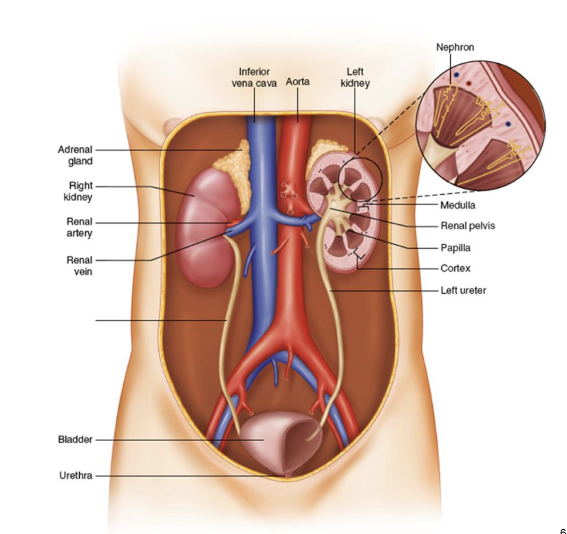
main structure of the urinary system: general characteristics of the urethra
excretory passage
transports urine from the bladder to the outside
contains internal (involuntary) and external (voluntary) sphincters that control urination
main structure of the urinary system: general characteristics of the urinary bladder
storage organ
hollow, muscular organ that temporarily stores urine
capable of expanding due to its transitional epithelium
avg. capacity ~400-600 mL in adults
main structure of the urinary system: general characteristics of the ureters
transport
muscular tubes that carry urine from each kidney to the bladder
peristaltic waves ensure one-way movement of urine
main structure of the urinary system: general characteristics of the kidneys
primary organ
bean-shaped organs located retroperitoneally
contain nephrons (functional units) that filter blood and form urine
responsible for excretion, reabsorption, secretion, and endocrine activity
what are the two main sections of the urinary tract?
upper urinary tract → kidneys and ureters
lower urinary tract → bladder and urethra
what are the parts of the lower urinary tract
bladder and urethra
what are the parts of the upper urinary tract?
kidneys and ureters

Urinary Tract Infections (UTI)
upper and lower require different treatment
upper → may require antibiotics administered intravenously
lower → can usually be treated with oral antibiotics
more prevalent in women
most common infection
how are upper and lower UTIs treated differently?
upper → may require antibiotics administered intravenously
lower → can usually be treated with oral antibiotics
what are signs/symptoms that indicate a UTI of the upper urinary tract?
Symptoms: dysuria; urgency; acute flank/back/groin pain nausea; fever; chills; extreme fatigue
Physical: same as lower
Chemical: same as lower
Microscope: same as lower (WBCs, RBCs, microorganisms, transitional epithelial cells), but also renal tubular epithelial cells & WBC incorporated casts
what are signs/symptoms that indicate a UTI of the lower urinary tract?
symptoms: dysuria, urgency, lower abdominal pain, low-grade fever
physical: possible blood tinged color (pink, red, brown), green from rare Pseudomonas infections, strong odor, cloudy-turbid
chemical: positive leukocyte esterase and nitrite from gram-negative bacteria; small amts of blood and protein are possible
microscopic: many WBCs, RBCs, microorganisms, transitional epithelial cells, no casts or few normal hyaline casts
what is the anatomical position of the kidney?
retroperitoneal, between T12 and L3 vertebrae
-right kidney lower due to the liver

what are the external kidney borders
convex lateral border
concave medial border
-hilum - vessel and ureter passage

what are the protective layers surrounding the kidneys starting from innermost to outermost?
Renal capsule (fibrous)
thin, tough fibrous capsule
bound to the renal surface
perirenal fat
cushioning layer
renal fascia (Gerota’s fascia)
encloses the kidney and the adrena gland
pararenal fat
outermost layer, provides additional cushioning

renal capsule
innermost protective layer surrounding the kidney
thin, tough fibrous capsule
bound to the renal surface
perirenal fat
protective layer surrounding the kidney → between renal capsule and renal fascia
cushioning layer

renal fascia (Gerota’s fascia)
protective layer surrounding the kidney → between perirenal fat and pararenal fat
encloses kidney and the adrenal gland

pararenal fat
outermost protective layer surrounding the kidney
provides additional cushioning

what are the two parts the kidney is divided into?
renal cortex and medulla

renal cortex
inside of the kidney, outermost layer beneath the renal capsule
houses renal corpuscles (glomeruli and bowman’s capsules) and convoluted tubules
main site of filtration

renal medulla
inner portion of the kidney
organized into renal pyramids
appears striated due to collecting ducts and loops of henle
Function:
concentration of urine by reabsorption and secretion

renal pyramids
cone-shaped structures (~8-18 per kidney) inside the medulla of the kidney
apex points inward to form the renal papilla
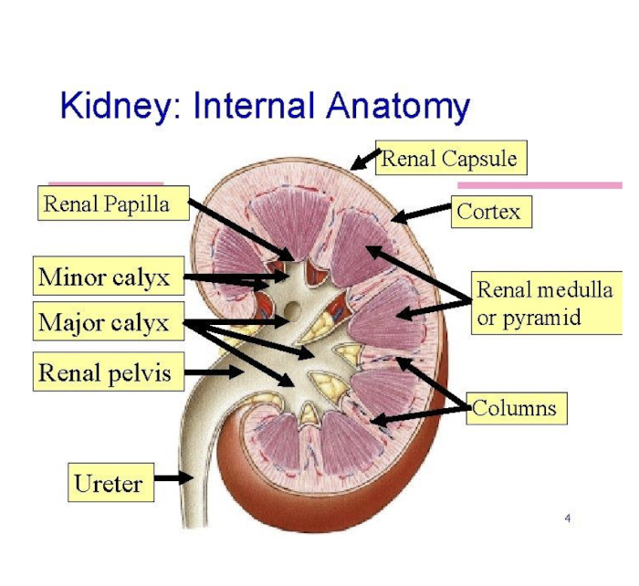
renal columns
extensions of cortical tissue between renal pyramids inside the medulla of the kidney
contain interlobar arteries and veins
provide structural support and pathway for blood vessels

Calyces of the kidneys
urine collection chambers
Minor:
cup-shaped structures
one per each renal pyramid
major:
receives urine from 2-3 minor calyces
both funnel urine toward the renal pelvis
