Cavities, Bones, Joints and Muscles of the Thorax
 Call Kai
 Learn
Learn Practice Test
 Spaced Repetition
 Match
Match Flashcards
 Knowt Play
1/43

There's no tags or description
Looks like no tags are added yet.
Name | Mastery | Learn | Test | Matching | Spaced | Call with Kai | Chat |
|---|
No analytics yet
Send a link to your students to track their progress
44 Terms
Thorax (General)
houses and protects
heart
lungs
conduit/pathway for structures passing between neck+abdomen
role in breathing
Apertures of Thorax
superior throacic aperture→narrow
open
contain AVNs
pass over rib 1
go inferior to/under clavicle
major A+V+brachial plexus→through axillary inlet
interior thoracic aperture→wider
closed→by diaphragm
Axillary Inlet
lies on each side of superior throacic aperture of thorax
superior to rib 1
inferior to clavicle
gateway to upper limb
margins
superior margin of scapula
clavicle
lateral margin of rib 1
contains
A
V
brachial plexus
Thoracic Cavity Compartments
3 compartments
pleural cavities→2
contain→lungs
mediastinum
Mediastinum (General)
soft tissue partition→thick but flexible
median sagittal position
separates pleural cavities
extends from
sternum→vertebral bodies
superior thoracic aperture→diaphragm
contains
trachea
esophagus
heart
major Ns
systemic blood vessels
clinical significance
can access heart without opening pleural cavities
abnormalities in one cavity→don’t always affect other cavities
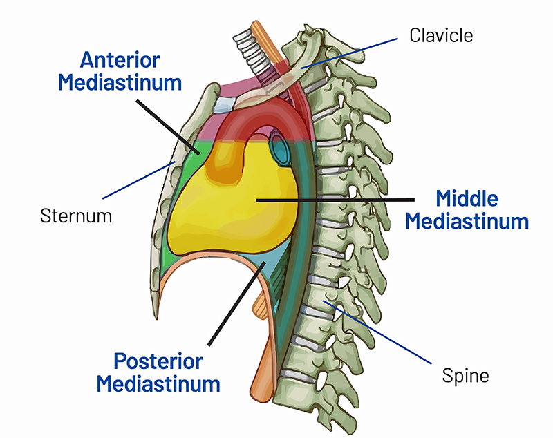
Superior+Inferior Portions of the Mediastinum
horizontal plane
passes thorugh sternal angle+IVD between T4-T5
superior mediastinum (red)
large vessels of heart
trachea
inferior mediastinum
subdivided by pericardium:
anterior mediastinum (green)
between sternum+pericardium
middle mediastinum (yellow)
pericarium
heart
posterior mediastinum (blue)
between pericaridum+thoracic vertebrae
Thoracic Wall
musculoskeletal wall
posteriorly
12 thoracic vertebrae
IVDs
laterally
ribs
intercostal muscles
anteriorly
sternum
Bony Thorax Components
thoracic vertebrae
ribs
sternum
Ribs (General)
12 pairs
all terminate→anteriorly with costal cartilages
all ribs→articulate with vertebral column posteriorly
not all articulate with→sternum
True Ribs vs False Ribs
true ribs
ribs 1-7
articulate directly with sternum→through own costal cartilage
false ribs
ribs 8-12
do not have own costal cartilage articulating with sternum
two types:
ribs 8-10→articulate w sternum via costal cartilages of superior ribs
ribs 11-12→no anterior connection to sternum (floating ribs)
General Rib Osteology (Positioning)
head
posterior side
superior+inferior articular facets on head
articulates with vertebrae:
superior articular facet of rib below
inferior articular facet of same rib
neck
connects head to angle
tubercle
articular facet on tubercle→articulates with vertebral transverse costal facet
angle
connects neck to body of rib
general components
internal surface
external surface
costal cartilage
Costal Groove
inferior margin on internal surface
contains
intercostal V
intercostal A
intercostal N
V→superiorly placed
N→inferiorly placed
-not protected by bone
-perforation→often injured
Rib 1 (General)
tubercle
articulates→with transverse costal facet of of T1
head
only 1 articular surface
articulates→only with body of T1
scalene tubercle
I: scalenus anterior
separates 2 grooves:
anterior groove→subclavian V
posteiror groove→subclavian A
Rib 10 (General)
has tubercle
head
only 1 articular surface
articulates→only with body of T10
Ribs 11+12 (General)
short
pointed anteirorly
no tubercles
no necks
articulate→with bodies of own vertebrae
still tipped with costal cartilage
Typical Rib Articulation
articulations→3
inferior articular facet of head of rib→superior costal facet of body of own vertebra
superior articular facet of head of rib→inferior costal facet of body of superiorly-placed vertebra
tubercle→transverse process of own vertebra
Rib 1 Articulation
articulation→2 points
head→single facet→articulates with body of T1
tubercle→articulates with transverse costal facet of T1
Rib 10 Articulation
articulation→2 points
head→single facet→articulates with body of T10
tubercle→articulates with transverse costal facet of T10
Ribs 11+12 Articulation
articulation→1 point
heads→articulate with facets on bodies of own vertebrae
no tubercle
Sternum Components
manubrium
body of sternum
xiphoid process
Manubrium Articulation Points
contains articular facets+notches
clavicular notch→sternal end of clavicle
costal cartilages of rib 1
costal cartilages of rib 2 (with body of sternum)
Body of Sternum Articulation Points
contains articular facets
costal cartilages of rib 2→manubrium+body of sternum
costal cartilages of ribs 3-6→body of sternum
costal cartilages of rib 7→body of sternum+xiphoid process
Xiphoid Process Articulation Points
contains articular facets
costal cartilages of rib→xiphoid process+body of sternum
Rib Articulation Components
thoracic vertebrae
costal cartilages
sternum (some)
Costovertebral Joints
synovial joint
between bodies of adjacent vertebrae+head of one rib
Costotransverse Joint Articulation+Stabilization
between transverse processes of thoracic vertebrae+rib
stabilized by
costotranverse ligament
-between neck+transverse processlateral costotransverse ligament
-between tip of transverse process+tuberclesuperior costotransverse ligament
-betweeen neck+transverse process of vertebra above
Sternocostal Joints
between upper 7 costal cartilages+sternum
sternocostal joint between costal cartilage of rib 1+manubrium
not a synovial joint
fibrocartilaginous connection
sternocostal joint between costal cartilage of ribs 2-7
synovial joints
thin joint capsules
reinforced by→sternocostal ligaments
Interchondral Joints
synovial joint
between costal cartilages of adjacent ribs
primarily→between ribs 7-10
provide indirect anchorage to sternum
reinforced by→interchondral ligaments
Manubriosternal Joint
between manubrium+body of sternum
slight movements during respiration
Xiphisternal Joint
between body of sternum+xiphoid process
over time→can fuse together
Pectoral Region (General)
external to→anterior thoracic wall
anchors upper limb to trunk
two components
superficial compartment
-skin
-superficial fascia
-breastsdeep compartment
-muscles
Breasts Components
skin
connective tissue
ligaments
fascia
adipose tissue
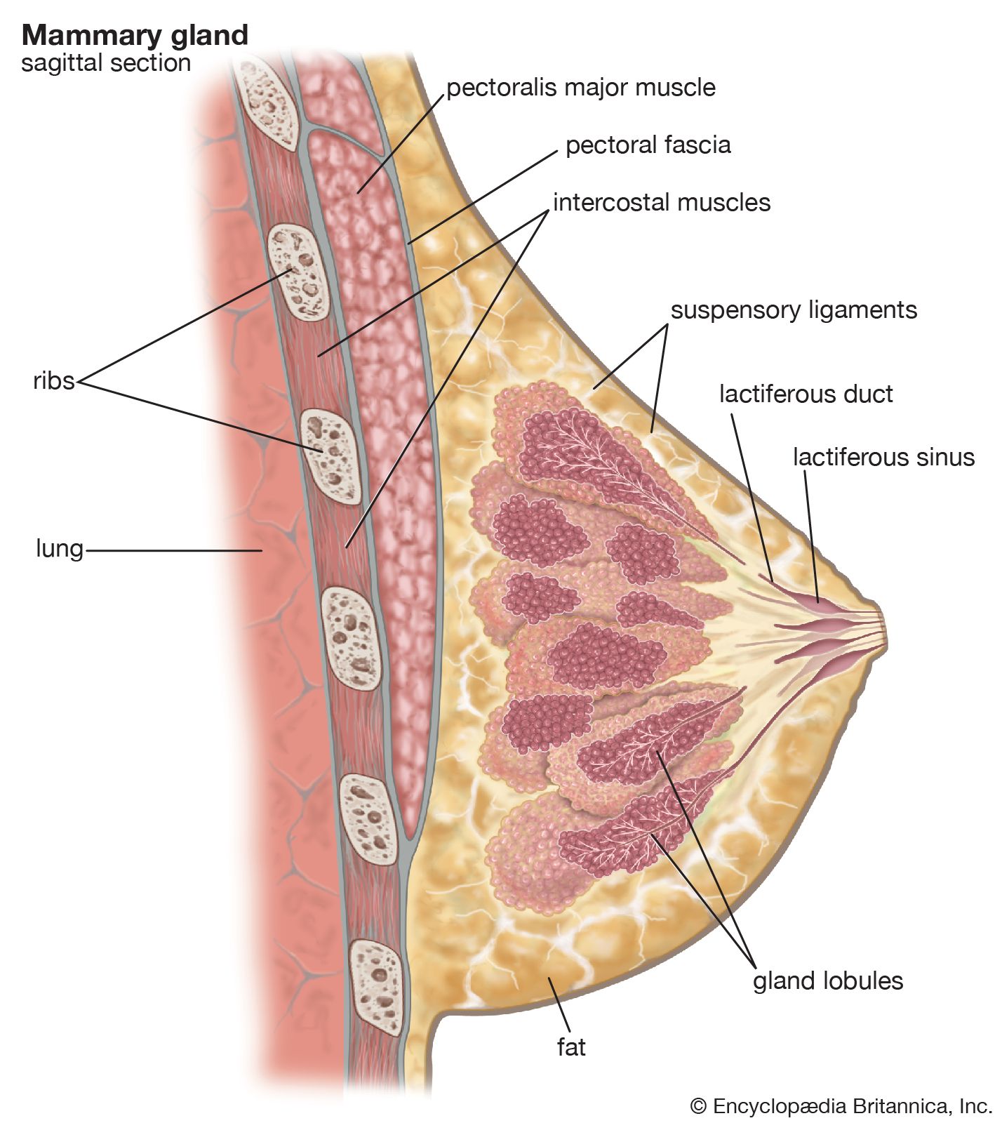
mammary glands
Mammary Glands
modified sweat glands
-series of ducts+secondary lobules→converge+form 15-20 lactiferous ducts→each open up to nipple
-nipple→surrounded by areola (pigmented skin around nipple)
located in
superficial fascia
anterior to pectoral muscles+anterior thoracic wall
Suspensory Ligaments of Breast
connective tissue→surrounds ducts+lobules→condense→form suspensory ligaments
continuous with dermis
support the breast
clinical importance
breast cancer→causes tension on ligaments→pitting of skin
orange peel texture (peau d’orange)
Breast (General) Margins
base of breast
attached to thorax
vertical/superior extent→ribs 2-6
lateral extent→midaxillary line
axillary process
clinical importance (axillary process)
breast pathologies→may present into axilla/along lateral margin of pectoralis major
Lymphatic Drainage
primarily (75%)
drainage from superolateral portions of breast→axillary lymph nodes
can also drain into
parasternal nodes
-drainage from medial partabdominal nodes
-drainage from inferior part/portionsupraclavicular nodes
lymph nodes associated with opposite breast
clinical importance
can be difficult to contain nodal metastatic breast cancer
Breast Tissue Margins
breast→superficial to deep fascia of pectoral muscles
fascia→just superficial to pectoralis major
retromammary space (additional space separating breast tissue from fascia)
Retromammary Space
additional layer of loose connective tissues
between breast+deep fascia
separates breast+pectoral muscles+fascia
clinical importance
mastectomy→removal of breast tissue to pectoralis major+fascia
Male Breasts
in males→rudimentary
connective tissue
small ducts→usually do not extend beyond areola
clinical importance
breast cancer→can occur in males
Muscles of the Thoracic Wall
fill in+support intercostal spaces
pass between sternum+ribs→cross attachments of ribs
include
external intercostal
internal intercostal
innermost intercostal
subscostales
transversus thoracis
Intercostal Muscles
located between adjacent ribs
3 layered muscles (best seen→lateral view between ribs)
external intercostal
-will see until costal cartilages→transitions to thin CT aponeurosis (external intercostal membrane)internal intercostal
-deep to external intercostals anterolaterally+laterally
-runs to→angles of ribs posteriorly→transitions to internal intercostal membrane→vertebraeinnermost intercostal
-best viewed inside thorax
-separated from internal intercostal by→intercostal AVN (key for distinguishing)
Subcostales
same level as innermost intercostals
key to ID→ jump over ribs
best viewed→inside of low thorax→muscles that span more than 1 rib
lower in thorax→more numerous
Transversus Thoracis
internal surface of anterior thoracic wall
over internal thoracic AVN
spider muscles on ribcage
Respiratory Diaphragm
musculotendinous diaphragm
separates thorax from diaphragm
closes inferior thoracic aperture