Prokaryotic Structure and Function
 Call Kai
 Learn
Learn Practice Test
 Spaced Repetition
 Match
Match Flashcards
 Knowt Play
1/180

There's no tags or description
Looks like no tags are added yet.
Name | Mastery | Learn | Test | Matching | Spaced | Call with Kai | Chat |
|---|
No analytics yet
Send a link to your students to track their progress
181 Terms
bacteria shape
mostly round and rods because microscopes can only see them with little resolution so basic shapes
Cell morphology of prokaryotes
coccus, rod/bacillus, spirillum, spirochetes, appendaged bacteria, filamentous bacteria
The term used to describe bacteria with round shape is
coccus
Coccus
spherical or ovoid

Rod/bacillus
cylindrical

spirillum
long, curved or spiral

spirochetes
tightly coiled & long
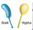
appendaged bacteria
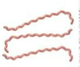
filamentous bacteria
cyanobacteria

Arrangement in prokaryotes
diploid, tetrads of Sarcina, chains (Streptococcus), grapelike clusters of Staphylococcus
Size range for prokaryote diameter
0.2 μm to > 700μm
cultured rod-shaped bacteria width
0.5 to 4.0 μm
cultured rod-shaped bacteria length
< 15 μm
examples of very large prokaryotes
Epulopiscium fishelsoni & Thiomargarita namibiensis
size range for eukaryotic cell diameter
2 to > 600 μm
advantages to being small
higher surface area / volume ratio, less energy required to multiply, more nutrient and waste product exchange
high surface area to volume ratio
more surface/membrane than volume so more transport membranes on surface for faster transportation of materials with less energetic requirements → multiples faster
ultramicrobacteria
0.2-0.4 μm in diameter; usually in open oceans; genomes highly streamlined & missing functions that must be supplied by other microbes/hosts
cytoplasmic membrane
surrounds cytoplasm, separates inside from environment, composed of lipids & proteins, selective permeability
selective permeability
proteins in the membrane transport nutrients into the cell and waste products out of the cell
Role of Proteins in cytoplasmic membrane
protein anchor, transport proteins, energy conservation and consumption
Protein anchor
holds transport proteins in place
transport proteins
accumulate solutes against the concentration gradient; proteins in the membrane transport nutrients into the cell and waste products out
Roles of proteins in bacterial cell membrane
protein anchor, transport proteins, energy conservation and consumption
Energy conservation and consumption
electron transport chain which generates the proton motive force and ATP through oxidative phosphorylation
mitochondria?
NO! in the membrane of bacteria for energy
Bacteria & fatty acids
they can change the length and saturation of fatty acids
fatty acid lengths change with
temperature
Increasing temperature, fatty acid lengths are
longer
Cholesterol in eukaryotes
stabilize membranes
hopanoids in bacteria
stabilize membranes
Archaea membranes
ether linkages in phospholipids, have isoprenes (no fatty acids), phosphoglycerol diethers with phytanyl C20 side chains to form lipid bilayers; diphosphoglycerol tetraethers with biphytanyl C40 side chains to form lipid monolayers
Bacteria and Eukarya linkages in phospholipids
ester
Archaea linkages in phospholipids
ether
Archaeal lipids have
isoprenes (no fatty acids)

archaeal lipid bilayers
phosphoglycerol diethers with phytanyl C20 side chains
Archaeal lipid monolayers
diphosphoglycerol tetraethers with biphytanyl C40 side chains → lots of stability with rings bc one phospholipid
Bacterial Cell Walls
located outside cytoplasmic membrane & protects it; can be targets for some antibiotics, makes cell shape, contributes to overall charge of the microbe, some components can elicit the production of antibodies (recognized by immune system)
Without the cell wall, bacteria would
all be circles
Gram stain
separates species of bacteria into two groups
Gram-negative cell wall
aka cell envelope; at least two layers: LPS (aka outer membrane) and peptidoglycan; may serve as barrier against antibiotics and other harmful agents
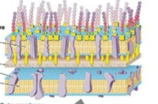
gram-positive cell wall
thicker, primarily one layer of peptidoglycan (90%); simpler; pentaglycine interbridge; teichoic acids bound to peptidoglycan which gives negative charge

Peptidoglycan
alternating modified glucose (N-acetylglucosamine and N-acetylmuramic acid) in β-1,4 glycosidic linkages; sheets are cross-linked differently in gram-positive or negative
Lysozyme
enzymes that cleave beta 1,4-glycosidic bond between sugars in peptidoglycan; major human defense against bacterial infection; cannot destroy Archaeal cell walls but destroys pre-existing peptidoglycan;
Crosslink for gram-negative bacteria
L-alanine, D-glutamic acid, L-lysine, D-alanine
Crosslink for gram-negative bacteria
L-alanine, D-glutamic acid, diaminopimelic acid (DAP), D-alanine
Gram-negative link between peptidoglycan sheets
Diaminopimelic acid (DAP) and D-alanine
Gram-positive link between peptidoglycan sheets
L-lysine and glycine (5 of them, then attached to D-alanine)
glycosidic linkages are in
carbohydrates
peptide bonds are in
amino acids (proteins)
L prefix for amino acids
usually used for enzymes
D prefix
usually not used
contains diaminopimelic acid (DAP)
most likely gram-negative cell wall
Which glucose monomer do you see peptide bonds from?
N-acetylmuramic acid (M)
The diether molecule within the membrane structure of the _____ yields a lipid ______
Archaea / bilayer
Negatively charged molecules that are partially responsible for the negative charge of the gram-positive bacterial cell surface are
teichoic acids
Penicillin
cannot destroy Archaeal cell walls; inhibits transpeptidation (cross linkage synthesis); compromises strength of peptidoglycan; destroy peptidoglycan
Pseudomurein amino sugars
N-acetylglycosamine & N-acetyltalosaminuronic acid
Pseudomurein linkage
beta 1,3-glycosidic linkage
What are the peptides that link pseudomurein?
L-lysine and L-glutamic acid
Why are there so many species of prokaryotes small?
higher SA:V ratio which supports more nutrient and waste exchange + less energy requirements → faster to divide

1 in bacterial membranes
glycerophosphates

2 in bacterial membranes
fatty acids

3 in bacterial membranes
integral membrane protein
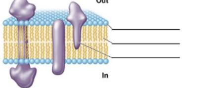
Is the bacterial membrane a lipid bilayer or monolayer?
bilayer
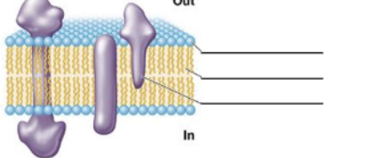
What is the name of the chemical linkage between the hydrophobic and hydrophilic group in bacterial membrane?
ester linkage

1 in archaeal membrane
glycerophosphates
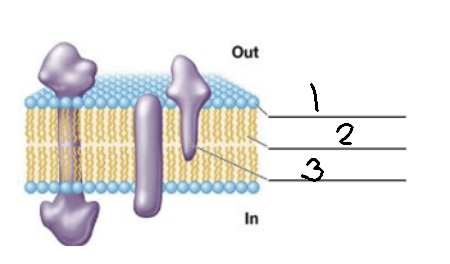
2 in archaeal membranes
phytanyl

3 in archaeal membranes
membrane protein
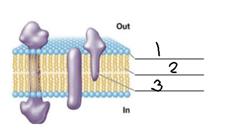
Is this archeael membrane a lipid bilayer or monolayer?
bilayer

What is the name of the chemical linkage between the hydrophobic and hydrophilic group in the archaeal membrane?
ether linkage
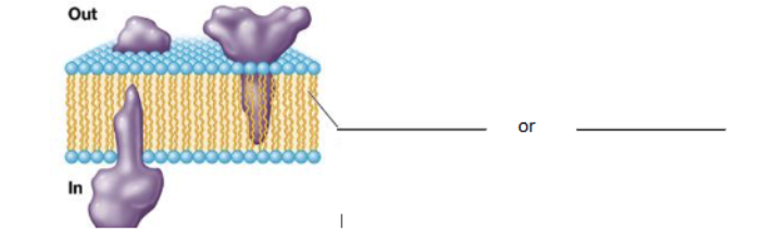
fill in the blank for thsi archaeal membrane
biphytanyl or crenarchaeol
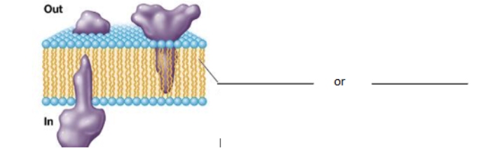
Is this archaeal membrane a lipid bilayer or monolayer?
monolayer
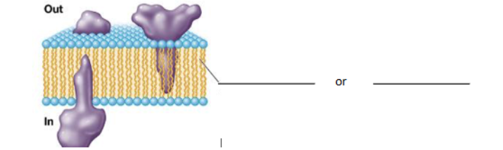
What is the name of the chemical linkage between the hydrophobic and hydrophilic group in the archaeal membrane?
ether linkage
Peptide order in gram-positive bacteria
L-alanine, D-glutamic acid-NH2, L-Lysine, D-alanine where L-lysine is bonded to 5 glycines, D-alanine, L-lysine, D-Glutamic acid-NH2, L-alanine

Peptide order in gram - bacteria
L-alanine, D-glutamic acid, Diaminopimelic acid (DAP), D-alanine with DAP bonded to D-alanine, L-lysine, D-glutamic acid, L-alanine

Full name for NAM
N-acetylmuramic acid
Full name for NAG
N-acetylglucosamine
What is the active site of lysozyme?
β 1,4-glycosidic linkage between NAG & NAM
How does penicillin affect cell walls?
inhibits the transpeptidation (cross linkage synthesis) to lessen the strength and formation of peptidoglycan

1
o-specific polysaccharide

2
core polysaccharide

3
lipid A

4
phospholipid
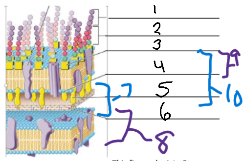
5
Braun lipoprotein

6
peptidoglycan
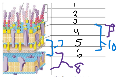
7
periplasm

8
cytoplasmic membrane

9
outer membrane

10
cell wall
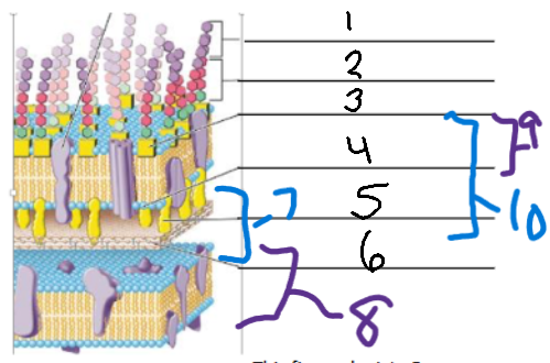
This figure depicts Gram ____ cell membrane and cell wall.
negative
Considering the structure of gram-negative cells, would you expect lysozyme to work?
yes bc there is a β 1,4-glycosidic linkage in gram - cells

Label the black and orange (one part)
lipid A

blue part
core polysaccharide

red part
o-specific polysaccharide
Which part of the lipopolysaccharide is an endotoxin?
Lipid A
When does lipid A become an endotoxin?
if it is released
Which part of the lipopolysaccharide elicits an antibody response?
o-specific polysaccharide

1
phospholipid