UWEC BIO 221 (Lab 3 questions)
1/39
There's no tags or description
Looks like no tags are added yet.
Name | Mastery | Learn | Test | Matching | Spaced | Call with Kai |
|---|
No study sessions yet.
40 Terms
Which is the platform onto which slides are placed and viewed?
The stage
Which part is used to move the slide?
The stage control
Which part or parts are used the adjust the amount of light passing through the sample?
Diaphragm
Which part or parts are used to raise/lower the slide and bring the specimen into focus?
Fine and course adjustment
Which part is the lens through which the sample is viewed?
Ocular lends
Which part collects the light passing through the sample and produces a magnified image?
Condenser lens
Write a brief description on each of the 3 basic principles of microscopy
Magnification:
the lenses of the microscope bend the light to produce an enlarged image
TM= M(ob) x M(oc)
unit: X
Resolution/Resolving Power:
the smallest distance between 2 objects that allows you to distinguish the two objects as separate
RP= wavelength / NA(cond) + NA(obj)
unit: nm
Contrast:
the difference between the object you are looking at and the background
Dyes
What information do you need to calculate total magnification? What is the formula for calculating total magnification?
TM= M(ob) x M(oc)
What is the total magnification of a 10X ocular lens and a 45X objective lens?
10(45) = 450X
If the total magnification is 2000X with the use of a 10X ocular lens, what is the magnification of the objective lens?
2000/10 = 200X
An object is 20 micrometers in diameter. If viewed through a 10X ocular lens and a 40X objective lens, how large will the object appear?
(what are the magnified dimensions of the object)?
10(40) = 400
400 x (20)
8000X
Consider the following microscope:
ocular power = 10X
low power objective = 20X
high power objective = 50X
What is the highest magnification you could get using this microscope? If 10 cells can fit end to end in the lower power field of view, how many of those cells would you see under high power?
highest magnification
10(50) = 500X
20(low)/50(high) = .4
.4 x 10 (amount of cells that can fit end to end)
= 4 cells
hint
higher magnification : less cells
lower magnification : more cells
*** think of zooming in on your iPhone. the more you zoom the less words you see.
What do each of the following symbols represent when calculating the resolving power of a microscope?
λ = wavelength
NAc = aperture of the condenser lends
NAob = aperture of the objective lens
What is the formula used to calculate the limit of resolution for a light microscope?
RP = λ / NAc + NAob
Look at your microscope to answer the following questions
What is the numerical aperture of the 10X objective?
What is the numerical aperture of the 40X objective?
(look at the 2nd number)
What is the limit of resolution for each objective on your microscope? Assume you are using blue light of 500 nm wavelength.
160/-
160/0.17
RP = 500/ 1.25 + 1.25
= 200
How many nanometers are in a micrometer?
How many micrometers in a millimeter?
1 micrometer = 1000 nanometers
1 millimeter = 1000 micrometers
You are viewing a bacterial cell that is 0.4 micrometers in length and width using the 100X objective on your microscope with yellow light of 600 nm. What is the limit of resolution? Will you be able to see an individual bacterial cell? Why or why not?
You are viewing two bacterial cells that are 0.5 micrometers apart. Assume you are using the 100X objective and blue light of 500 nm wavelength. What is the limit of resolution? Will you be able to distinguish the cells as two separate objects? Why or why not?
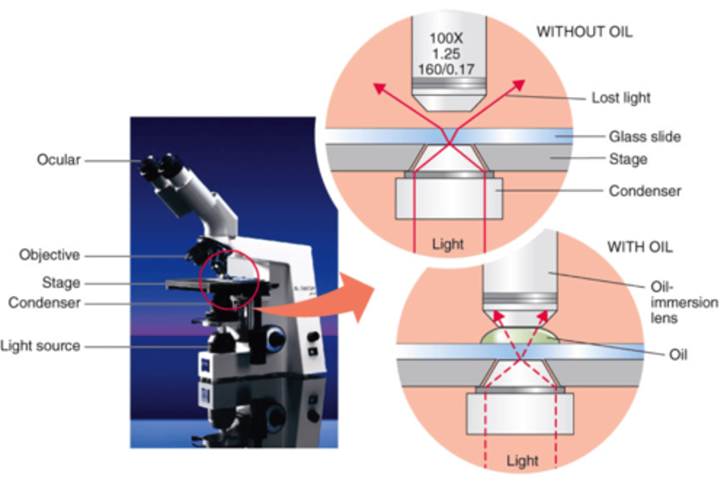
Why does immersion oil improve resolution with the 100X objective (only one that uses it)
It increases numerical aperture and maintains a uniform light speed
the oil reduces the loss of light & has the same refractive index as glass
What are the two groups of dyes most commonly used to stain microorganisms? How do they differ?
Basic Dyes (positive charge)
stain cells
Acidic Dyes (negative charge)
stains background
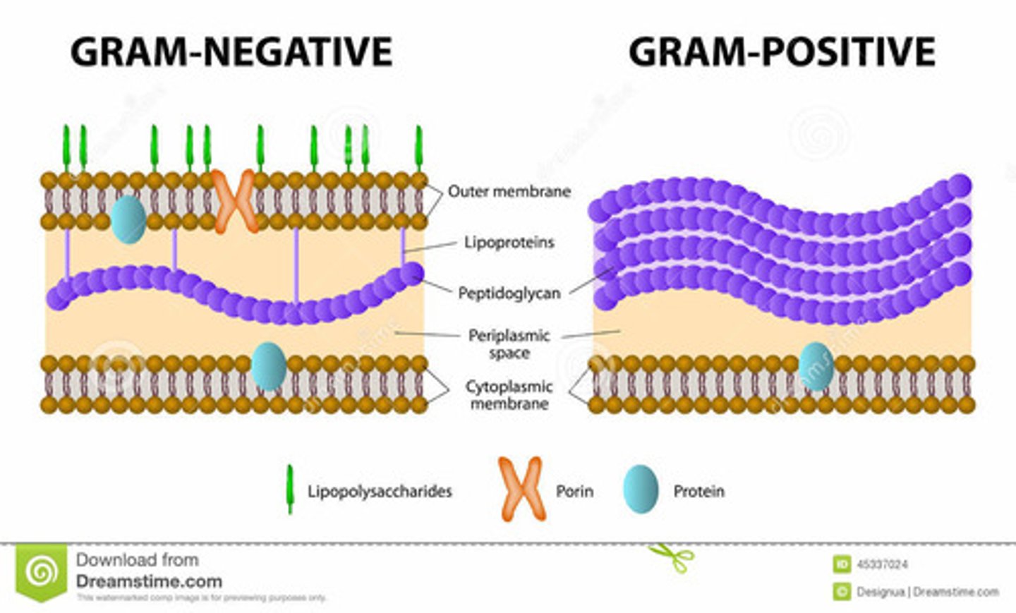
Write a short description of the structural and chemical differences between gram-positive & gram-negative cells walls
Gram-positive
- purple
an abundance of peptidoglycan on the exterior of the membrane
Gram-negative
-pink
thin layer of peptidoglycan that is found in between the interior and outer membrane
Describe the function of each of the following components in the gram stain procedure (know the order)
Crystal Violet:
basic dye, stains ALL bacteria (blue/violet)
Grams Iodine:
mordant; forms a complex with the crystal violet & peptidoglycan of gram + cell walls
Ethanol:
decolorizer; solubilizes the outer membrane of gram - cells. removes crystal violet from gram + cells that resist decolorization
Safranin:
basic dye that functions as a counterstain to dye the gram - cells pink.
What if the differential stain? What makes the Gram stain a differential stain and how does it differentiate bacteria?
Gram Stain considered a differential stain The Gram Stain differentiates two types of bacteria based on the composition of their cell walls, Gram Positive bacteria with a thick pectidoglycan cell wall, and Gram Negative bacteria that have only a thin layer of pectidoglycan.
Were you able to differentiate between M luteus & E. Coli using the Gram stain when they were in a mixed culture? Explain.
E. coli colonies are off- white, round (larger than M. luteus) with undulate edges and a rough appearance
M. luteus are yellow, round (small), raised (convex) with smooth edges
Would you be able to differentiate between M luteus & E.coli if you had only used crystal violet on the mixed culture? Explain
No, you would not be able to differentiate between the two because crystal violet stains ALL bacteria.
What information can you get by performing a Gram stain on bacteria? how is this information useful?
If your doctor suspects you have an infection, they may order a culture and gram stain to check for bacteria. If bacteria are present, this test can also help your doctor learn if the bacteria are gram negative or gram positive. The difference between gram-negative and gram-positive bacteria can affect their recommended treatment plan.
Gram stains can be performed on various types of specimens, including:
blood
tissue
stool
urine
sputum
What would the consequences be if you forgot to add the Gram's Iodine during the Gram stain procedure?
Iodine (Gram's iodine) solution (1% iodine, 2% potassium iodide in water) acts as a mordant and fixes the dye(Crystal violet) , making it more difficult to decolorize and reducing some of the variability of the test.
So without it the Crystal violet stain may decolorize. Meaning everything may look red due to the red stain (safranin or carbol fuchsin)
Under what circumstances would it be important to determine the Gram reaction of a bacterial species?
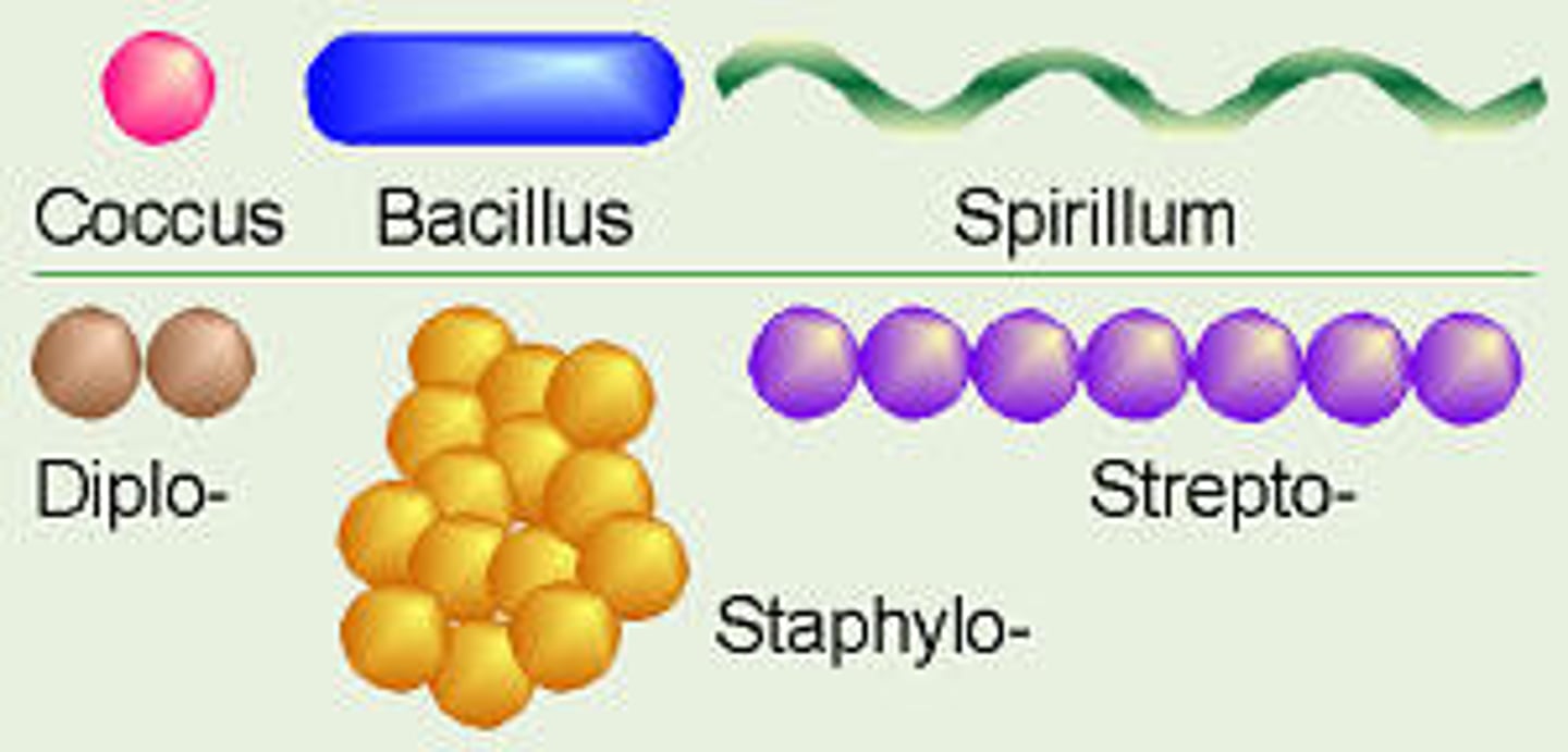
Different types of shapes of bacteria
Magnification
the ability to see the microbes, the lenses of the microscope bend the light to produce an enlarged image
Resolution
the ability to see the microbes as one or separately
Contrast
the difference between the object you are viewing and the background
The Gram stain procedure involves a series of steps. Number the steps in chronological order
1) Application of crystal violet
2) Application of Iodine
3) Application of safranin/ethanol
What is the purpose of adding the safranin/ethanol in the Gram stain procedure?
After alcohol is added to remove the crystal violet complexes from the peptidoglycan, the safranin acts as a counter stain
Gram Positive
stains blue/violet because there is a think peptidoglycan layer which takes longer for the alcohol to remove the crystal violet complexes. Therefore when safranin is added, it adds to the color already present from the crystal violet dye
Gram Negative
stains pink because when alcohol is added, it destroys the outer membrane and can remove the crystal violet complex more easily since it has a think layer of peptidoglycan. Therefore when safranin is added it will stain pink
Ethanol
decolorizer, solubilizes the outer membrane of Gram negative cells, removes the crystal violet, gram positive cells resist decolorization
Grams Iodine
Mordant, forms a complex with the crystal violet and peptidoglycan of gram positive cell walls
crystal violet
basic dye, stains all bacteria blue/violet
Briefly describe the difference between magnification & resolution
Magnification is the ability to see the microbes
Resolution is the ability to see the microbes as one or separately