📝 Ch. 19 - Likely Asked Questions
 Call Kai
 Learn
Learn Practice Test
 Spaced Repetition
 Match
Match Flashcards
 Knowt Play
1/227

There's no tags or description
Looks like no tags are added yet.
Name | Mastery | Learn | Test | Matching | Spaced | Call with Kai | Chat |
|---|
No analytics yet
Send a link to your students to track their progress
228 Terms
The visceral layer of the serous pericardium:
is the outer layer of the heart
The thickest wall in the heart due to its increased work load is the:
left ventricle
What is the correct pathway of blood starting at the right atrium?
right atrium, right ventricle, pulmonary trunk, pulmonary veins, left atrium, left ventricle, aorta
A skeletal muscle may undergo tetanus but cardiac muscle cannot.
True
What is occurring during isovolumetric contraction?
All four valves are closed
The walls of the atria are thicker because the atria receive blood under pressure.
False
The walls of the ventricles are thicker because the ventricles pump blood under higher pressure over greater distances.
True
The wall of the right ventricle is thinner than that of the left ventricle because the right side of the heart pumps blood a shorter distance at lower pressure.
True
Although the right side of the heart has a smaller workload, the right and left ventricle simultaneously eject the same amount of blood.
True
Prolapse of the atrioventricular valves is prevented by
contraction of the papillary muscles
The semilunar valves open when pressure in the right and left ventricles exceeds that in the pulmonary trunk and aorta, respectively.
True
Which represents the correct flow of excitation through the cardiac conduction system?
SA node, AV node, bundle of His, right and left bundle branches, Purkinje fibers
Which of the following places the phases of the cardiac cycle in correct order?
atrial contraction, isovolumetric contraction, ventricular ejection, isovolumetric relaxation, ventricular filling, atrial contraction
The plateau seen in action potentials recorded from ventricular fibers is due to inflow of Ca+2.
True
Isovolumetric contraction and isovolumetric relaxation both occur when all four valves are closed.
True
Purpose of the structure indicated is to: (right auricle?)
slightly increase the capacity of the atrium (see diagram)
The pericardium consists of a fibrous and serous layer. The visceral layer of the serous pericardium may be referred to as the epicardium.
True
The right atrium receives blood from the superior and inferior vena cava and the coronary sinus while the left atrium receives blood from the 4 pulmonary veins.
True
Autorhythmic fibers are self-excitable
True
Autorhythmic fibers: are pacemakers
True
Autorhythmic Fibers: Form the conduction system of the heart
True
The left ventricular pressure must be greater than the aortic pressure during ventricular ejection so that blood will move out through the pulmonary arteries.
False
The membrane that surrounds and protects the heart is called the
pericardium
Which of the following is used to reduce friction between the layers of membranes surrounding the heart?
pericardial fluid
Which layer of the heart wall consists of cardiac muscle tissue?
myocardium
Contraction of the ventricles of the heart leads to blood moving directly
into arteries
Which of the following chambers of the heart contains deoxygenated blood?
right atrium and right ventricle
Cardiac muscle fibers are electrically connected to neighboring fibers by
gap junctions
Which network of specialized cardiac muscle fibers provides a path for each cycle of cardiac excitation to progress through the heart?
cardiac conduction system
Which term refers to the period of time during a cardiac cycle when contraction of a chamber occurs and pressure within the chamber rises?
systole
Which wave in an electrocardiogram represents repolarization of the ventricles?
T wave
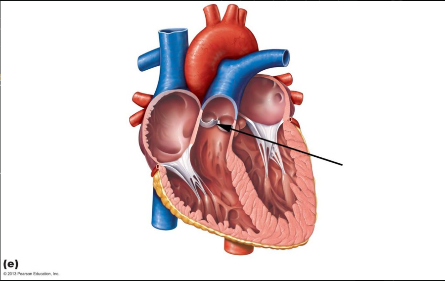
Identify the structure indicated in this diagram
pulmonary valve
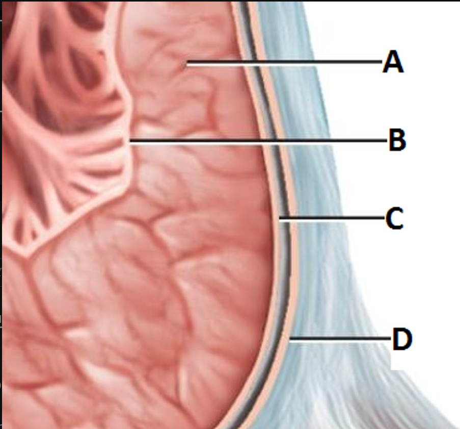
The structure indicated is the:
endocardium (C)
A = Myocardium
B = Endocardium
C = Epicardium
D = Serous pericardial/ Fibrous pericardium
Contractility can be decreased by _____________________.
-increased end-diastolic volume.
-presence of positive inotropic substances.
-decreasing inflow of calcium ions.
-increased end-diastolic volume and presence of positive inotropic substance.
-increased end-diastolic volume, presence of positive inotropic substance, and decreasing inflow of calcium ions.
decreasing inflow of calcium ions.
The amount of blood ejected from either ventricle every minute is called:
-stroke volumn
-end-diastolic volumn
-cardiac output
-after load
-heart rate
cardiac output
Which vessel does not supply the myocardium with blood?
-coronary sinus
-left coronary artery
-circumflex branch
-marginal branch
-posterior interventricular branch
coronary sinus
Which of the following statements is false in reference to heart regulation?
-at rest, parasympathetic stimulation predominates
-baroreceptors measure the amount of sodium ions present in the blood
-sympathetic stimulation triggers the release of norepinephrine
-the movement of limbs is monitored by proprioceptors
-parasympathetic fibers travel via the vagus nerve
barboreceptors measure the amount of sodium ions present in the blood
The intercalated discs seen in cardiac myocytes
1. have desmosomes which help to hold the muscle fibers together.
2. have gap junctions which allow action potentials to conduct from one muscle fiber to the next.
3. are the result of actin/myosin overlap.
1 only
2 only
3 only
1 and 2 are correct.
1, 2 and 3 are correct.
1 and 2 are correct
The term used to describe a backflow of blood from the left ventricle into the left atrium is:
-aortic stenosis
-mitral/bicuspid valve prolapse
-semilunar valve prolapse
-mitral/bicuspid valve stenosis
-aortic insufficiency
mitral/bicuspid valve prolapse
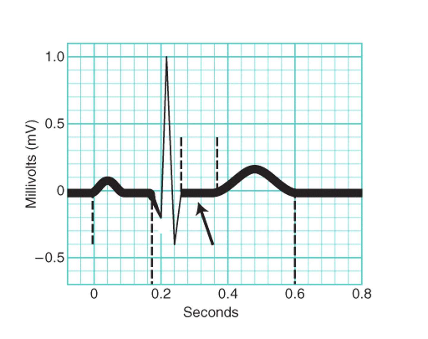
What occurs after the QRS phase of the ECG?
-the atria contract.
-the atria relax.
-the ventricles contract.
-the ventricles relax.
-diastole occurs.
The ventricles contract
Which of the following statements is NOT correct?
-The walls of the ventricles are thicker because the ventricles pump blood under higher pressure over greater distances.
-The walls of the atria are thicker because the atria receive blood under pressure.
-The wall of the right ventricle is thinner than that of the left ventricle because the right side of the heart pumps blood a shorter distance at lower pressure.
-Although the right side of the heart has a smaller workload, the right and left ventricle simultaneously eject the same amount of blood.
-All of these choices are correct.
The walls of the atria are thicker because the atria receive blood under pressure
Which wave is matched correctly with the heart's activity?
-QRS complex - atrial repolarization
-P wave - ventricular repolarization
-T wave - ventricular depolarization
-S-T segment - the ventricles are depolarized
S-T segment - the ventricles are depolarized
The ventricles must produce enough force to move the residual blood in the aorta before it can eject the blood from the ventricle. The aortic pressure is called
-preload
-afterload
-contractility
-relaxation
afterload
A patient with high blood pressure is prescribed a calcium channel blocker to lower blood pressure. This drugs acts as a
-parasympathetic inhibitor
-sympathetic accelerator
- positive inotropic agent
-negative inotropic agent
negative inotropic agent
All of the following statements concerning cardiac output (CO) are true except
-decreased contractility will increase CO
-if preload decreases, CO is decreased
-if a person hemorrhages, heart rate will increase to try and maintain CO
-if blood pressure rises, hormonal influence will attempt to compensate by decreasing blood volume and decrease CO
decreased contractility will increase CO
Frank-Starlings law of the heart explains a relationship between
- preload and contractility
-afterload and contractility
-stroke volume and volume of blood ejected
-preload and afterload
preload and contractility
The sympathetic nerve extends from the medulla oblongata to
-interventricular septum
-Purkinje fibers
-SA (sinoatrial node)
-bundle of HIS
SA (sinoatrial node)
All of the following are true of sympathetic nerves extending from the cardiac accelerator nerve of the medulla oblongata except
- it is composed of two neurons- a short and a long one
-it innervates the SA node, AV node, and myocardium
-innervation of the heart speeds up the rate of depolarization at the SA node
-the neurons release epinephrine which binds the beta 1 receptors
the neurons release epinephrine which binds the beta 1 receptors
Parasympathetic nerve fibers that innervate the SA node release
- norepinephrine
-epinephrine
-dopamine
-acetylcholine
acetylcholine
Which of the following is true?
-the majority of the blood filling the ventricles enters during atrial systole.
-More blood is ejected from the right heart verses the left heart.
-The same volume of blood is ejected from the right and left ventricles.
-More blood is ejected from the left heart verses the right heart.
The same volume of blood is ejected from the right and left ventricles
After a frightening experience is over, how does the heart restore normal heart rate?
- SA node fires at its own innate rate.
-The adrenal medulla adjusts the heart rate by releasing epinephrine.
-The cardiac inhibitor activates parasympathetic impulses to the SA node.
-The cardiac accelerator activates sympathetic impulses to the SA node.
The cardiac inhibitor activates parasympathetic impulses to the SA node
During isometric contraction, which of the following occurs?
-All of the choices are correct.
-Atrioventricular valves are closed.
-Ventricular pressures rises.
-Blood volume in the ventricles remain the same.
All of the choices are correct
One sign of hyperthyroidism is tachycardia. Which of the following can occur because of tachycardia?
- an increase in cardiac output
-an increase in stroke volume
-an increase in contractility
-decreased EDV (end diastolic volume)
decreased EDV (end diastolic volume)
All of the following are indicators of increased afterload except
-increase in ESV
-decrease in stroke volume
-decrease in ESV
-hypertension
decrease in ESV
When heart rate exceeds 160 BPM, all of the following occurs except
-stroke volume decreases
-venous return increases
-cardiac output decreases
-end diastolic volume decreases
Venous return increases
An analysis of the electrocardiograph can reveal all except
- stroke volume
-damaged areas in the heart
-an enlarged heart
-abnormalities in the conduction pathway
stroke volume
Desmosomes function to
-anchor fibers together
-transmit ions from cell to cell
-bind the sarcolemma of cardiac cells together
-act as receptors for neurotransmitters
anchor fibers together
Which component of the conduction system provides the only electrical connection between the atria and the ventricles?
-AV node
-bundle of HIS
-Purkinje fibers
-SA node
bundle of HIS
All of the following modify the timing or rate of the heart, except
- parasympathetic nerve
-sympathetic nerve
-sinoatrial node
-hormones
SA (sinoatrial) node
Autorhythmicity of the cardiac pacemaker cells occurs because
- the SA node is innervated by the sympathetic nervous system
-the SA node has an unstable resting potential
-the SA node has a stable resting potential
-the SA node is innervated by the parasympathetic nervous system
the SA node has an unstable resting potential
The P wave of the electrocardiogram is closely followed by __________.
a) contraction of both atria
b) contraction of the left atrium
c) relaxation of both atria
d) contraction of the right atrium
a) contraction of both atria
The volume of blood remaining in the ventricle as diastole begins is called the __________.
a) end-diastolic volume
b) end-systolic volume
c) cardiac reserve
d) stroke volume
b) end-systolic volume
If you know both the heart rate and stroke volume, you can calculate the __________.
a) ejection fraction
b) end-systolic volume
c) cardiac output
d) end-diastolic volume
c) cardiac output
The epicardium is also known as the __________.
a) visceral pericardium
b) myocardium
c) endocardium
d) parietal pericardium
a) visceral pericardium
Which of these is an important difference between cardiac muscle and skeletal muscle?
a) Skeletal muscle is striated.
b) Cardiac muscle lacks troponin.
c) Cardiac muscle contracts spontaneously.
d) Cardiac muscle can't be tetanized.
d) cardiac muscle can't be tetanized
Blood returning to the heart from the pulmonary circuit first enters the __________.
a) pulmonary valve
b) left atrium
c) pulmonary trunk
d) right atrium
b) left atrium
The papillary muscles function to __________.
a) close the AV valves
b) push blood from the atria into the ventricles
c) prevent the AV valves from reversing into the atria
d) eject blood from the ventricles
c) prevent the AV valves from reversing into the atria
The __________ valve prevents backward flow of blood into the left ventricle.
a) tricuspid
b) bicuspid
c) aortic
d) pulmonic
c) aortic
Blood flowing in the vena cava will next enter the __________.
a) left ventricle
b) right ventricle
c) left atrium
d) right atrium
d) right atrium
Excess fluid in the ________ causes cardiac tamponade.
a) both atria
b) left ventricle
c) pericardial cavity
d) apex of heart
e) visceral pericardium
c) pericardial cavity
Intercalated discs serve to transfer ________ from cell to cell.
a) action potentials
b) electrical signals
c) the force of contraction
d) ionic currents
e) All of the answers are correct.
e) All of the answers are correct
Cardiac muscle cells and skeletal muscle cells differ in a few ways. Which of the following is not one of them?
a) Cardiac muscle cells branch.
b) Skeletal muscle cells lack intercalated discs.
c) Cardiac muscle cells are smaller in size.
d) Cardiac muscle cells have a single, centered nucleus.
e) Cardiac muscle cells lack transverse tubules.
e) Cardiac muscle cells lack transverse tubules.
Blood returning to the heart from the systemic circuit first enters the
a) conus arteriosus.
b) left atrium.
c) left ventricle.
d) right ventricle.
e) right atrium.
e) right atrium
The cusps (leaflets) of atrioventricular valves attach directly to
a) interatrial septa.
b) papillary muscles.
c) trabeculae carneae.
d) coronary sulci.
e) chordae tendineae.
e) chordae tendineae
The right ventricle pumps blood to the
a) right and left lungs.
b) aorta.
c) left atrium.
d) right atrium.
e) left ventricle.
a) right and left lungs
The left ventricle pumps blood to the
a) pulmonary circuit.
b) right ventricle.
c) aorta.
d) right atrium.
e) lungs.
c) aorta
When a blood clot forms on a ruptured plaque in a coronary artery, the condition is referred to as a(n)
a) myocardial infarction.
b) coronary spasm.
c) angina pectoris.
d) coronary thrombosis.
e) pulmonary embolism.
d) coronary thrombosis
The first heart sound is heard when the
a) AV valves open.
b) semilunar valves close.
c) blood enters the aorta.
d) AV valves close.
e) atria contract.
d) AV valves close
The pulmonary arteries carry blood to the
a) heart.
b) liver.
c) intestines.
d) brain.
e) lungs
e) lungs
The pulmonary veins carry blood to the
a) liver.
b) intestines.
c) brain.
d) lungs.
e) heart.
e) heart
The foramen ovale in the fetal heart is located in the
a) left atrium.
b) interatrial septum.
c) left ventricle.
d) right atrium.
e) right ventricle.
b) interatrial septum
Heart rate is controlled by __________.
a) the phrenic nerves
b) both the right vagus and sympathetic nerves
c) sympathetic nerves in the cardiac plexus
d) the right vagus nerve
b) both the right vagus and sympathetic nerves
The rapid depolarization phase in the ventricular action potential appears in the electrocardiogram as the __________.
a) QRS complex
b) P wave
c) PR segment
d) T wave
a) QRS complex
The plateau phase of the ventricular action potential corresponds to the __________ phase of the electrocardiogram.
a) PR interval
b) ST segment
c) T wave
b) ST segment
During the plateau phase of the cardiac action potential, which ion is entering the cardiac muscle cell?
a) hydrogen
b) potassium
c) calcium
d) sodium
c) calcium
The conducting system in the heart begins in the __________.
a) AV bundle
b) AV node
c) SA node
d) Purkinje fibers
c) SA node
The ________ deliver(s) blood to the myocardium.
a) carotid arteries
b) coronary sinus
c) superior vena cava
d) cardiac veins
e) coronary arteries
e) coronary arteries
Blood is supplied to the left atrium by the
a) phrenic arteries.
b) right coronary artery.
c) brachiocephalic artery.
d) left coronary artery.
e) pulmonary arteries.
d) left coronary artery
The bicuspid or mitral valve is located
a) in the opening of the aorta.
b) where the venae cavae join the right atrium.
c) between the left atrium and left ventricle.
d) between the right atrium and right ventricle.
e) in the opening of the pulmonary trunk.
c) between the left atrium and left ventricle
The function of an atrium is to
a) collect blood.
b) pump blood to the lungs.
c) collect blood then pump it to the ventricle.
d) pump blood to the ventricle.
e) pump blood into the systemic circuit.
c) collect blood then pump it to the ventricle
The normal pacemaker of the heart is located in
a) the sinoatrial node.
b) the Purkinje fibers.
c) both the left and right ventricles.
d) the atrioventricular node.
e) the wall of the left ventricle.
a) the sinoatrial node
If the connection between the SA node and AV node becomes blocked,
a) the ventricles will beat more slowly.
b) the atria will contract more forcefully.
c) the ventricles will beat faster.
d) the ventricular beat will remain unchanged.
e) cardiac output will increase.
a) the ventricles will beat more slowly
Depolarization of the ventricles is represented on an electrocardiogram by the
a) QRS complex.
b) S wave.
c) P wave.
d) T wave.
PR complex.
a) QRS complex
What is the relaxed state of the ventricle called?
a) systole
b) ventricular filling
c) ventricular ejection
d) diastole
d) diastole
The one-way nature of the left AV valve prevents blood flow from _________.
a) the left ventricle to the aorta
b) the left atrium to the left ventricle
c) the left ventricle to the left atrium
d) the aorta to the left ventricle
c) the left ventricle to the left atrium
The closing of the left AV valve occurs near the beginning of __________.
a) atrial systole
b) ventricular systole
c) isovolumetric relaxation
d) ventricular diastole
b) ventricular systole
The majority of ventricular filling occurs while the ventricles and atria are in what state(s)?
a) ventricular diastole and atrial systole
b) ventricular systole and atrial systole
c) ventricular and atrial diastole
d) ventricular systole and atrial diastole
c) ventricular and atrial diastole
Which of the following most correctly describes end-diastolic volume?
a) the volume of the ventricle when it is most full
b) the volume of the ventricle when it is least full
c) the increase in ventricular volume during atrial systole
d) the volume of the ventricle at the end of atrial diastole
a) the volume of the ventricle when it is most full
Left ventricular filling occurs __________.
a) while the AV valve is open
b) during isovolumetric relaxation
c) during ventricular systole
d) while the semilunar valve is open
a) while the AV valve is open
Heart valves are in what state during isovolumetric contraction?
a) The AV valves and semilunar valves are open.
b) The AV valves and semilunar valves are closed.
c) The AV valves are closed, and the semilunar valves are open.
d) The AV valves are open, and the semilunar valves are closed.
b) The AV valves and semilunar valves are closed