Liver Doppler, TIPS Evaluation, and Liver Transplant Assessment
 Call Kai
 Learn
Learn Practice Test
 Spaced Repetition
 Match
Match Flashcards
 Knowt Play
1/27

There's no tags or description
Looks like no tags are added yet.
Name | Mastery | Learn | Test | Matching | Spaced | Call with Kai | Chat |
|---|
No analytics yet
Send a link to your students to track their progress
28 Terms
normal flow within the portal veins should be
hepatopetal, continuous, monophasic, and have low velocity
velocity of the normal portal vein
between 20 and 40 cm per second
because of their close association with the left atrium, the hepatic veins typically demonstrate
a triphasic flow pattern
deep inspiration may result in — of hepatic vein pulsatility
blunting or complete loss
the hepatic artery should demonstrate
a continuous, low resistance wave form pattern, with a quick upstroke, and a gradual deceleration with diastole
the normal resistive index of the hepatic artery is
between 0.5 and 0.8
in patients with advanced cirrhosis and portal hypertension, a TIPS stent may be inserted via the
jugular vein
a TIPS stent is usually placed between
a hepatic vein and intrahepatic portion of the portal vein
most often, the shunt is created between the
right portal vein and the right hepatic vein, and a bridging stent is left in place
a stent reduces the amount of
blood flow to the liver by rerouting the blood coming from the portal vein to the hepatic vein, ultimately sending the blood back to the heart
the primary goal of TIPS
is to prevent the rupture and hemorrhage of gastroesophageal and other varices, an occurrence that is often fatal
on sonography, TIPS appears as a
highly echogenic tube within the liver
it may be noted that flow within the right and left portal veins typically
reversed after stent placement
the normal flow velocity within the shunt ranges between
90 and 190 cm per second
the signs of TIPS failure include
signs of clot within the shunt, stenosis of the shunt, stenosis of the hepatic vein, reversal of intrahepatic flow, flow void next to the stent, reversal of hepatic venous flow, drop in shunt velocity between examinations, and abnormally high or low shunt velocity
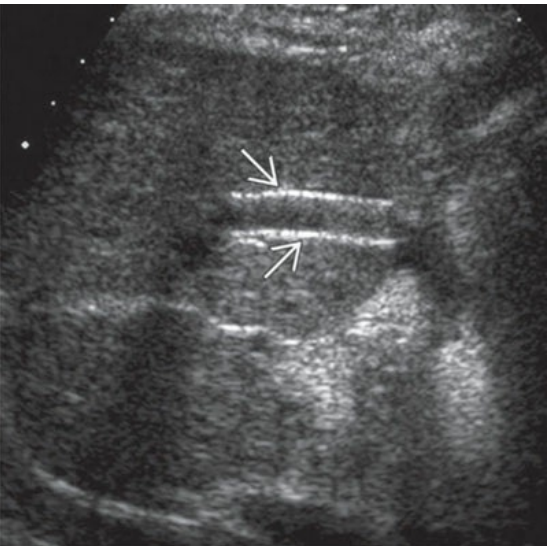
TIPS sonographic appearance. the wall of the TIPS appears highly echogenic. TIPS, transjugular intrahepatic portosystemic shunt
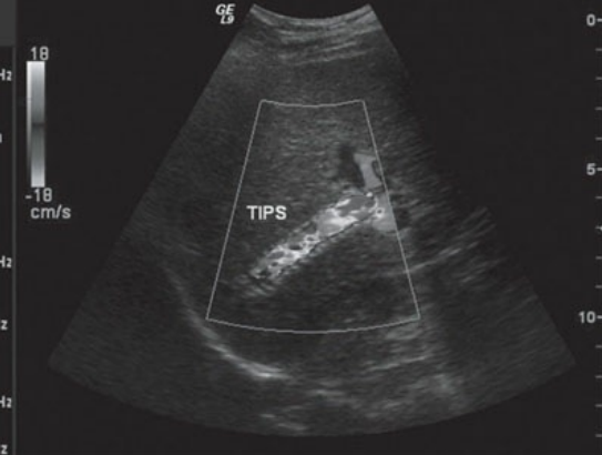
color image of a normal TIPS
— is the most common liver disease requiring a liver transplant, closely followed by alcoholic liver disease and cirrhosis
hepatitis C
the transplanted liver is attatched end to end to the native — & — termed — and a “—” attachment is made between the donor and recipient IVC
portal vein & hepatic vein; piggy back
a liver transplant should appear similar to
a normal native liver
although often with acute rejection the liver appears normal, suspicion should arise when the liver appears
diffusely heterogeneous
is the most common vascular complication of a liver transplant
hepatic artery thrombosis
when hepatic artery thrombosis is suspected, the sonographer should closely evaluate the liver for signs of
infarction
infarction appears as
hypoechoic wedge-shaped area scattered throughout the periphery of the liver
other complications of liver transplants include
biliary strictures, cholangitis, biliary sludge and stones, hepatic artery stenosis, hepatic artery pseudoaneurysms, celiac artery stenosis, portal vein stenosis and thrombosis, and fluid collections, including ascites, hematomas, bilomas, and abscesses
if present, stenoses, strictures, adn pseudoaneurysms are often discovered at
anastomotic site
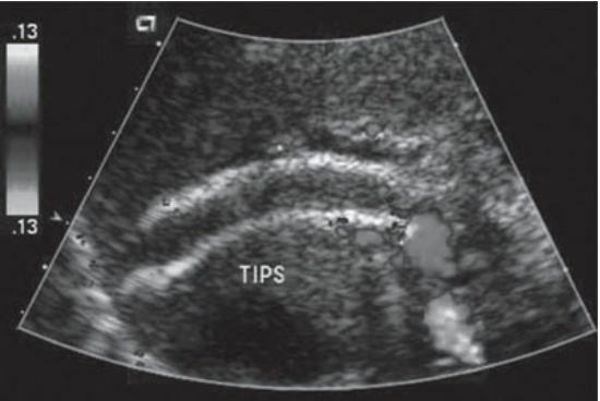
TIPS failure. this occluded TIPS lacks color flow filling and echogenic material (clot) can be noted within the shunt
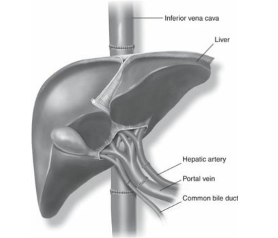
liver transplant. diagram demonstrating the most common surgical anatomy following liver transplantation with an interposition IVC graft. IVC, inferior vena cava