Fetal Head,Spine,Face, and Neck anomalies
 Call Kai
 Learn
Learn Practice Test
 Spaced Repetition
 Match
Match Flashcards
 Knowt Play
1/116

There's no tags or description
Looks like no tags are added yet.
Name | Mastery | Learn | Test | Matching | Spaced | Call with Kai | Chat |
|---|
No analytics yet
Send a link to your students to track their progress
117 Terms
What are the different kinds of head shapes?
Doli-chocephaly
• Bra-chycephaly
• Cranio-synostosis
• Micro-cephal
What is the Calvarium?
Bone around the entire skull
What can the Calvarium be associated with?
With skeletal dysplasia due to the minerilization around it
What are the 5 sutures?
sagittal
lamboid
squamous
Posterior fontanele
Coronal
What is a Dolichocephal head?
flattened head
What is a Brachycephaly head?
Rounded and overestimated
What are the 8 bones of the skull
Ethmoid
Sphenoid
Fontal
2 Occipital
2 Parietal
2 Temporal
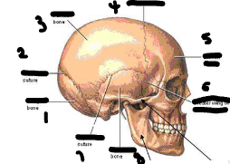
Lable this image
1-lamboidal
2-parietal
3-Coronal
4-Frontal bone
5-Great sphenoid bone
6-Temporal
7-Squamous suture
What is Cranio-Synostosis?
congenital anomaly caused by the closure of 1 or more than 5 sutures.
Most prominant in M:F (2,1)
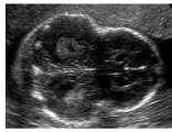
What image is this?
Craniosynostosis
What does patients with Craniosynostosis need?
Require a surgical release and cranial valt remodeling
What is Microcephaly?
determined by small BPD
fetal head is much smaller than expected
IS Microcephaly a common condition?
No it is not
What is Microcephaly caused by?
Infections: Rubella,Toxoplasmosis,zika,cytomegalovirus
Exposure of harmful substance(Alcohol,drugs,toxic chemicals)
Interuptions of blood supply

What does this image represent?
Microcephaly
What are the neural tube defects?
NEURAL TUBE DEFECTS
Anencephaly
Acrania/Exencephaly
Encephalocele
Vein of Galen Aneurysm
Arnold Chiari Malformation
Spina Bifida
Agenesis of the Corpus Callosum
Dandy-Walker
Holoprosencephaly
Ventricular System Related
Ventriculomegaly
Hydrocephaly
Hydrancephaly
Aqueductal Stenosis
Choroid plexus cyst
Porencephaly
What is Anencephaly?
Absence of cranium and prosencephalon due to failure of nerural tube.
Who are most likely to be affected by Anencephaly?
Females(4:1)
Cause unknown
What is Anecnephaly associated with?
Spina bifida
Cleft lip
Clubfoot
Omphalocele
polycephaly
What so we see on U/S in patient with Aneccephaly?
Little to no brain tissue
Have polyhydraminos
Orbit very shallow and causes protrusion of the eyes

what does this image show?
Anencephaly
What is Acrania/Excephaly?
parts of the Fetal skull not formed
-Brain tissue is exposed to amniotic fluid and risk of friction on wall and degration
What is Encephalocele?
Parts of the intracranial content through the bony part of skull, Caused by failure of the neural tube to form.
Who is Encephalocele most common in?
Common in Women
May be genetic or due to environmental problem
Associated with Meckle-Gruber syndrome
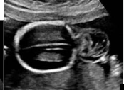
What does this image represent?
Encephalocele
What are Encephalocele associated with?
Meckel-Gurber syndrome
Encephalocele
PCKD(polycistic kidney disease)
Polydactyly-extra digits
What are Veins of Galen Malformation?
The areteries of thebrain are conected directly to the veins instead of the capillaries causing high blood pressure
What occurs when the Veins of Galen are malformed?
The blood rushes into the vein- and causes too much force and little resistance and the veind dialate
What can a large Malformation result in?
Great amount of blood circulation causing Cardiomegaly,pulmonary hypertension, hydrops
What does the vein of Galen appear as?
Anechoic cystic space, located midline and posterior superior to the 3rd ventricle

What does this image indicate?
Vein of Galen Malformation

Lable this image?
1- superior sagittal sinus
2-Coronal view
3-Inferior sagittal sinus
4-Internal Cerebral vein
5-Vein of Galen
6-Superior we,Ian vein
7-Precentral vein
8Straight sinus
9-tercular Heroph
What are Arnold Chiari Malformation?
Structural defects in the Cerebellum
What are the types of Arnold Chiari malformations?
Type 1* – displacement of the cerebellum posteriorally obliterating
the cisterna magna
Type 2- accompanies a meningomyelocele
Type 3- occurs with an encephalocele
Type 4- no cerebellum develops at a
What is the most common Arnold Chiari Malformation?
Chiari 1 is the most common type thant occurs in the region wher the brain and spinal cord join
What does Arnold Chiari Malformation cause?
The cerebellum and brainstem to lie lower than usual
-a portion of the cerebellar tonsil and protudes out the base of skull into spinal canal.
What is Syringomyelia?
Chronic disorder that involves the spinal cord
What causes Syringomyelia to occur?
Cerobrospinal fluid forund outside the Spina cord and enters the inferior spinal cord known as syrinx
In what patient are Arnold Chiari Malformation seen?
In children with myelomeningocele
What can Arnold Chiari Malformation led to abnormal developent of?
Cerebellar vermis
Medulla oblongata descend into for men magnum
What is frequently present in patient with Arnold Chiari Malformation?
Hydrocephalus
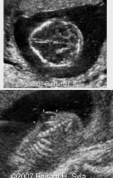
What does this image represent?
Arnold Chiari Malformation
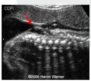
What does this image represent?
Spina Bifida

What does this image represent?
Obliterated Cisterna Magna/Banana sign
When there is Spina Bifida what does we see?
Head sign
hydrocephalus
Obliterated ,Cisterna magna, banana sign,lemon sign
What kind of vertebrae shape do we seen with spine bifida?
U shaped
What is Spina Bifida divided into ?
Occulta(15% covers skin
Aperta (85% exposure of neural canal

What does this image represent?
Spina Bifida
What is a indication of Spina bifida?
High MSAFP
Found in lumbosacral spine
In posterior fossa of head

What does this image represent?
Spina Bifida Meningomyelocele
What are the U/S finding of Spina Bifida?
Banana sign-Cerebellum flattens
Lemon Sign- Frontal bone cave in
Ventriculomegaly

What is this image indicating?
Banana sign

What does this image indicate?
lemon sign

What does this image represent?
Ventriculomegaly
What is the corpus collosum?
Thick band of fibers that connect the RT and LT hemispheres
How does the corpus collosum develop?
Ant- Post
1. the rostrum develops first
2. followed by the genu
3. then the body
4. and lastly the splenium

What does this image indicate?
corpus callosum
What does agenesis of the corpus callosum represent?
IT represents the left and agenesis of corpus collosum.
When corpus is absent the ventricals are set apart and Cavum Septum not normally seen
What is the corpus callosum closely related to?
CC closely related to the Cavum Septum pellicidum which is absent and 3rd Ventrical is higher in the head.
When is the Corpus Callosum formed and best seen?
Formed by the 17th week and best seen after the 22nd week
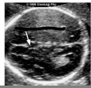
What is this image representing?
cavum septum pellucidi
What does the agenesis of the corpus callosum represent?
Colpocephaly
What does colpocephaly refer to ?
When the ant and lat horns become narrower and posterior/occipital horn become wider and enlarged
What are the key features of Dandy Walker Syndrome?
Enlargement of the 4th ventrical which allows fluid to flow freely between the upper and lower areas of the brain and spinal cord

What does this image represent?
Dandy Walker syndrome
What are the key features of Dandy Walker Syndrome?
Enlargement of the 4th ventrical
Partial or complete absence of area between the 2 cereabellar hemisphere
Cyst formation near the lower part of skull
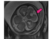
What does this image represent?
Dandy Walker syndrome
With dandy walker Syndrome how big is the Cisterna Magna?
Cisterna magna > 1 cm
• 50-70 % have associated
malformations such as absent
corpus callosum and
malformations of the heart, face,
limbs, fingers and toes.
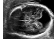
What does this image represent?
Cerebellum posterior fossa

What does this image indicate?
Normal Cerebellum and posterior fossa
What is Holoprosencephaly?
disorder that caused by failure of prosencephalonn to divide into double lobe of cerebral hemisphere
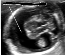
What does this image represent?
Holoprosencephaly
What are the 3 classification of Holoprosencephaly?
Alobar
Semilobar
Lobar
What is Alobar?
Brain not divided and associated with facial deformities
What is semilobaar?
The brain hemisphere is divided and causes an intermediate form of the disorder
What is Lobar holoprosencephaly?
evidence the brain hemisphere is seperated, in some cases the baby can be normal
What do we see in Alobar and Semilobar Holoprosencephaly baby?
Single ventricle
• Fused thalami
• Absent CSP
• Agenesis corpus callosum
What are the associated anomalies of Holoprosencephaly?
Median cleft lip
proboscis on forehead
Cyclopia

What does this image show?
Proboscis
Do most babies die with Holoprosencephaly?
Yes most cases they do die with severe birth defects
What are some environmental causes of Holoprosencephaly?
maternal diabetes mellitus,
maternal alcoholism,
inutero infections with CMV,
rubella or toxoplasmosis, some drug
What are some associated anomalies of Holophosencephaly?
Sever skull and facial defect
What is the common term for the ventricular system?
ventriculomegaly(mildly enlarged)
What term is used when the Ventricular system measures more than >15mm?
Hydrocephalus
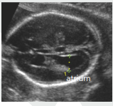
What does this image represent?
Dialated lateral ventricle

What does this image show?
Hydrocephalus

What does this image represent?
Dialation of the 3rd and 4th ventricle
Who is Ventriculomegaly most common in?
More in men
What are the sizes?
11-15=mild ventriculomegaly
>15mm= hydrocephaly
Normal width=<1cm

What is this image?
Dangling choroid
Who is Ventriculomegaly most common in ?
Females more than males

What does this image represent?
Ventriculomegaly

What does this image represent?
Dialated 3rd ventricle

What does this image indicate?
Clot within Hydrocephalus
What is Hydranencephaly?
rare condition.
brains cerebral hemispheres are absent and replaced with sac filled cerebrospinal fluid
What is Hydranencephaly thought to be?
Severe hydrocephalus and alobar holoprosencephaly
What is Hydranencephaly caused by?
caused by ischemic lesion due to occlusion of both corotid arteries
What are the causes of Hydranencephaly?
Bilateral occlusion of carotid arteries
Toxoplasmosis
Cytomegalovirus
Cocaine use
What is Aqueductal stenosis a form of?
Form of no communicating hydrocephalus and complete/ partial obstruction of the CSF flow at aqueductsylvian
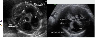
What does this image indicate?
Aqueductal stenosis