CARDIOVASCULAR SYSTEM
 Call Kai
 Learn
Learn Practice Test
 Spaced Repetition
 Match
Match Flashcards
 Knowt Play
1/57

There's no tags or description
Looks like no tags are added yet.
Name | Mastery | Learn | Test | Matching | Spaced | Call with Kai | Chat |
|---|
No analytics yet
Send a link to your students to track their progress
58 Terms
CARDIOVASCULAR SYSTEM
The organs and tissues involved in circulating blood and lymph through the body.
CARDIOVASCULAR SYSTEM PRIMARY PURPOSES
delivery of O2 and substrates for metabolism, growth and repair
removal of by products of cellular metabolism (CO2)
CARDIOVASCULAR SYSTEM SECONDARY PURPOSES
fast cell communication (hormones)
heat transfer
inflammatory and defence responses to foreign organisms
CARDIOVASCULAR SYSTEM COMPONENTS
heart
blood and lymph
vessels
SYSTEMIC CIRCULATION
high pressure
perfuses most of the tissues and organs with blood
PULMONARY CIRCUIT
low pressure
takes blood to and from the lungs
CARDIAC OUTPUT RULE
The cardiac output of the right side of the heart = cardiac output of the left side of the heart.
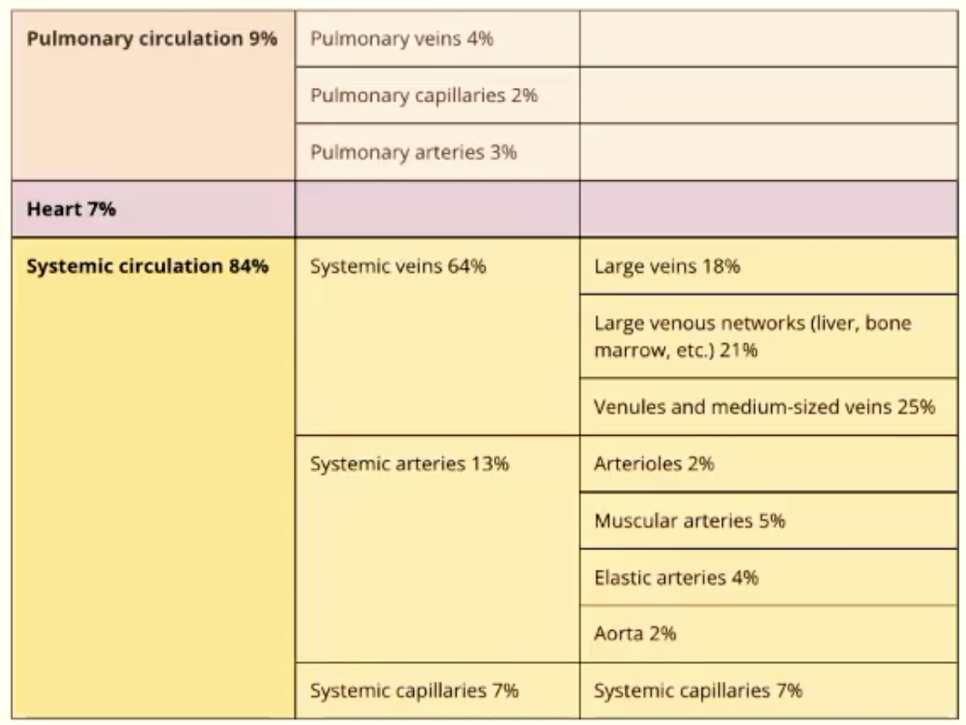
BLOOD DISTRIBUTION
Pulmonary circulation = 9%
Heart = 7%
Systemic circulation = 84%
Veins = 64%
Capillaries = 13%
Arteries = 7%
systemic veins are essentially blood volume reservoirs
the ‘reserve’ can be utilised when needed
BLOOD FLOW
The pressure gradient drives blood flow.
PRESSURE = FORCE / AREA
FLOW = PRESSURE (1) - PRESSURE (2) / RESISTANCE
Highest velocity of blood flow is in the middle.
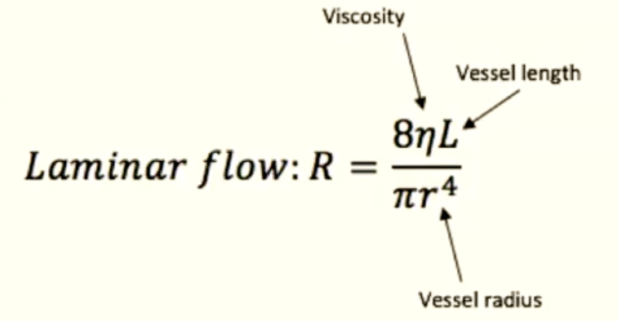
RESISTANCE FACTORS
geometry of blood vessels and type of flow
blood viscosity
vessel length
vessel width
LAMINAR FLOW
Laminar flow: R = 8(viscosity)(vessel length) / π(radius)^4
THEREFORE…
FLOW = Change in pressure / Resistance
THEREFORE…
FLOW = π(pressure difference)(radius)^4 / 8(viscosity)(length)
IMPLICATIONS OF POISEUILLE’S LAW:
fluid must be compressible
tube must be simple geometry - straight, rigid, cylindrical and unbranched (NOT ALL VESSELS ARE RIGID)
Velocity of fluid at wall must be zero
flow must be laminar not turbulent
blood flow must be steady (IT IS ACTUALLY PULSATILE)
Blood viscosity must be constant
CARDIAC OUTPUT
The amount of blood that the heart pumps through the circulatory system in a minute. Approx. 5L.
CO = STROKE VOLUME X HEART RATE
HAEMODYNAMICS
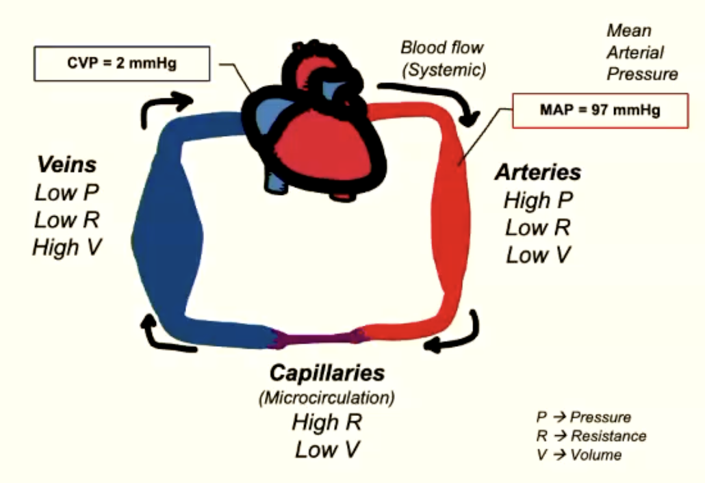
BLOOD FLOW SYSTEMIC
Mean Arterial Pressure (MAP) = 97mmHg
Central Venous Pressure (CVP) = 2mmHg
Difference in pressure drives blood flow
CO (of whole systemic circuit) = △P (MAP-CVP) / TPR (total peripheral resistance)
TOTAL PERIPHERAL RESISTANCE
TPR = Rarteries + Rarterioles + Rcapillaries + Rvenuoles + Rveins
MEAN ARTERIAL PRESSURE
MAP = average pressure through one cardiac cycle.
CO = (MAP - CVP) / TPR
But CVP is close to zero so disregard…
CO = MAP / TPR
Rearrange…
MAP = CO x TPR
BLOOD VESSEL STRUCTURE
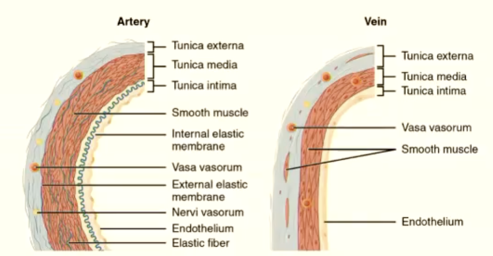
Inner lumen - passageway for blood flow
Surrounded by endothelium (TUNICA INTIMA)
Smooth muscle layer of varying thickness (TUNICA MEDIA)
Outer fibrous layer (TUNICA EXTERNA)
Variable amount of elastic connective tissue
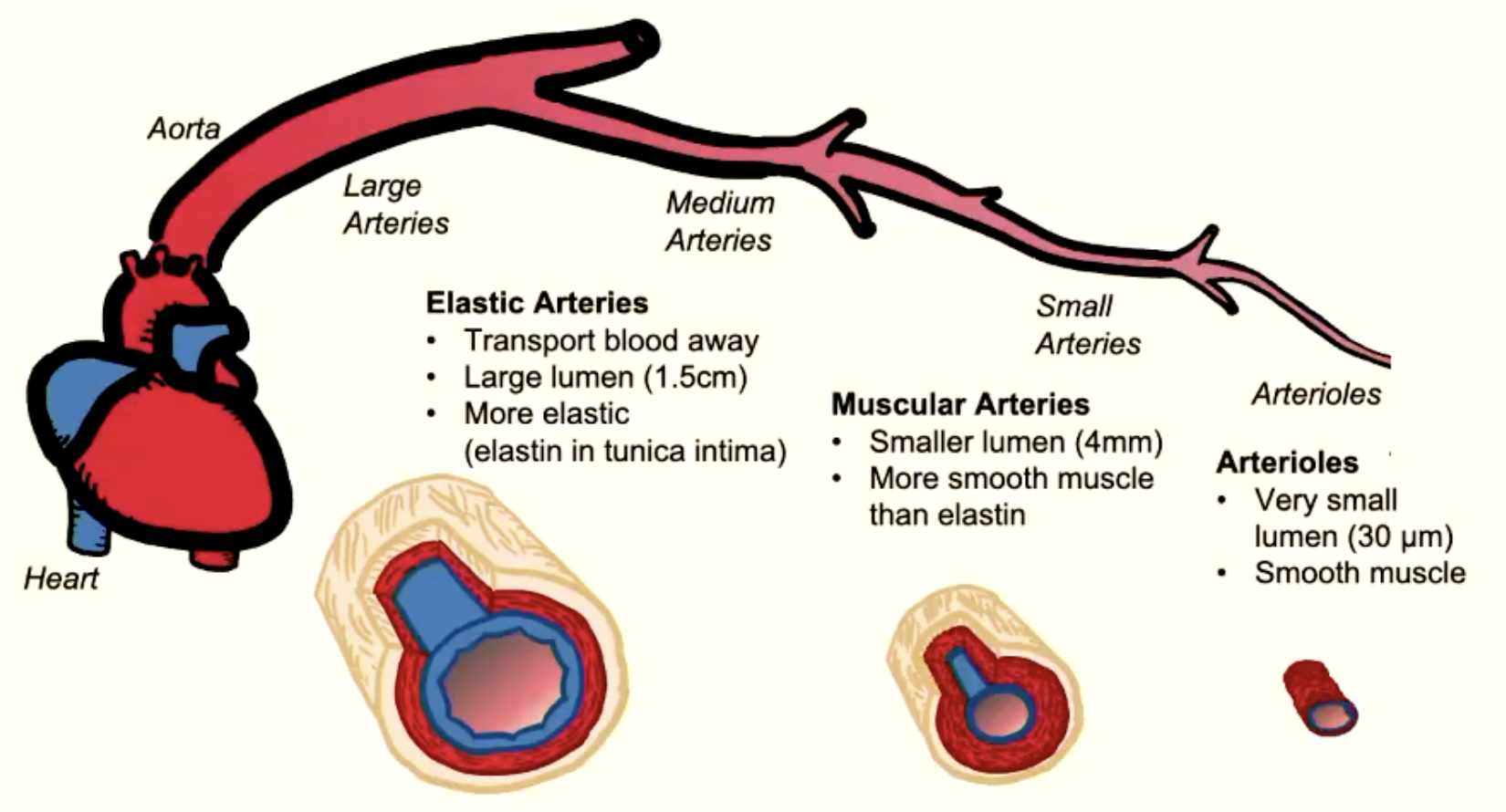
ARTERY + ARTERIOLES
transport blood FROM heart to capillaries
thicker walls than veins
more connective tissue and muscle than veins
little resistance due to large radius
pressure fluctuates in relation to systole and diastole
arterial pressure doesn’t drop to 0mmHg during diastole
arterioles are main resistance vessels
VASCULAR TONE
Vasoconstiction = smaller lumen (pathway)
Vasodilation = larger lumen (pathway)
EXTRINSIC CONTROL OF TONE:
autonomic nervous system
endocrine system
INTRINSIC CONTROL OF TONE:
metabolic factors
local signals
local temperature
stretch
can override extrinsic
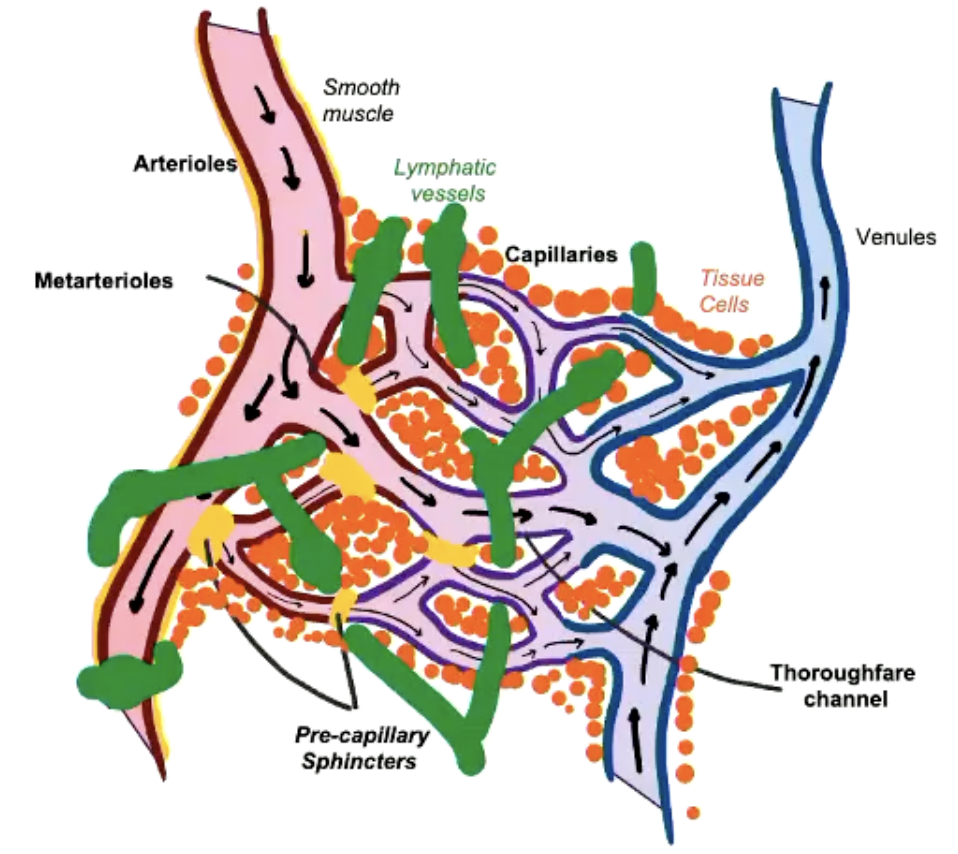
CAPILLARIES
Microscopic lumen
supply blood to tissue via perfusion
one endothelial cell thick
site of nutrient, waste and fluid exchange
CAPILLARY COMPONENTS
Pre-capillary sphincters
smooth muscle that controls the flow into the capillary beds
regulated by local factors
Interstitial fluid:
between plasma and cells
Lymph vessels:
important for fluid uptake
CAPILLARY FLUID MOVEMENT
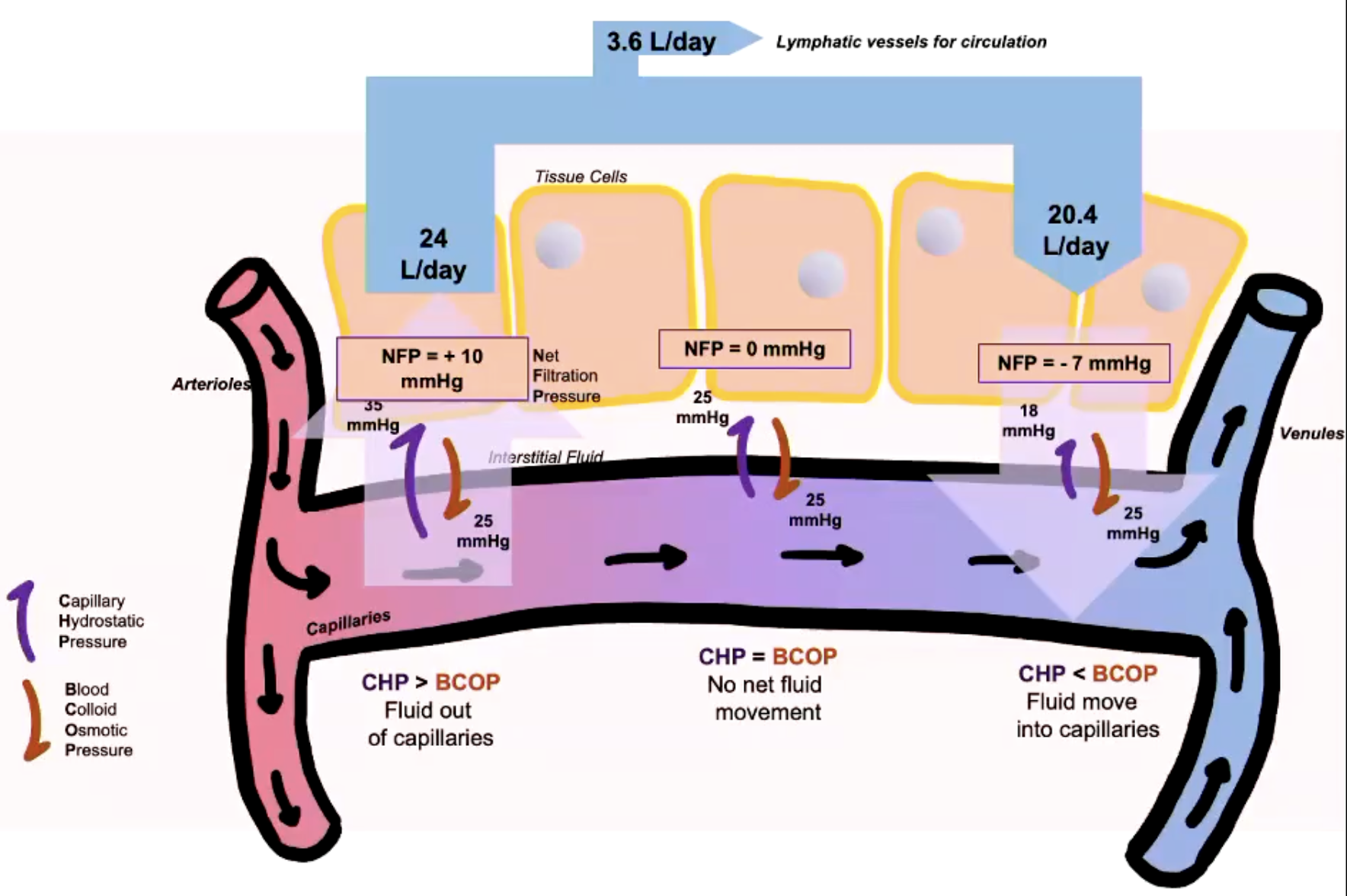
Driven by opposing pressure gradients:
Capillary Hydrostatic Pressure = blood pressure of capillaries
Blood Colloid Osmotic Pressure = due to presence of proteins in blood
CHP > BCOP = fluid out of capillaries
CHP = BCOP = no net fluid movement
CHP < BCOP = fluid moves into capillaries
LYMPHATICS
Lymph = fluid that flows through the lymphatic system
part of ECF
similar composition to interstitial fluid
branched network of ducts which terminate in small blind-ended (open) capillaries in the tissue
LYMPH FUNCTIONS
return excess fluid into circulation
immune defence
transport of lipids from GI tract
LYMPH FLOW
Interstitial fluid enters the lymphatic system via pores in the lymph capillaries
lymph is propelled by smooth muscle contractions and external pressure from skeletal muscle squeezing lymph vessels
they have valves to prevent back flow
OEDEMA
Swelling in soft tissue as a result of fluid accumulation - shift in the balance in the capillaries.
Excess interstitial fluid → increased distance between blood and cells → decreased rate of diffusion → inadequate nutrient supply.
VEINS AND VENUOLES
bring blood BACK to the heart
large lumen, thin walls
valves to prevent backflow
low resistance, low pressure
less smooth muscle and elastin than arteries
stretchy but no recoil
expand when filled with blood
VENOUS RETURN REASONS
Valves to prevent backflow
Skeletal muscles contractions squeeze veins
Lung pressure acts like a pump
Cardiac suction due to atrial enlargement
+VE EFFECTS ON HEART RATE
Sympathetic Nervous System
Adrenaline
Noradrenaline
Hormones
Adrenaline
Thyroid T3 and T4
Body temperature
fever
Ions
hypercalcemia
-VE EFFECTS ON HEART RATE
Parasympathetic Nervous System
acetylcholine
Ions
Hypocalcemia
CARDIAC OUTPUT - HEART RATE
Controlled by SA node
at rest parasympathetic NS is more dominant
CHRONOTOPIC AGENTS = drugs that affect heart rate
CARDIAC OUTPUT - STROKE VOLUME
Stroke volume is the volume of blood ejected in each ventricular contraction.
usually 70ml/beat @ rest
SV = EDV (end diastolic vol.) - ESV (end systolic vol.)
CARDIAC LENGTH TENSION RELATIONSHIP
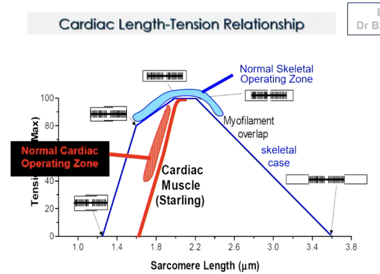
similar to skeletal muscle in that it has a length-tension relationship
sarcomere length determines tension developed = stroke volume
normal cardiac operating zone lower than skeletal
no negative relationship (no descending limb)
heart failure does not equal too much stretch (unlike skeletal)
FACTORS AFFECTING STROKE VOLUME
Preload
Afterload
Contractility
PRELOAD
The myocardial sarcomere length just prior to contraction.
A function of:
ventricular filling
ventricular & pericardial compliance
ventricular wall thickness
Frank-Starling Law.
AFTERLOAD
The force against which the ventricles must act in order to eject blood.
sum of elastic and kinetic forces
‘resistance’ to flow
Main opposing forces:
arterial blood pressure
vascular tone
CONTRACTILITY
Inherent vigour of contraction of the heart muscle during systole OR pumping strength.
independent of pre and after load
shift in Frank Starling curve
All impact Ca2+
Shift to left
Positive inotropic agents
Shift to right
Negative inotropic agents
BARORECEPTOR REFLEX
Receptors in aortic arch and carotid sinus = arteriol baroreceptors
measure high pressure (MAP)
impulses relayed to CVS control centres in medulla
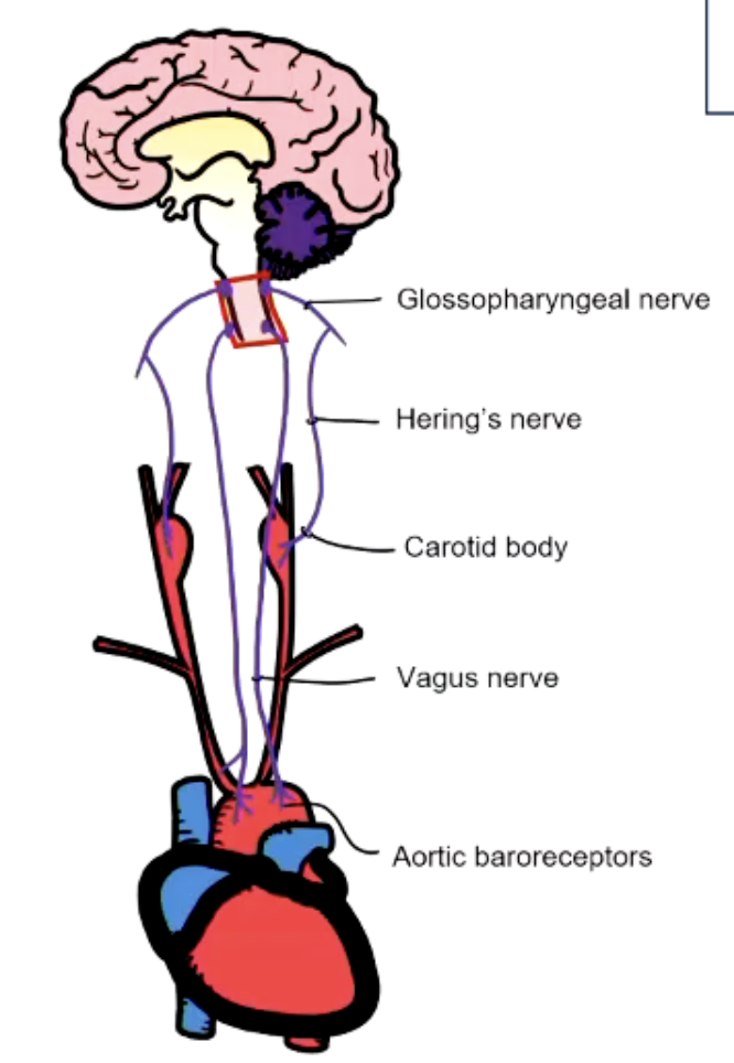
BARORECEPTOR PATHWAYS
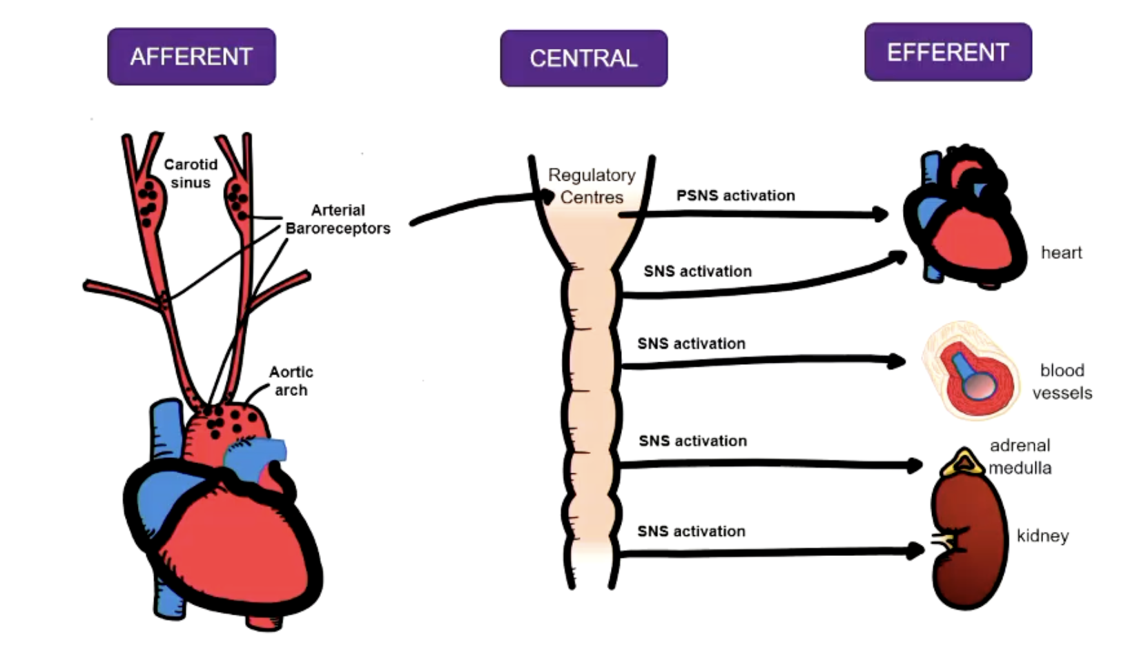
CHEMORECEPTOR REFLEX
mainly regulates ventilation but also has CVS effects
arterial not venous detection
CENTRAL CHEMORECEPTORS
within medulla oblongata
detect changes in cerebral spinal fluid
respond to high PCO2 - low pH
PERIPHERAL CHEMORECEPTORS
carotid and aortic bodies
respond to low pH in blood
CVS RESPONSES
Hypocapnia / hypexia (increase CO2 and peripheral resistance)
Hypercapnia (decrease CO2, bradycardia)
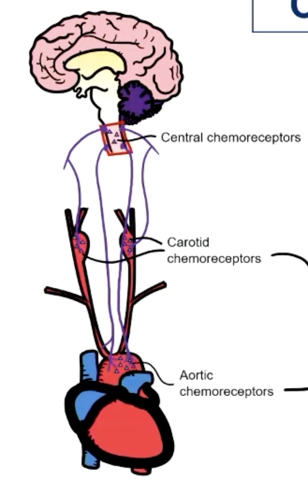
CARDIOVASCULAR CONTROL
SENSOR (AFFERENT NEURONS)
baroreceptors, chemoreceptors, proprioreceptors
INTEGRATION CENTRE
CVS centres in medulla oblongata
EFFECTOR (EFFERENT NEURONS)
Autonomic nervous system and hormones (adrenaline)
MAP REGULATION
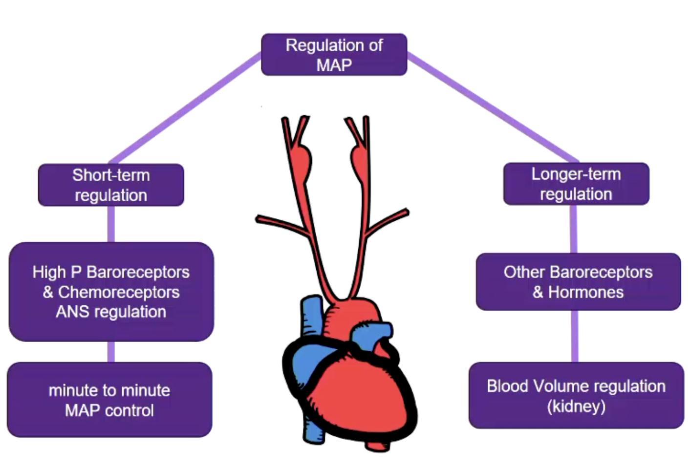
OTHER BARORECEPTORS
CARDIOPULMONARY BARORECEPTORS
in atria @ junction between large veins and pulmonary artery
impulses sent via vagus nerve to CVS centers in medulla
low pressure ‘stretch’ baroreceptors
measures venous return and blood volume
efferent effects include vasopressin
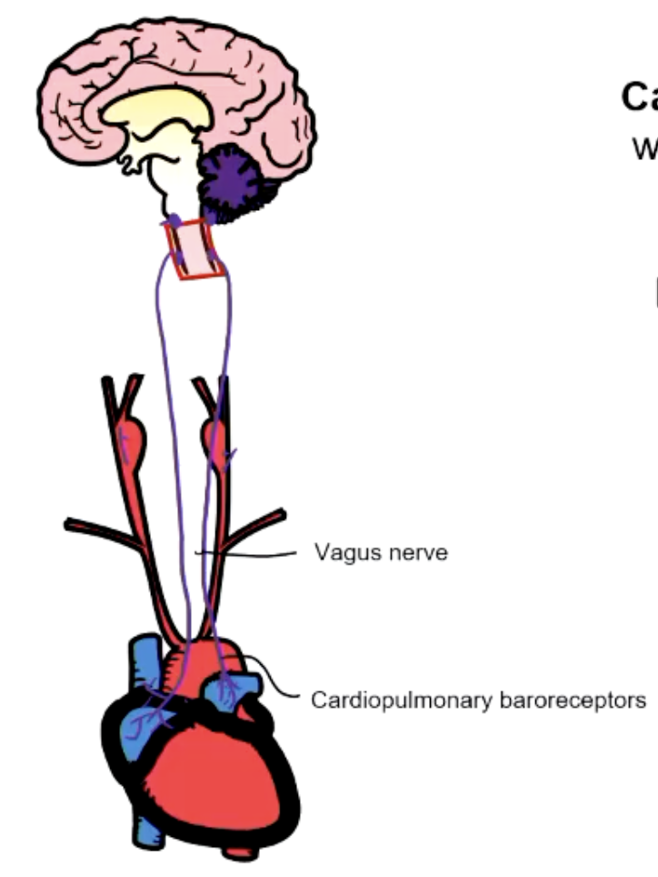
OTHER BARORECEPTORS PATHWAY
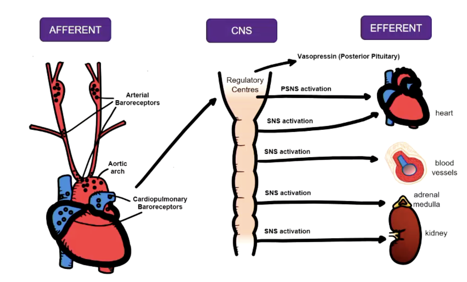
VASOPRESSIN
Neurohormone released from posterior pituitary
decrease water excretion from kidneys
vasocontriction
Secretion regulators:
osmoreceptors in ECF
cardiopulmonary baroreceptors in blood
Increase solute concentration:
Osmoreceptors in brain → increase vasopressin
Decrease solute concentration:
Active low pressure baroreceptors → decrease vasopressin
RENIN ANGIOTENSIN ALDOSTERONE SYSTEM
Targets longer-term blood volume regulation
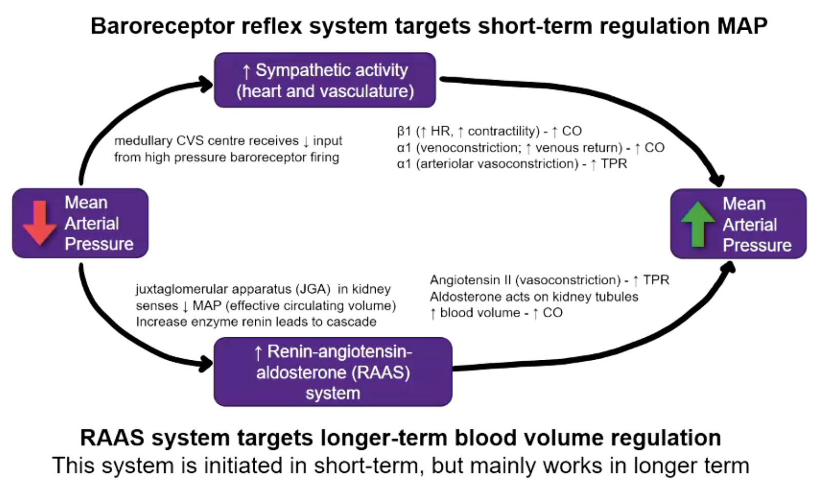
BLOOD PRESSURE REGULATION OVERVIEW
SENSORS
baroreceptors and chemoreceptors at different locations
INTEGRATION
medulla oblongata
EFFECTORS
autonomic nervous system and hormones
adrenaline, vasopressin, angiotensin, aldosterone
*baroreceptor reflex = short-term control
*blood volume regulation = long-term control
BLOOD LOSS
During mild/medium blood loss a reduction in capillary hydrostatic pressure causes the movement of interstitial fluid into the blood vessel to partially restore plasma volume.
CENTRAL ISCHEMIC REFLEX
Emergency back-up reflex activated when cerebral blood flow is critically compromised.
SENSED:
increased CO2 and metabolite that cause direct stimulation of medullary vasomotor centres
RESPONSE:
massive SNS outflow to increase cardiac output and vasocontriction
REALITY:
drives extreme tachycardia (heart rate increase) to prevent BP decline
kicks in at <60mmHg systolic BP
PULMONARY CIRCULATION
purpose = respiratory gas exchange
blood passes through lungs
low pressure circuit
relatively short circuit
branches immediately = increase exchange area = decrease resistance
less muscle = increase compliance
vascular response to hypoxia = vasoconstriction
HYPO AND HYPERTENSION
HYPOTENSION
too low MAP = inadequate blood flow
not enough blood for normal function
organ failure → shock → death
HYPERTENSION
too high MAP
extra work for heart and tissue damage
cardiac, vascular, renal failure → shock → death
PRIMARY VS SECONDARY HYPERTENSION
PRIMARY
multi-factorial pathogenesis
not one distinct cause
SECONDARY
has an identifiable cause
HYPERTENSION
BLOOD PRESSURE:
Systolic BP > 140mmHg
Diastolic BP > 90mmHg
RISK FACTORS:
diet
obesity
excessive alcohol consumption
inadequate exercise
CONSEQUENCES:
stroke
heart failure
heart disease
chronic kidney disease
ADAPTATION TO HYPERTENSION
Baroreceptors adapt/reset operate/maintain a higher MAP
they dont bring BP down but buffers acute BP fluctuations
HYPERTENSION PATHOPHYSIOLOGY
Hypertension = increase pressure = increase flow x resistance
causes too much volume → increase pressure → potential damage
HYPERTENSION CONSEQUENCES ON VASCULAR ARCHITECTURE
increased shear stress
endothelial cell damage
fibrotic scar tissue
atherosclerotic plaque
HYPERTENSION CONSEQUENCES ON BLOOD VESSELS
Stenosis = narrowing vessels → myocardial infarct
Thrombosis = clot from plaque formation & rupture → stroke
Aneurysm = bulge due to weakened wall → stroke
HYPERTENSION CONSEQUENCES ON HEART
Muscle hypertrophy → change in shape → more muscle → less space → less efficient
less compliant
less prelaod
lower EDV
lower SV