Normal Abdominal Wall
1/78
Earn XP
Description and Tags
UT 302 - Abdomen 1
Name | Mastery | Learn | Test | Matching | Spaced | Call with Kai |
|---|
No analytics yet
Send a link to your students to track their progress
79 Terms

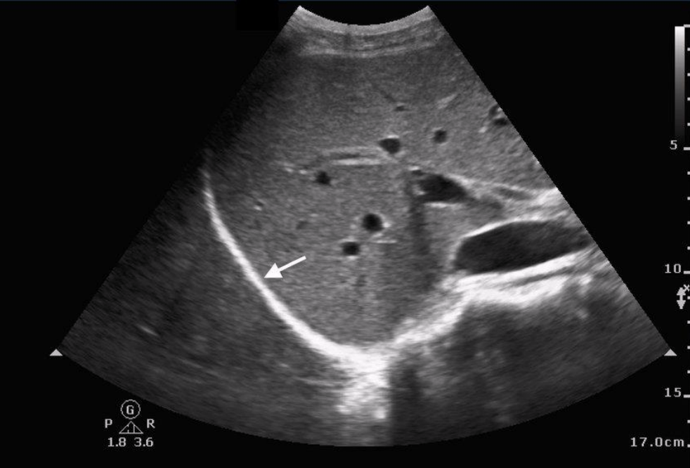
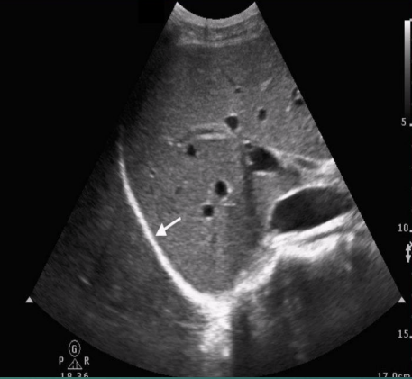
what is the white arrow pointing to? describe its echogenicity
diaphragm
hyperechoic

what type of artifact happens on the opposite side (in the lungs)?
mirror image artifact
what are inferior and superior to the diaphragm?
inferior: liver and abdominal organs
superior: lungs and heart
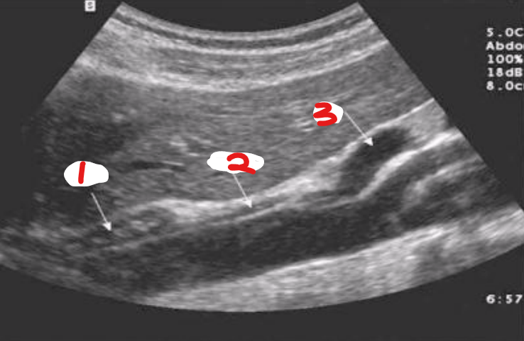
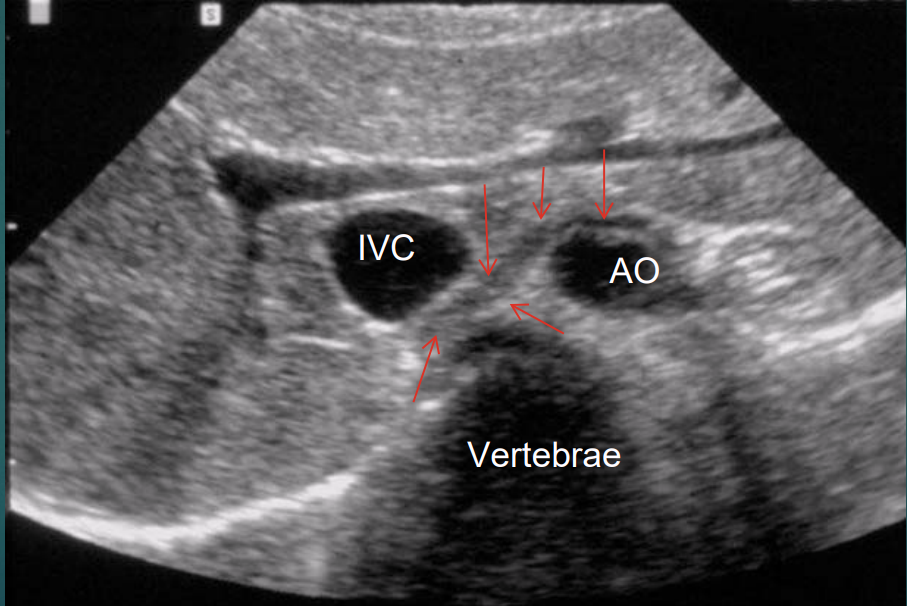
which arrow is pointing to the crus?
2
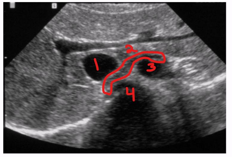
label the structures
IVC
crus of diaphragm
aorta
vertebra
indications for ultrasound of abdominal wall
palpable lump
pain
pain with an associated intermittent mass
inflammation
post surgical complication
trauma
which probe should be used to scan the abdominal wall?
use the highest frequency TDR possible
might be necessary to use the curvilinear TDR on some patients
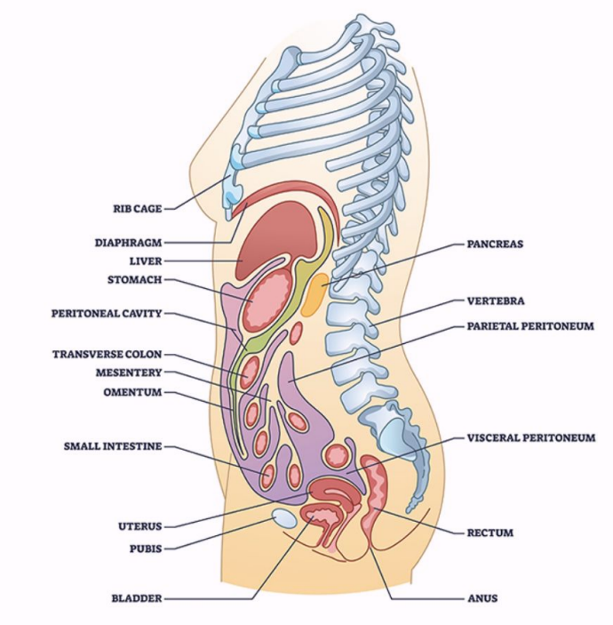
what are the two major body cavities?
dorsal cavity (posterior)
ventral cavity (anterior)
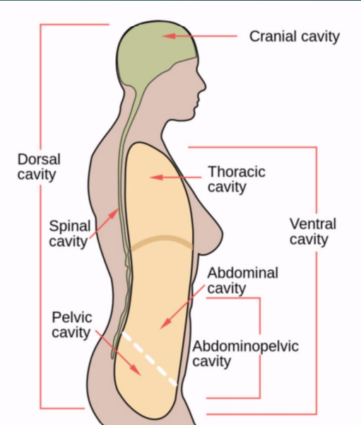
the dorsal cavity is divided into the …
cranial and spinal cavity
the ___ separates the thoracic cavity from the abdominopelvic cavity
diaphragm
the abdominopelvic can be separated into the
abdomen (superior)
pelvis (inferior)
abdominopelvic cavity
nine regions of the abdomen
R hypochondriac region
epigastric region
L hypochondriac region
R lumbar region
umbilical region
L lumbar region
R iliac region
hypogastric region
L iliac region
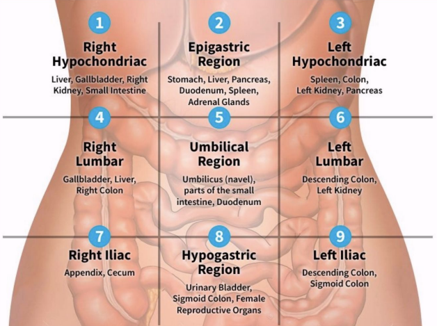
R hypochondriac region organs
liver
gallbladder
R kidney
small intestine
epigastric region organs
stomach
liver
pancreas
duodenum
spleen
adrenal glands
L hypochondriac region organs
spleen
colon
L kidney
pancreas
R lumbar region organs
gallbladder
liver
R colon
umbilical region organs
umbilicus
part of the small intestine
duodenum
L lumbar region organs
descending colon
L kidney
R iliac region organs
cecum
appendix
hypogastric region organs
bladder
sigmoid colon
female reproductive organs
L iliac region organs
descending colon
sigmoid colon
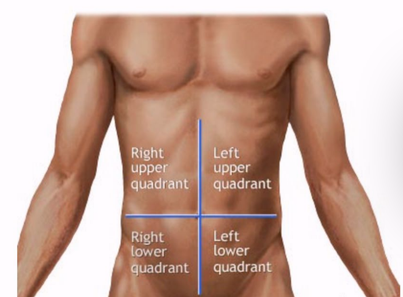
four abdominal quadrants
right upper quadrant (RUQ)
left upper quadrant (LUQ)
right lower quadrant (RLQ)
left lower quadrant (LLQ)
fascia
a fibrous tissue network located between the skin and the underlying structures
aponeuroses
large, sheet-like layers of connective tissue with a similar composition to tendons
can also attach to bone, as in the scalp, and to the fascia of other muscles or tissues
their large form and shape provides structure and distributes tension across a wider area or large number of muscle groups
aponeurosis vs. tendon
tendons connect muscle to bone
aponeuroses connect muscle to muscle, muscle to bone, muscle to fascia, or muscle to skin
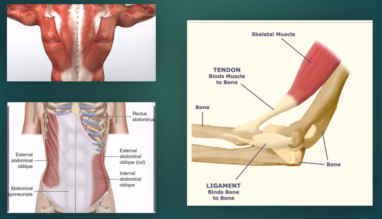
abdominal fascia can be divided into
superficial: attached to skin
deep: covers muscles and partitions them into groups
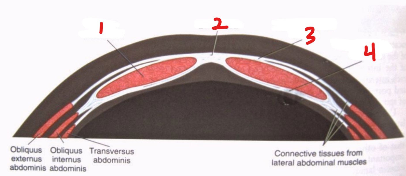
anterolateral abdominal wall layers
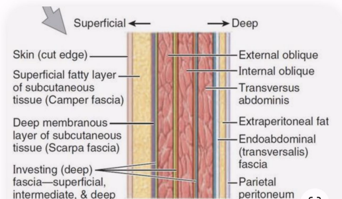

1
investing (deep) fascia divided into
superficial (external oblique muscle fascia)
intermediate (internal oblique muscle fascia)
deep (transverse abdominis muscle fascia)

2
deep membranous layer of subcutaneous tissue (Scarpa fascia)
3
superficial fatty layer of subcutaneous tissue (Camper fascia)

4
skin

5
parietal peritoneum

6
endoabdominal (transversalis) fascia

7
extraperitoneal fat

8
transverse abdominis muscle

9
internal oblique muscle

10
external oblique muscle
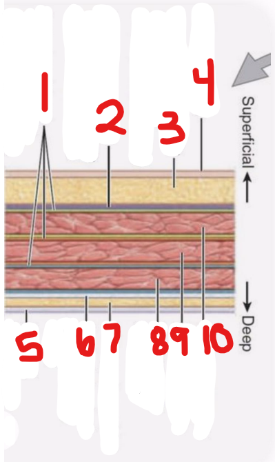
anterior abdominal wall has 5 bilaterally paired muscles
rectus abdominis
external oblique
internal oblique
transverse abdominis
pyramidalis
rectus abdominus muscle
bilaterally-paired, long, broad, vertical, strap-like muscle that is mostly enclosed in the rectus sheath
medial and anterior

the 3 flat, bilaterally paired muscles of the anterolateral group are the …
external oblique muscle (most superficial)
internal oblique muscle (middle)
transverse abdominis muscle (deepest)

pyramidalis muscle
small triangular muscle, missing in 20% of the population
rectus sheath
strong, fibrous compartment for the rectus abdominis and pyramidalis muscles as well as for some arteries, veins, lymphatic vessels and nerves
the ___ ___ separates the rectus muscles from the peritoneum
transversalis fascia
linea alba
oriented vertically and courses the length of the anterior abdominal wall
it separates the bilateral rectus sheaths
"white line"
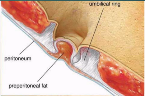
umbilicus
the area where all layers of the anterolateral abdominal wall fuse
umbilical ring
A defect in the linea alba and is located under the umbilicus
This is the area where the umbilical vessels passed to and from the umbilical cord and placenta

1
rectus abdominis

2
linea alba

3
anterior rectus sheath
4
posterior rectus sheath
1
subcutaneous tissue

2
linea alba

3
rectus abdominis muscle
4
anterior and posterior rectus sheaths

5
parietal peritoneum

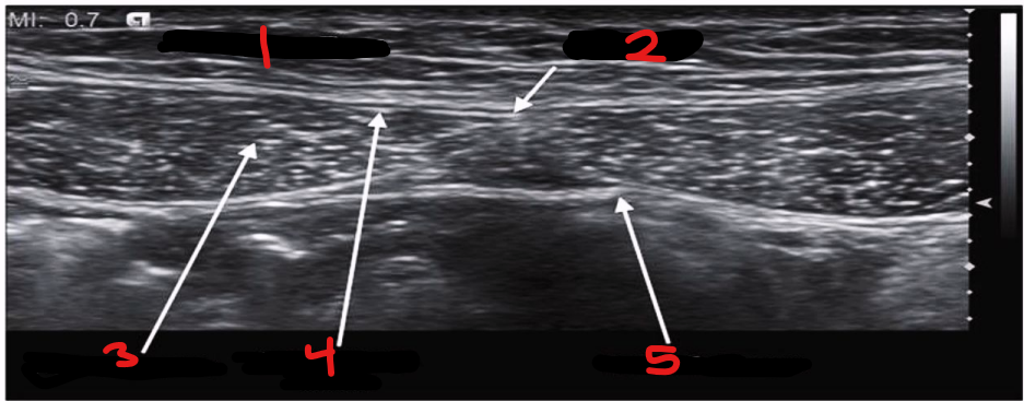
1
adipose layer (subcutaneous fat)

2
external oblique muscle

3
internal oblique muscle

4
transverse abdominis muscle

5
abdominal cavity

6
small bowel

7
fascia transversalis

8
internal oblique muscle-transverse abdominis muscle fascia

9
external oblique muscle-internal oblique muscle fascia

10
external oblique muscle fascia
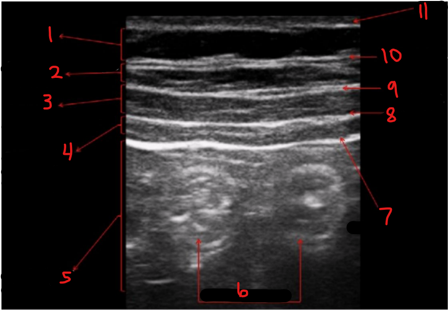
11
skin
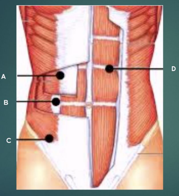
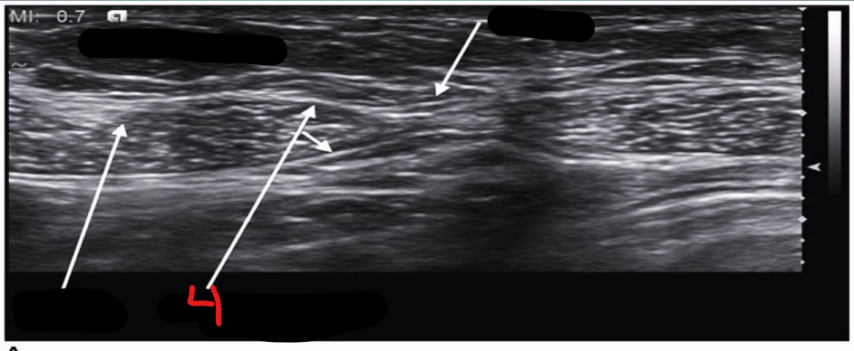
label the image
A - transverse abdominis
B - internal oblique muscle
C - external oblique muscle
D - rectus abdominis muscle
diaphragm
double domed, musculotendinous partition separating the thoracic cavity from the abdominal cavity
“parachute”-shaped
pleural effusion
fluid collection between the lungs and the chest wall
superior to the diaphragm

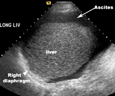
ascites
fluid collection in the peritoneal cavity
inferior to the diaphragm
movement of the diaphragm during respiration
can be imaged with m-mode
during inspiration, the diaphragm contracts and moves downward (creating negative pressure in the thoracic cavity)
during expiration, the diaphragm relaxes and moves upward (creating positive pressure in the thoracic cavity)
crus of the diaphragm
musculotendinous bands that arise from anterior surface of the first 3 lumbar vertebrae
right and left crura
right is longer and passes anteriorly to the aorta and posterior to IVC
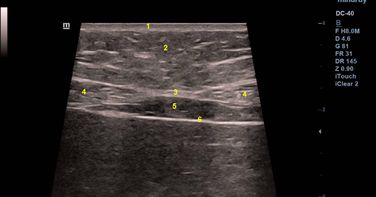
1
skin

2
subcutaneous fat

3
linea alba

4
rectus abdominis muscle

5
extraperitoneal fat

6
parietal peritoneum