The Heart and Blood Vessels
1/211
Earn XP
Description and Tags
Chapter 19 and 20
Name | Mastery | Learn | Test | Matching | Spaced |
|---|
No study sessions yet.
212 Terms
Where is the heart located?
In the thoracic cavity. In the middle of the chest, between the lungs in the mediastinum
What is the shape of the heart?
Conical in shape, with a broad base that points to the right shoulder and a narrow apex that points to the left hip
What is the size of the heart?
About 12cm long, 8cm wide, and 6cm thick.
Weighing between 250 and 350 grams (about the size of your fist)
What is the fibrous pericardium?
A tough, non-elastic outer layer of the pericardium (the sac that encases the heart)
What is the fibrous pericardium made of?
Connective tissue that prevents your heart from expanding too much
Contains collagen and elastic fibers
Fused with a parietal layer of the serous pericardium
What does the fibrous pericardium attach to?
Attaches to your great vessel (at the top of your heart) and to the central tendon of your diaphragm (at the bottom of your heart)
What is the parietal pericardium?
The outer layer that’s firmly attached to your fibrous pericardium (there is no space between them)
What is the job of the parietal pericardium?
Protects the heart from physical trauma and infection
Reduces friction between the heart and surrounding structures
Proves a space for the heart to expand and contract
What is the visceral pericardium? (also known as epicardium)
A serous membrane that forms the innermost layer of the pericardium and the outer surface of the heart
What does the visceral pericardium represent?
The portion of the pericardium that covers the heart and the blood vessels arising directly from the heart
What is the pericardial cavity?
A thin, fluid-filled space located between the heart and the pericardium, a sac-like membrane that encloses the heart
What is the function of the pericardial cavity?
Lubrication: the pericardial fluid within, reduces frictions between the heart and the pericardium, allowing the heart to move smoothly
Protection: The fluid acts as a cushion, protecting the heart from impact and injury
Stability: The cavity helps to keep the heart in place within the chest cavity
Immune Function: The cavity contains WBCs that help fight infection
What is the epicardium?
Outer most layer of the heart, surrounds the heart and roots of the coronary vessels
What is the function of the epicardium?
Protects the heart
Provides signals for embryonic development
Secretion of factors for proliferation/survival
Helps with heart injury response
What is the myocardium?
The muscle layer of the heart, makes up the bulk of the heart walls
What type of muscle is the myocardium made of?
Striated muscle
What is the role of the myocardium?
Plays a large role in heart function
What is the endocardium?
The innermost layer of tissue that lines the chambers of the heart
What is the function of the endocardium?
Provides protection to the valves and heart chambers and controls myocardial function
What is the endocardium made up of?
Primarily made up of endothelial cells
What are the right and left side of the heart separated by?
The interatrial and interventricular septum
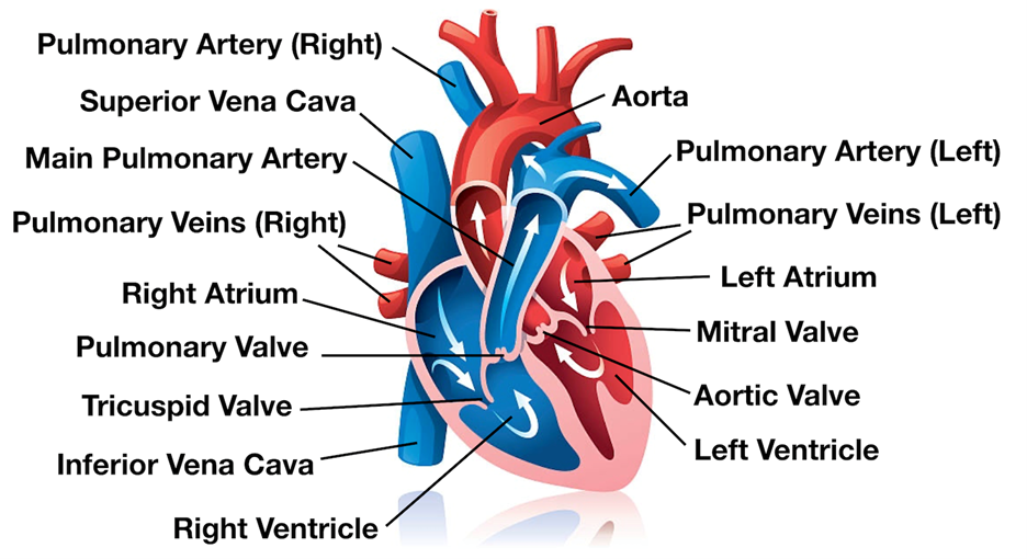
What is the atria?
The two upper chambers of the heart that receive blood from the body and lungs
What is the role of the atria?
A crucial role in maintaining the proper flow of blood through the heart (contracts and pumps the blood into the lower chambers of the heart called ventricles)
What do the atria have that prevent blood from flowing back into the veins?
Valves
What are auricles?
Ear-shaped projections in the atria, or upper chambers of the heart
What is the shape of the auricles?
Right: broad and triangular
Left: longer and more hooked
What are ventricles?
The two lower chambers of the heart, one on the right and one on the left
Where do the ventricles receive blood from?
The hearts upper champers (atria)
Where do the ventricles pump blood to?
The right ventricle pumps blood into the lings
The left ventricle pumps blood into the rest of the body
What ventricle has a thicker wall?
The left, it needs to push the blood into the rest of the body
What is the purpose of the tricuspid and bicuspid valves?
To prevent blood from backflowing into the atria
Where is the tricuspid valve located?
In the right side of the heart, between the right atrium and the right ventricle
How many leaflets/cusps are in the tricuspid valve?
3, the anterior leaflet, posterior leaflet and septal leaflet
Where is the bicuspid valve located?
The left side of the heart, between the left atrium and the left ventricle
How many leaflets does the bicuspid have?
2
What is the pulmonary valve?
A semilunar valve located in the heart that controls the flow of blood from the right ventricle to the pulmonary artery (which carries blood to the lungs for oxygenation)
When does the pulmonary valve open?
It opens during ventricular systole (contraction) to allow blood to flow from the RV into the pulmonary artery
When does the pulomary valve close?
During ventricular diastole (relaxation) to prevent backflow
What is the aortic valve and what is the responsibility?
A crucial component of the heart’s circulatory system, it is responsible for regulating blood flow from the left ventricle into the aorta
Where is the aortic valve located?
It exits the heart and branches out into smaller blood vessels (helping oxygen-rich blood flow through the body)
What vital role does the aortic valve play?
Maintaining proper blood circulation though the body
What can a damaged/dysfunctional aortic valve cause?
Serious heart problem
Potentially life-threatening complications
What are papillary muscles?
Piller-like muscles located within the ventricles
What is the role of papillary muscles?
Proper valve function, by attaching to the atrioventricular (AV) valve leaflets via chordae tendinae, preventing the valve from prolapsing during contraction (“anchors” for the valves)
What are chordae tendineae?
Thin strands of connective tissue that anchor the leaflets of each AV valve so that they cannot open into the atrium (prevent backflow)
What are trabeculae carneae?
Muscular ridges that line the inside of the heart’s ventricles
When is a valve replacement done?
When valves are severely damaged
What are some reasons for a valve replacment?
Rheumatic fever
Congenital reasons: Mitral valve prolapse, valve stenosis, missing cusps, regurgitation
What are the two types of valve replacements?
Biological: Processed, modified human, sheep or cow valve (lasts 8-10 years)
Mechanical (last 30+ years)
What are fibrous rings in the heart and what is it made up of?
A part of the heart’s fibrous skeleton, made of dense connective tissue
Where are fibrous rings located?
The rings surround the heart’s valves
What is the role of fibrous rings?
Providing support and prevent excessive stretching
What are some heart attachements?
Pericardium
Interventricular septum
Valves
A double-layered membrane (pericardium) surrounds your heart like a sac
the outer layer surrounds the roots of your heart’s major blood vessels and it attached to your spinal column, diaphragm and other parts of your body
What is the function of the heart’s skeleton?
Prevents dilation of orifices during ventricular contraction
Blockers the direct spread of electrical impulses from atrial to ventricular muscle
Anchors valve cusps
Insertion point for the bundles of cardiac his in atria and ventricles
Acts as a barrier against infection
Know the path of blood through the heart

What are coronary arteries?
Immediate branches off aorta just above aortic valve
What is the right coronary artery?
A blood vessel that supplies blood to the right side of the heart
Where does the right coronary artery originate from and where does it go?
The aorta and branches off into smaller arteries
What is the marginal artery and what is it’s role?
The marginal branch of the right coronary artery that provides blood supply to the lateral portion of the right ventricle
What is the role of the posterior descending artery branch?
Supply blood to the inferior aspect of the heart
What is the posterior interventricular artery and what is it’s role?
A large coronary artery, also called posterior descending artery (PDA)
Supply blood to its posterior (bottom) portion
Where is the posterior interventricular artery located?
Runs lengthwise along the back of the heart
What is the role of the left coronary artery?
Supply blood to the left side of the heart muscles (LV and LA)
What is the circumflex artery?
A coronary artery that supplies oxygenated blood to the back and outer side of the heart. It branches off from the left coronary artery and winds around the heart’s left side
What is the anterior interventricular artery?
A branch of the left coronary artery that supplies blood to the fron of the heart
What can a blockage in an artery cause?
A myocardial infarction (heart attack)
What is the coronary sinus?
The largest vein in the heart (main vein drain) a muscular tube that runs along the back of the heart between the left atrium and ventricle
What is the role of the coronary sinus?
Draining deoxygenated blood from the heart muscle into the right atrium
What is the great cardiac vein?
A large blood vessel found on the anterior (sternocostal) surface of the heart
When does the anterior interventricular vein become the great cardiac vein?
Once it has left the anterior interventricular sulcus and has entered the coronary sulcus
What is another name for the middle cardiac vein?
Inferior interventricular vein
What is the Inferior interventricular vein responsible for?
Draining the ventricular septum and the diaphragmatic portions of the walls of the cardiac ventricles
Where do the Inferior interventricular vein empty into?
The coronary sinus near the coronary sinus ostium
What is another name for the small cardiac vein?
Right coronary vein
What is the right coronary vein?
A small vein in the heart that drains deoxygenated blood from the right ventricle
What is the cardiac cycle?
A repeated series of events in which the atria contact while the ventricles relax - then the ventricles contract while the atria relax (one heartbeat)
What does the cardiac cycle dpend on?
Pressure, valves and elctrical conduction
What are the steps during Ventricular systole?
Isovolumetric (isovolumic) contraction
Ventricular ejection
What are the steps in Ventricular diastole?
Ventricular filling
Atrial Systole
When does the cycle start for a heartbeat?
When the blood fills the relaxed atria and relaxed ventricles
What are they listening for in a heartbeat? (with a stethoscope)
The sounds of the valves closing (“Lub-Dub”)
What creates the “lub” sound in the heart, and what does it signal?
When the mitral and tricuspid valves close, signals the beginning of ventricular systole
What creates the “dub” sound in the heart, and what does it signal?
When the aortic and pulmonary valves close at the end of the heartbeat, signals that the ventricles are relaxing, and the next contraction cycle is about to begin (short, high-pitched sound)
What is a murmur?
An extra or unusual sound made by blood flowing through the heart (can sound like a whoosh or vibration)
What can cause a heart murmurs?
Leaky, narrow, or tight heart valves
Increased heartrate due to nervousness
What is aortic stenosis?
A narrowing of the aortic valve that occurs during the contraction phase of the heart (systole), obstructing the flow of blood from the left ventricle to the aorta, causing resistance to blood ejection
What are some symptoms of aortic stenosis?
Chest pain
Shortness of breath
What is mitral regurgitation?
Blood flowing backwards from the left ventricle to the left atrium through the mitral valve during contraction phase
What is the cause for mitral regurgitation?
The mitral valve not closing properly, causing leakage of blood (considered an abnormal heart condition)
What is aortic regurgitation?
Blood flowing backwards from the aorta into the left ventricle during the relaxtion pahse
What causes aortic regurgitation?
A faulty aortic valve, causing a leak in the valve that allows blood to flow in the wrong direction
What is mitral stenosis?
A narrowing of the mitral valve opening (occurs during the relaxation phase of the heart cycle), significantly hindering the flow of blood from the left atrium to the left ventricle
What does mitral stenosis cause?
A pressure gradient between the two chamber and often producing a characteristic low-pitched rumbling murmur
What holds the connection in cardiac muscle fibers?
Intercalated disks (via gap junctions and desmosomes)
What is functional Syncytium?
A group of cells that work together as a single unit while each cell maintains its own role
What are gap junctions?
They connect the cells, allowing ions and electrical signals to pass between them (allowing them to work as a unit)
What works together as a unit?
Atrial and Ventricular
What is the role of the fibrous skeleton?
Provides structural and functional support for the heart’s valves, insulates the heart electrically
What is the cardiac conduction system?
Network of specialized cells in the heart that send electrical signals to make the heartbeat
What are the steps in cardiac conduction system? (CCS)
The sinus node generates electrical impulses
The impulses spread through the atria, that upper chambers
The atria contract, pumping blood into the ventricles
The impulses travel through the atrioventricular bundles and the bundle branches
The impulses reach the Purkinje fibers, which case the ventricles to contract
The ventricles relax