CH. 25 DIGESTIVE Exam 3 Rebecca Effler SUMMERTIME
 Call Kai
 Learn
Learn Practice Test
 Spaced Repetition
 Match
Match Flashcards
 Knowt Play
1/107

There's no tags or description
Looks like no tags are added yet.
Name | Mastery | Learn | Test | Matching | Spaced | Call with Kai | Chat |
|---|
No analytics yet
Send a link to your students to track their progress
108 Terms
Digestive system has two subdivisions:
digestive tract and accessory organs
Digestive tract
-30 ft long muscular tube extending from mouth to anus
-Mouth, pharynx, esophagus, stomach, small intestine, and large intestine
-Gastrointestinal (GI) tract is the stomach and intestines
Accessory organs
Teeth, tongue, salivary glands, liver, gallbladder, and pancreas
Digestive system
-Organ system that processes food, extracts nutrients, and eliminates residue
-Five stages of digestion: ingestion, digestion, absorption, compaction, defecation
Mechanical digestion
-The physical breakdown of food into smaller particles
-Cutting and grinding action of the teeth
-Churning action of stomach and small intestines
-Exposes more food surface to digestive enzymes
Chemical digestion
-A series of hydrolysis reactions that breaks dietary macromolecules into their monomers
-Carried out by digestive enzymes produced by salivary glands, stomach, pancreas, and small intestine
4 Layers of the Digestive Tract
1. Mucosa- epithelium, lamina propria, muscularis mucosae
2. Submucosa
3. Muscularis externa- inner circular layer, outer longitudinal layer
4. Serosa- areolar tissue ,mesothelium
Mucosa
lines the lumen!!!! (mucous membrane)
Epithelium
-Stratified squamous from mouth through esophagus, and in lower anal canal
-Simple columnar in most of digestive tract
Lamina propria
loose connective tissue layer
Muscularis mucosae
-Thin layer of smooth muscle
-Tenses mucosa creating grooves and ridges that enhance surface area and contact with food
-Improves efficiency of digestion and nutrient absorption
Submucosa
-Thicker layer of loose connective tissue
Composed of:
-Blood vessels, lymphatic vessels
-Nerve plexus
-Glands that dump lubricating mucus into the lumen
Muscularis externa
-Two layers of smooth muscle
-Inner layer—cells encircle tract
-Outer layer—cells run longitudinally; propels food and residue through the digestive tract
Serosa
outermost layer with two layers (areolar tissue, simple squamous mesothelium)
Areolar tissue
-Loose connective tissue, consists of collagen and elastic fibers
-Almost all cells obtain nutrients from and release their waste into this tissue
-Found beneath all epithelial tissue
Simple squamous mesothelium
-Lining of cavity
-Begins in lower esophagus, ends just before the rectum -Pharynx, upper esophagus, and the rectum have no serosa but are surrounded by a layer called adventitia
The mouth and its functions
-The oral cavity, or buccal cavity
-Functions: ingestion (food intake); chewing and chemical digestion; swallowing, speech, and respiration
Tongue
contains lingual papillae that contain taste buds
Intrinsic muscles
-Contained entirely within the tongue
-Produce subtle tongue movements of speech
Extrinsic muscles
-Attachments outside in the tongue
-Produce stronger movements of food manipulation
Palate
-Separates oral cavity from nasal cavity
-Makes it possible to breathe while chewing food
Uvula
hangs down from the soft palate at the back of theoral cavity
Teeth
-Dentition
-32 adult teeth
-16 in mandible (lower jaw), 16 in maxilla (upper jaw)•
-2 incisors—chisel-like cutting teeth used to bite off a piece of food
-1 canine—pointed and act to puncture and shred food
-2 premolars- have two rounded bumps = bicuspid
-3 molars—4-5 cusps
-Premolars and molars-crushing, shredding, and grinding -Wisdom Teeth are the 3rd molar
Mastication
-Chewing—breaks food into smaller pieces to be swallowed and exposes more surface to digestive enzymes
-1st step in mechanical digestion
-Food stimulates oral receptors that trigger an involuntary chewing reflex
Functions of saliva:
-Moistens mouth
-Begins starch and fat digestion
-Saliva production begins when you see, smell, or think about food
-Vitamins, free amino acids, minerals, cholesterol, and water do not need to be digested (already in usable form)
There are two kinds of salivary glands:
intrinsic and extrinsic
Intrinsic (minor) salivary glands
-Small glands among oral tissues; secrete saliva at constant rate (whether we are eating or not)
-Lingual glands: in the tongue; produce
-Labial glands: inside of the lips
-Palatine glands: of the palate
-Buccal glands: inside of the cheek
-Secrete lingual lipase and lysozyme
Extrinsic (major) salivary glands
-Connected by ducts
-Parotid glands—front of earlobe
-Submandibular gland—beneath mandible, 70% of saliva
-Sublingual gland—located in the floor of the mouth
Saliva is a hypotonic solution of
97.0% to 99.5% water, a pH of 6.8 to 7.0, and the following solutes: mucus, electrolytes, lysozyme, immunoglobulin A (IgA), salivary amylase, lingual lipase
Mucus
binds and lubricates a mass of food and aids in swallowing
Electrolytes
salts of Na+, K+, Cl-, phosphate, and bicarbonate
Lysozyme
enzyme that kills bacteria
Immunoglobulin A (IgA)
antimicrobial antibody
Salivary amylase
enzyme that begins starch digestion inthe mouth, deactivated by stomach acid
Lingual lipase
enzyme that begins fat digestion(activated in stomach by HCl)
Pharynx
-Circular skeletal muscles that form pharyngeal constrictors (superior, middle, and inferior)
-Force food downward during swallowing
-When not swallowing, the inferior constrictor remains contracted to exclude air from the esophagus (upper esophageal sphincter)
Esophagus
straight muscular tube 25 to 30 cm long between pharynx and stomach
Lower esophageal sphincter (LES)
-At inferior end of esophagus
-Prevents stomach contents from regurgitating into the esophagus
-Protects esophageal mucosa from erosive stomach acid -Heartburn—burning sensation produced by acid reflux into the esophagus
Swallowing
-Deglutition—a complex action involving over 22 muscles in the mouth, pharynx, and esophagus
-Coordinated by swallowing center—a pair of nuclei inmedulla oblongata
3 phases of swallowing
1. Oral- the tongue forms a food bolus and pushes it into the laryngopharynx
2. Pharyngeal- the palate, tongue, vocal cords, and epiglottis block the oral and nasal cavities and airway while pharyngeal constrictors push the bolus into the esophagus
3. Esophageal- peristalsis drives the bolus downward, and relaxation of the lower esophageal sphincter admits it into the stomach
Stomach
Primarily functions as a food storage organ, mechanical digestion, chemical digestion of proteins and some fat
Results in chyme
acidic, soupy mixture of semi digested food that passes on to the small intestine
Mucosa and submucosa
are flat when the stomach is full. wrinkled when the stomach is empty
Rugae
increases surface area
4 regions of the stomach
cardiac, fundic, body, pyloric
Regenerative (stem) cells
produce new cells to replacecells that die
Parietal cells
-Secrete hydrochloric acid (HCl), intrinsic factor
-HCl - activates Pepsin and lingual lipase and helps to liquidize food
-Intrinsic factor needed for B12 absorption
Chief cells
-Secrete gastric lipase and pepsinogen
-Gastric lipase - fat in milk
-Pepsinogen is converted toPepsin by HCL
Enteroendocrine cells
Secrete hormones and paracrine messengers that regulate digestion
Gastrin
stimulates gastric juice production
Gastric juice
secretion of gastric glands- mostly water, HCL, and pepsin
The small intestine
-Duodenum• Jejunum• Ileum•
-The site of nearly all chemical digestion and nutrient absorption
-Receives secretions from the pancreas liver and gallbladder
Large intestine size and regions:
-Measures 5 ft long and 2.5 in
-Functions to absorb water and salts from indigested food before it is eliminated
-Begins with cecum - Appendix attached to cecum
-Appendix is densely populated with lymphocytes and is a significant source of immune cells
Colon
portion of large intestine between ileocecal junction and rectum
The motility and secretion of the digestive tract are controlled by
hormonal, paracrine mechanisms, and neural
Hormonal Controls
-Gastrin- stimulates the release of gastric juice
-Secretin- stimulates the pancreas to produce digestive enzymes
Paracrine Controls
-Cell to cell signaling; autocrine-same cell, paracrine-next cell or into tissue fluids
-Histamine-increases HCl
-Prostaglandins-inhibits gastric secretion
Neural Controls
long and short autonomic reflexes
Short-reflexes
-myentericnerve plexus - stretching or chemical stimulation of the digestive tract•
-Causes contractions in nearby regions of the muscularis externa (peristaltic contractions)
Long-reflexes
-(vagovagal) involve the CNS
-Regulates motility and secretion
-Sight, smell, and taste trigger long reflexes
Enteric Nervous System
regulates the digestive tract motility, secretion, and blood flow
Submucosa plexus (Meissner)
-In submucosa (CNS viavagus nerve)
-Controls movements in the muscularis mucosae and the glandular secretions of the mucosa
Myenteric plexus (Auerbach)
-In between the layers of the muscularis externa.
-Controls movements (peristalsis) and contractions of muscularis externa
Regulation of Gastric Function
-Gastric secretion and motility regulated by nervous and endocrine systems•
-Increase gastric secretion and motility when food is eaten; suppress them when the stomach empties
Gastric activity is divided into three phases:
-Cephalic phase: stomach being controlled by brain
-Gastric phase: stomach controlling itself
-Intestinal phase: stomach being controlled by small intestine Phases overlap and can occur simultaneously
chyme also stimulates duodenal enteroendocrine cells to release?
-Secretin and cholecystokinin (CCK)
-These stimulate the pancreas and gallbladder, but suppress gastric secretion
Liver
-Reddish brown gland
-The body's largest gland, weighs about1.4 kg (3 lbs)
-Huge variety of functions, but only one contributes to digestion: the secretion of bile
-Gallbladder adheres to a depression on the inferior surface of the liver
Hepatocytes functions between meals and after meals:
-After a meal, hepatocytes absorb from the blood: glucose, amino acids, iron, vitamins, and other nutrients for metabolism or storage•
-Remove and degrade: hormones, toxins, bile pigments, and drugs
-Between meals, hepatocytes break down stored glycogen and release glucose into the blood
-Secrete into the blood: albumin, lipoproteins, clotting factors ,angiotensinogen, and other products
Bile
-Is an emulsifier
-Assists in lipid digestion
-Hydrophilic and hydrophobic components (like soap)
-The liver produces bile acids from cholesterol
-Bile acids break down and suspend fat molecules into smaller droplets
-Pancreatic lipases have maximum surface area for digestion -Only way to get rid of excess cholesterol
Gallbladder
-Concentrates and stores bile
-Bile acids are reabsorbed (80%) by the ileum and then sent back to the liver to be reused
Gallstones
(biliary calculi)
-Hard masses in either the gallbladder or bile ducts -Composed of cholesterol, calcium carbonate, and bilirubin
-Most common in obese women over 40, but also men and even children
-Can occur when cholesterol becomes too concentrated, precipitates as crystals that grow in size
-Painful obstruction of ducts
-Result in jaundice (yellowing of skin), poor fat digestion, and impaired absorption of fat-soluble vitamins
-Gallstones usually removed by laparoscopic surgery
The Pancreas endocrine and exocrine portion
-Endocrine portion: pancreatic islets that secrete insulin and glucagon
-Exocrine portion: secretes pancreatic juice - alkaline mixture of water, bicarbonate, and enzymes
Duodenum
-Receives stomach contents, pancreatic juice, and bile
-Neutralizes stomach acid
-Emulsifies fats
-Inactivates pepsin (due to the elevated pH)
-Reactivates amylase
-Pancreatic enzymes take over chemical digestion
Jejunum
-Most digestion and nutrient absorption
-Has large circular folds for increased absorption
Ileum
-Contains Peyer's Patches -lymphoid nodules•
-Contents move by segmentation- circular constrictions along the intestine (time to churn and mix material)
-Pacemaker cells in the muscle of the intestines set the rhythm for segmentation
-When the majority of the nutrients have been absorbed, segmentation ends
Migrating motor complex
-Activates successive waves of peristalsis
-Moves chyme to colon (2 hours)
-Refilling the stomach at the next meal suppresses peristalsis and reactivates segmentation
Fecal Content
-Water make up the majority
-Bacteria make up the majority of the solid material
-Followed by fiber, fat, and proteins
Bacterial Flora
-Digest cellulose (absorb sugar)
-Produce B vitamins and Vitamin K (blood clotting)
Water involvement in digestion
-Digestive system is involved in water balance
-Digestive tract receives about 9 L of water/day
-Water is absorbed by osmosis following the absorption of salts and organic nutrients
-Diarrhea—occurs when large intestine absorbs too little water
-Feces pass through too quickly if intestine is irritated
-Feces contain high concentrations of a solute (such as lactose)
-Constipation—occurs when fecal movement is slow, toomuch water gets reabsorbed, and feces become hardened55
Haustra
pouches in the walls of the colon
Haustral contractions
-Occur every 30 minutes
-Distension of a haustrum stimulates it to contract
-Promoting water and salt absorption
Mass movements
-Stronger contractions that occur one to three times a day
-Moves further distance than haustral
-Filling of the stomach and duodenum stimulates motility of the colon
Stretching of rectum yields two defecation reflexes:
-Contraction of the rectum (myenteric plexus)
-Relaxation of the internalanal sphincter(parasympathetic)
External anal sphincter
remains contracted (voluntary control)
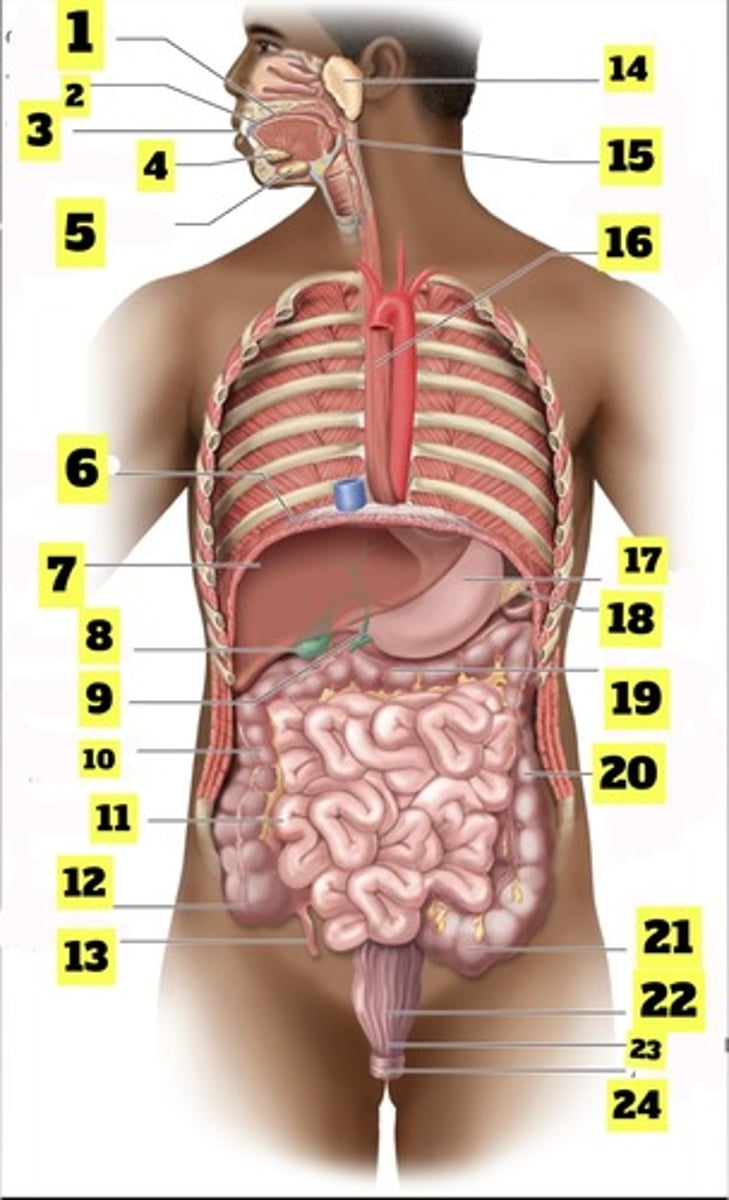
What is number 1?
oral cavity
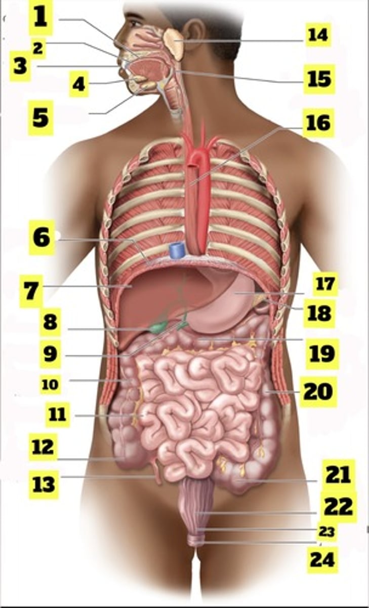
What is number 2?
tongue
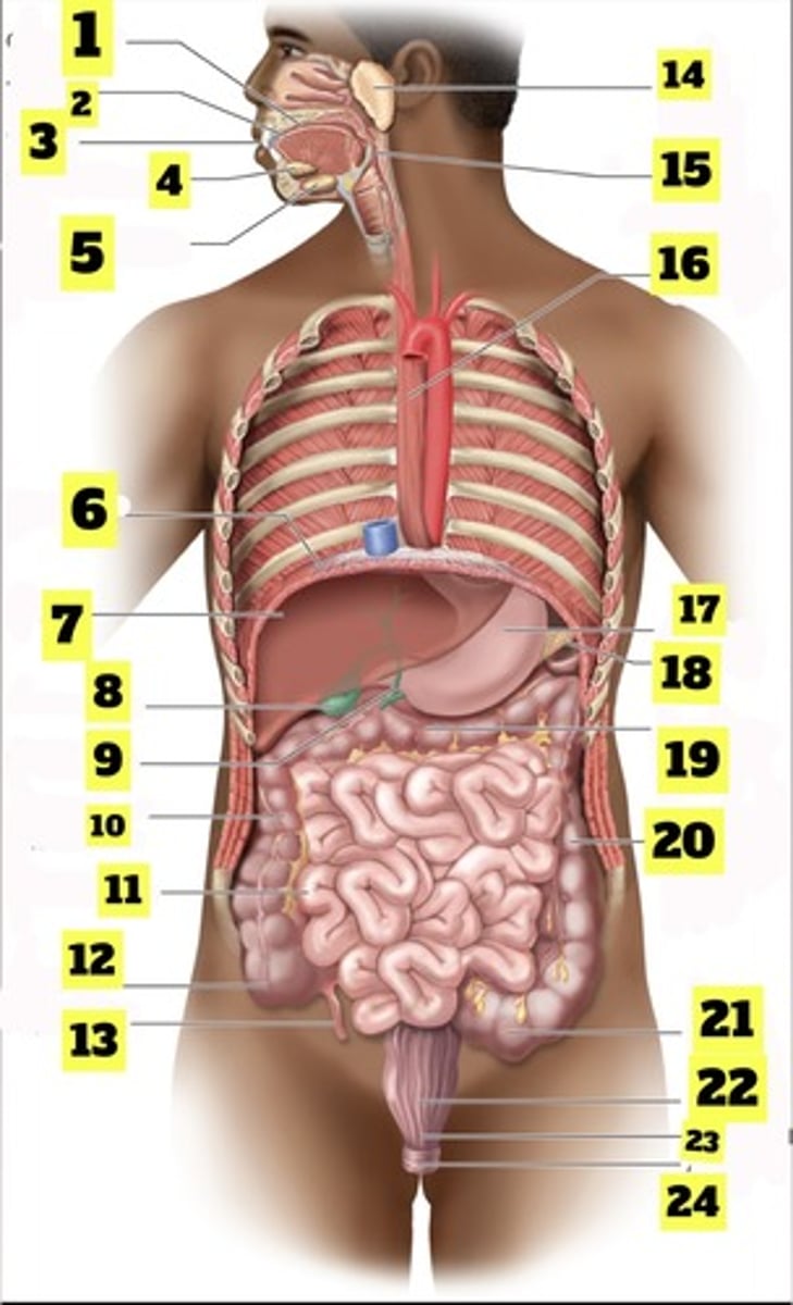
What is number 3?
teeth
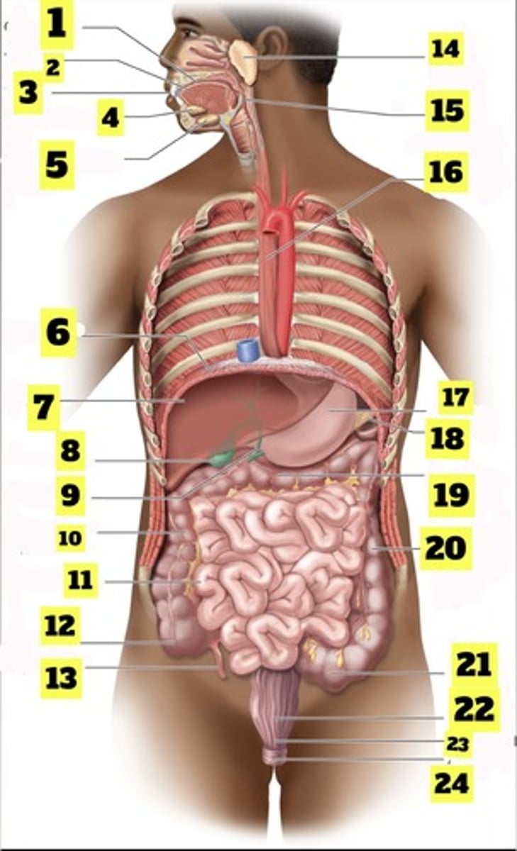
What is number 4?
sublingual gland
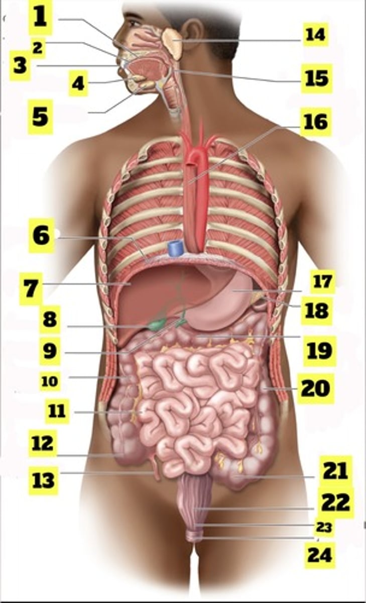
What is number 5?
submandibular gland
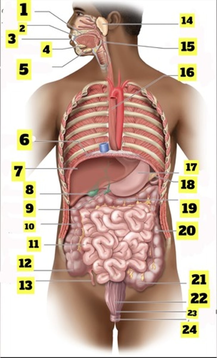
What is number 6?
diaphragm
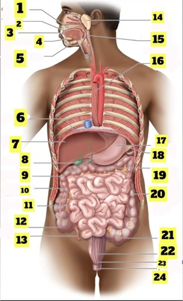
What is number 7?
liver
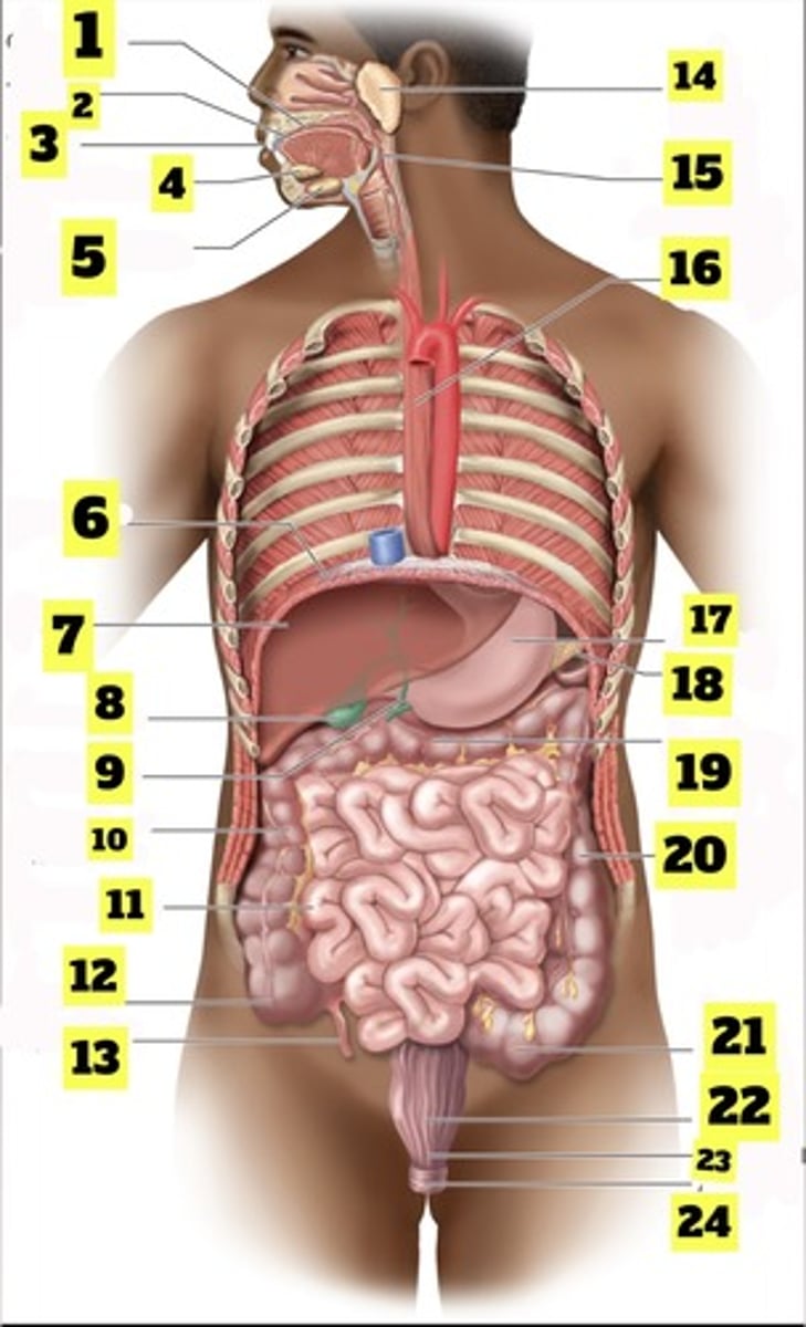
What is number 8?
gallbladder
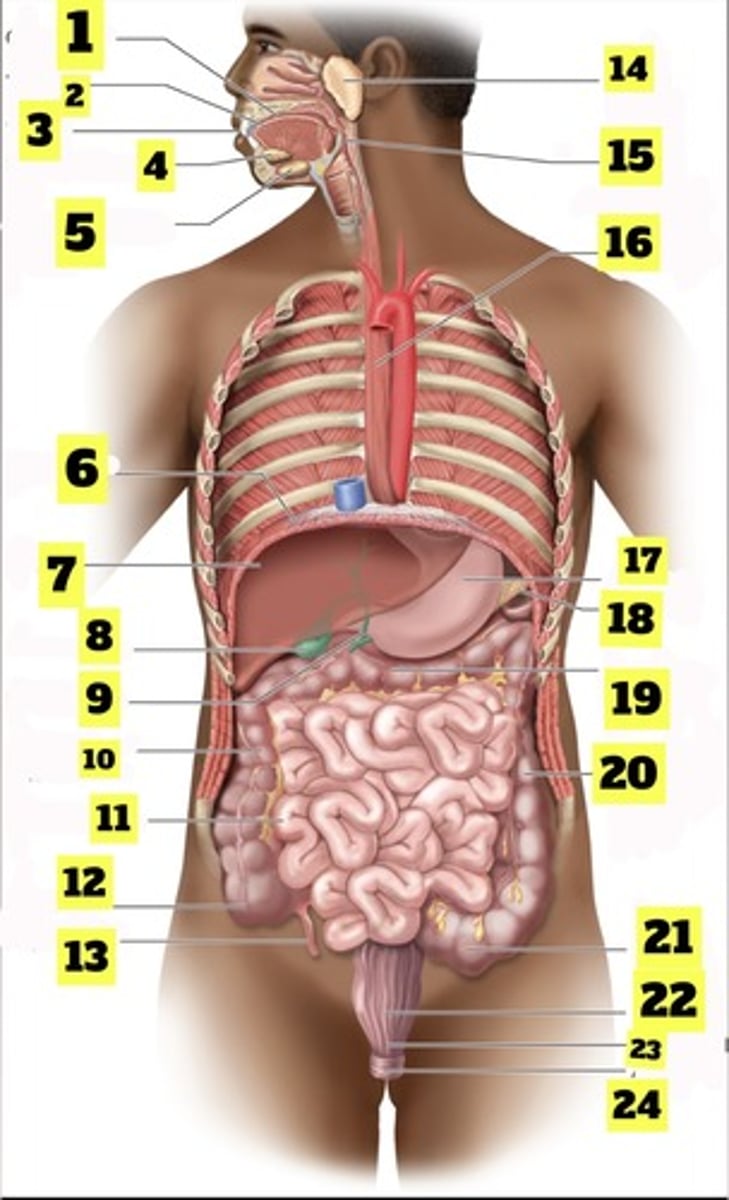
What is number 9?
bile duct
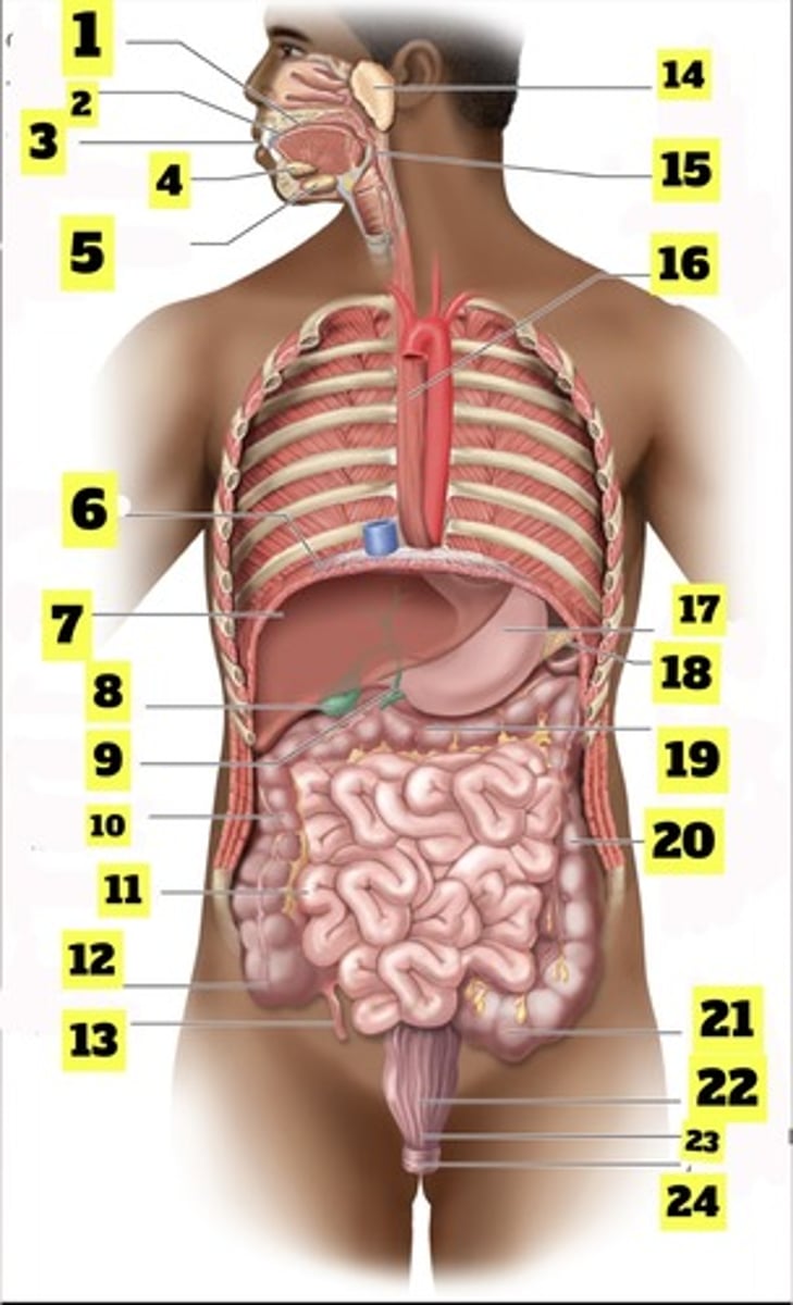
What is number 10?
ascending colon
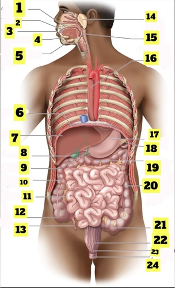
What is number 11?
small intestine
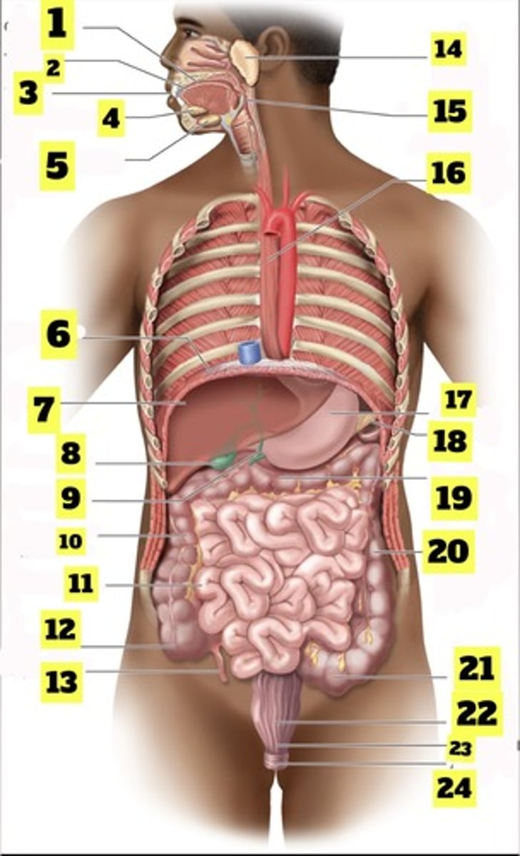
What is number 12?
cecum
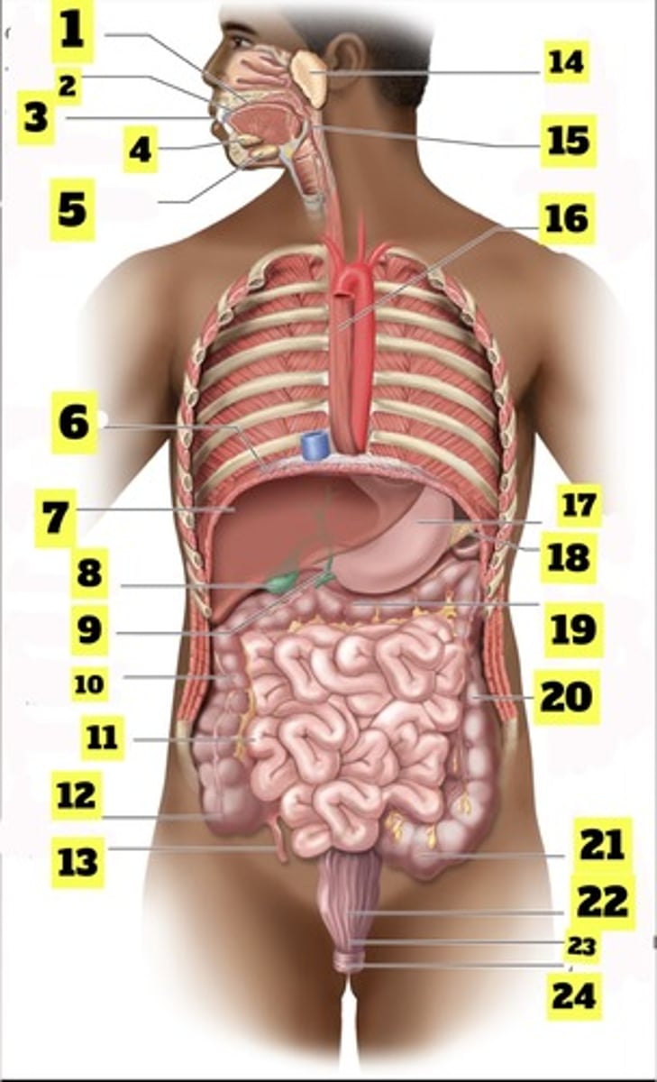
What is number 13?
appendix
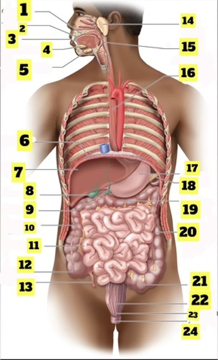
What is number 14?
parotid gland
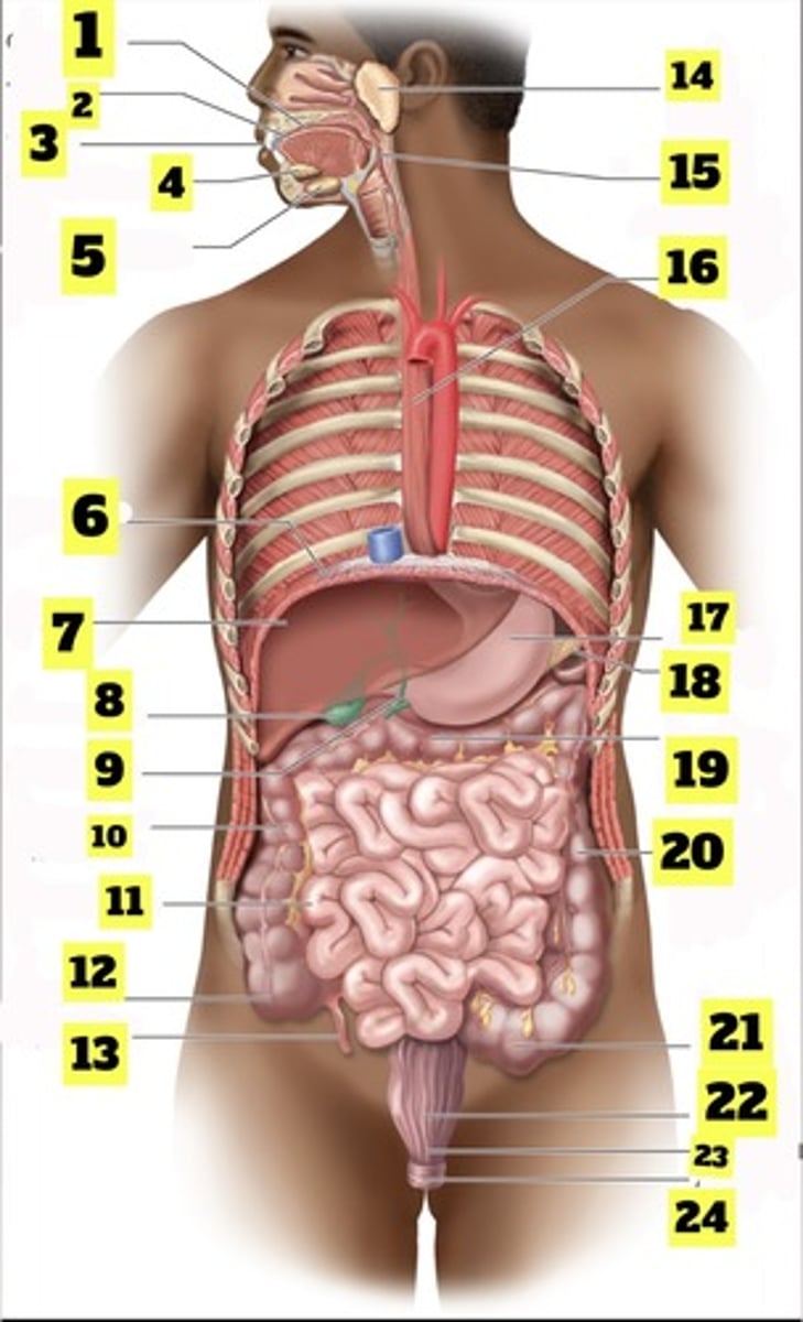
What is number 15?
pharynx
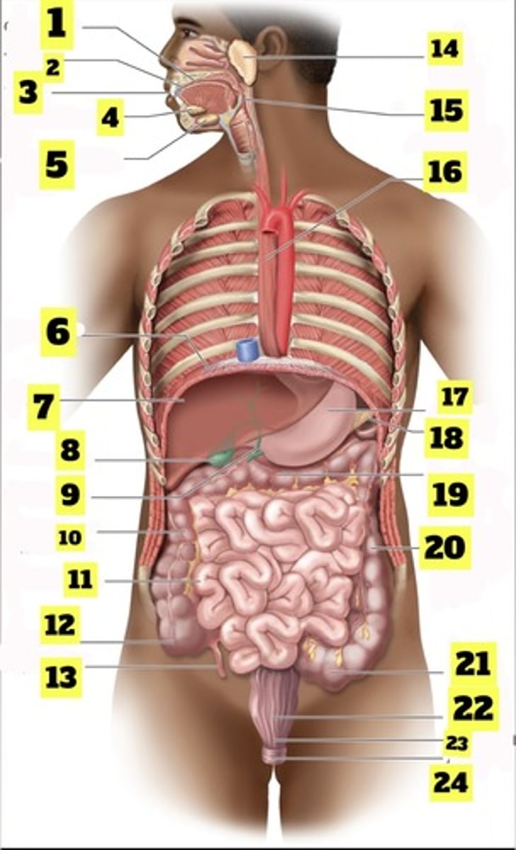
What is number 16?
esophagus