Axial skeleton Anatomy Practical
1/79
There's no tags or description
Looks like no tags are added yet.
Name | Mastery | Learn | Test | Matching | Spaced | Call with Kai |
|---|
No analytics yet
Send a link to your students to track their progress
80 Terms
ILOs
Use appropriate directional and locomotor terminology • Discuss the form and function of the vertebral column in a clinical context • Discuss the function and innervation of the epaxial and hypaxial muscle groups, and the ventral muscles of the neck. • Describe the pathways(s) of the facial and trigeminal nerves, with respect to skull features and musculature • Discuss structures of the head & face with respect to aural surgery and regional nerve blocks • Discuss the arterial supply and venous drainage of the head in a clinical context. • Demonstrate where to obtain a pulse and arterial/venous blood samples in the head in a variety of veterinary species • Describe the blood supply of the dental arcades and nasal cavity • Describe the locations of the lymph nodes and salivary glands of the head and neck and discuss in a clinical context • Describe the joints and ligaments of the skull, including the TMJ • Discuss the location, drainage, function and clinical relevance of sinuses in the skull • Describe the form, function and clinical significance of the guttural pouch • Describe the Innervation of the horn and the cornual nerve block for disbudding / dehorning • Describe important species differences in skull anatomy • Interpret standard diagnostic images of the vertebral column and skull, e.g. X-rays, identifying relevant bony landmarks
Basic movement terms
Flexion: Decreasing the angle between two bones1
•
Extension: Increasing the angle between two bones1
•
Abduction: Movement away from the midline1
•
Adduction: Movement towards the midline1
•
Supination: Turning the palm/sole upwards or forwards1
•
Pronation: Turning the palm/sole downwards or backwards
Cranial translation of the proximal tibia =
The proximal tibia moves cranially with the joint surface sliding in relation to the distal femur
Valgus deviation at the hock (tarsal joint)
The limb distal to the hock is deviated laterally
Varus deviation at the carpus
The limb distal to the carpus is deviated medially
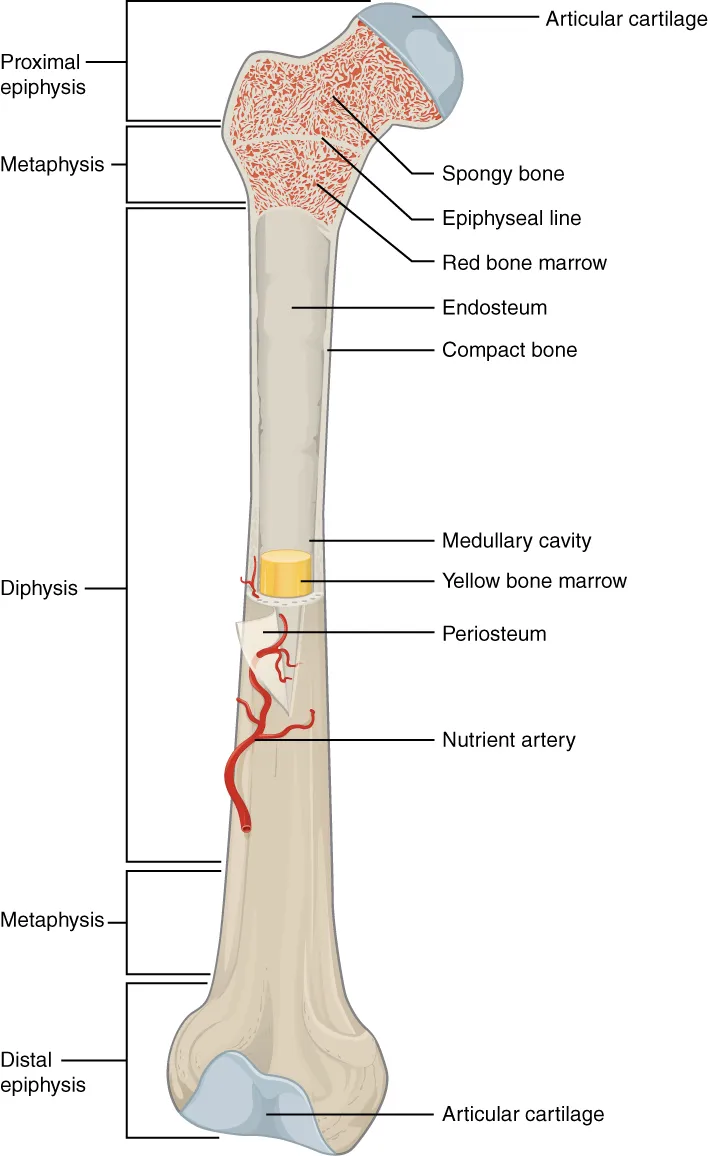
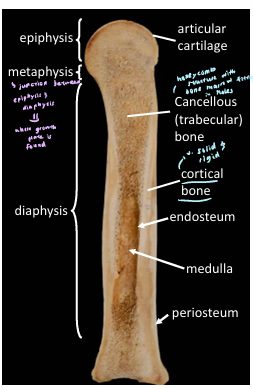
Bone terminology
Epiphysis = End of a long bone
Diaphysis = the shaft of a long bone
Metaphysis = Region between epiphysis and diaphysis (where growth plate is found)
Medullary cavity = central cavity containing bone marrow
Trabeculae = thin columns of bone that create spongy structure in cancellous bone
Cortical bone = dense outer layer of bone
Cancellous/trabecular bone = spongy inner layer of bone
Difference between origin and insertion of muscle?
Origin: More stationary attachment of muscle, more proximal
Insertion: Distal on the limb and moves more
Difference between ligament and tendon
Tendon connects muscle to bone or muscle to muscle/fascia
Ligament connects bone to bone
How do radiographs generate images of the body
Use X-rays that are absorbed differently by different tissues, creating a 2D image
X-ray images
White: Bone = Radio-opaque
Light grey: Soft tissue (kidney), Fluid (bladder)
Dark grey: Fat
Black: Air (lungs) = Radio-lucent
Ultrasound image
White: Bone & Air = Hyperechoic
Light grey: Ligaments & Tendons (soft tissue)
Dark grey: Soft tissue (spleen)
Black: Fluid (bladder) = Hypoechoic
The more “fluidy” it is, the more black
What is an orthogonal view
An additional view of the same structure in a different orientation
What does MRI use to generate images?
Magnetic fields are used to align molecular polarity. The energy release as the molecules return to their natural state is then measured.
What does CT use to generate images?
X-rays and computer
Why does MRI differentiate soft tissue structure better than CT
There is a greater difference in the energy released by different types of tissue which provides a greater contrast compared to the difference in amount fo X-rays absorbed by different types of soft tissue when undergoing radiography or CTs
Which muscle of mastication originates on the external sagittal crest and where does it insert? Would you expect this bony ridge to be more prominent in the carnivore or herbivore?
The temporalis muscle originates on the external sagittal crest and inserts on the coronoid process of the mandible. Temporalis acts to elevate the mandible and is more developed in the carnivore to allow them to bite down hard on prey.
Muscles of Mastication innervated by the mandibular branch (V2) of the trigeminal nerve
Temporalis
Pterygoids
Masseters
Digastricus (rostral belly)
Which muscle is situated in the masseteric fossa and what does it do?
Masseteric fossa: A depression located on the lateral surface of the ramus
Masseter muscle of mastication is situated in the masseteric fossa
When acting bilaterally, its vertical fibres are elevators of the jaw, closing jaws and its more horizontal fibres allow protraction (protrusion) of the jaw. When acting unilaterally, it allows lateral extrusion of the jaw, which is very limited in carnivores due to overlapping cheek teeth and conformation of the temporomandibular joint
In horses, what landmarks to locate infraorbital foramen? which direction to run fingers in live animal?
Infraorbital foramen lies approximately midway along a line connecting the naso-incisive notch and facial tubercle.
Run fingers rostral-caudally to feel edge of foramen as there is a tunnel-like opening to this hole (hard to feel if running fingers dorsal-ventrally). also must push levator nasolabialis out of the way.
Why are there no individual foraminae around the tympanic bulla in the horse? instead tympano-occipital fissure (fusion of holes around the tympanic bulla)
The holes are bigger around the bulla and everywhere around the skull to accommodate larger nerves/vessel, so the holes fuse to make a larger fissure '→ tympano-occipital fissure
How big is the frontal sinus in the cow & how to identify its caudal-most extent?
In ox, the frontal sinus extends to the very back of teh skull, pushing the parietal bone onto the caudal aspect of the skull. Palpating the crest of the back of the skull shows the caudal-most extent of the frontal bone and its sinus
Safest place to drain sinus in horses?
Maxillary sinus - dorsal facial crest/ridge, caudal to facial tubercle, rostral to medial corner of orbit and ventral to line of nasolacrimal duct
how does the frontal sinus drain in most species
in most species, the frontal sinus drains directly into the caudal nasal cavity. in the horse, the frontal sinus drains into the caudal part of the maxillary sinus then into the middle meatus of the nasal cavity.,
Clinical implications of thin alveolar bone for animals with large cheek teeth (esp. horses)
teeth roots of horses v long and only thing separating the maxillary sinus from the roots is the thin alveolar bone holding the roots to the socket
→ tooth infections can end up eroding the thin alveolar bone and lead to sinus infections or tooth root abscesses.
additionally,
the walls of the sinuses can be punctured by a blow to the side of the head, which can infect the sinus. sinus infections may also erode the alveolar bone and affect the teeth
how does the structure of a pig’s frontal sinus affect captive bolt stunning
captive bolt stunning: captive bolt gun fires metal rod into skull and penetrates brain
in pigs, the frontal sinus is well-developed and hollows out the entire dorsal surface of the skull behind the nasal bones. As a result, the brain is situated deep in the skull, protected by 2 plates of bone. This arrangement makes captive bolt stunning extremely unreliable because u have to be super accurate, and most slaughterhouses opt for electrocution or carbon dioxide stunning instead.
In bovine skull, the frontal sinus protrudes into the cornual process. Consequences?
Cows have a cornual diverticulum, which is a bit of caudal frontal sinus that protrudes into the horn.
Removal of the horns when the cow is >6months old would risk the frontal sinus being opened, increasing risk of haemorrhage and sinus infection. The sinus mucosa is innervated by sensory nerves which cannot be blocked, hence even if the cornual nerve is blocked, horn removal would still inevitably be painful.
Best site to block cornual nerve
Along temporal ridge, between the medial canthus of the eye and the base of the horn.
The orbit, horn base and temporal ridge are useful landmarks. Cornual nerve block is used for disbudding calves and dehorning adult cattle.
What does the trigeminal nerve innervate?
Sensation across face and muscles of mastication
Branches of Trigeminal Nerve (CNV)
Opthalmic (CNV1), sensory
passes through orbital fissure
Maxillary (CNV2), sensory
passes through infraorbital foramen, round foramen and maxillary foramen
Mandibular (CNV3), MIXED
passes through mandibular foramen, mental foramen and oval foramen
Common carotid artery branches into the external and internal carotid arteries. what areas of the head do these supply and what hole does the internal carotid artery pass through to reach the pterygopalatine fossa?
ICA passes through the foramen lacerum, a hole at the base of the skull, to get to the pterygopalatine fossa. In most mammals the Internal Carotid Artery contributes to the Circle of Willis, which supplies the brain. The external carotid artery supplies the neck and superficial structures of the head and face (branches: OACLFCSM, occipital, ascending pharyngeal, cranial laryngeal, lingual, facial, caudal auricular, superficial temporal, maxillary
what does the facial nerve innervate?, passes through stylomastoid foramen
muscles of facial expression, taste rostral 2/3 of tongue
branches of facial nerve
auriculopalpebral branch: innervates eyelid, ear, forehead
dorsal and ventral buccal branches: motor innervation to lips and cheeks
caudal auricular and coli branches: move ear caudally and coli branch helps neck muscle movement
also innervates caudal belly of digastricus
How to differentiate between the facial and trigeminal nerves?
Facial nerve is located superficially. The motor root of the nerve can be seen dividing in the substance of the parotid gland. The trigeminal nerve is better appreciated in a deeper dissection within the pterygopalatine fossa region.
Which nerve branches are desensitised during dental nerve blocks
upper arcades: branches of the maxillary nerve (CNV2)
lower arcades: branches of the mandibular nerve (CNV3)
what does the orbicularis oculi do?
closes the eyelids
what does the buccinator do
returns food from the cheek to the centre cavity of mouth
caudal auricular group
moves auricular cartilages caudally
salivary glands
parotid
submandibular
sublingual
Parasympathetic innervation of salivary glands
parotid (CNIX glossopharyngeal nerve)
submandibular (CNVII facial nerve)
sublingual (CNVII)
where do the salivary glands empty in the oral cavity?
parotid duct runs across the masseter → punches through the inner cheek muscle (buccinator) → and opens into the mouth (vestibule) near the 3rd upper premolar tooth to deliver saliva.
submandibular duct empties at the sublingual caruncle at the floor of the mouth (just beside frenulum)
sublingual gland empties via several ductules under tongue
which superficial lymph nodes in head are palpable in each species
dogs:
(sub)mandibular lymph node (normally and when enlarged)
horses
(sub)mandibular lymph node (normally and when enlarged), parotid and retropharyngeal (only when enlarged)
cattle:
submandibular (normally and enlarged)
Muscles of mastication and their functions
Temporalis: close the jaw
ipsilateral Masseter: close the jaw and move the jaw laterally (forms functional pair with pterygoids in herbivores)
contralateral Pterygoids: close and move jaw inward, with some protrusion
If only the left pterygoids contract, the jaw moves to the right.
If only the right pterygoids contract, the jaw moves to the left.
When both sides contract together, they help close the jaw and cause protrusion (moving the jaw forward).
digastricus: opens mouth, caudal belly innervated by facial nerve and rostral belly innervated by trigeminal nerve mandibular branch CNV3
masseter, temporalis, pterygoids and rostral belly of digastricsus innervated by CNV3
how are the muscles of mastication different in equine bovine (herbivores) vs canine
carnivores employ a ‘scissor-like’ jaw movement and have well developed temporalis muscles. herbivores use transverse jaw movements so they have well developed masseters and pterygoids which form a functional pair. herbivores have less developed temporalis muscles
what do the buccinators and tongue do
they maintain food between the teeth when chewing
prior to what procedures would blocking branches of the facial or trigeminal nerve be employed
dental procedures and ocular examination (paralyses the eyelids temporarily so animal cannot squeeze eye shut)
Dental nerve blocks
Lower incisor
target mental nerve of mandibular branch of trigeminal nerve
access via middle of mental foramen
Lower premolar or rostral molar
target inferior alveolar nerve of mandibular branch of trigeminal nerve
access via mandibular foramen
Lower caudal molar
target inferior alveolar nerve of mandibular branch of trigeminal nerve (CNV3)
access via mandibular foramen
Motor innervation to upper eyelid
target auriculopalpebral branch of facial nerve
access via crest of the zygomatic arch near the ear
upper incisors
target infraorbital nerve of maxillary branch of trigeminal nerve CNV2
access via infraorbital foramen
upper premolars and rostral molars
target infraorbital nerve of maxillary branch of trigeminal nerve CNV2
access via infraorbital foramen
caudal molars
target maxillary branch of trigeminal nerve CNV2
access just caudal to 2nd maxillary molar
How to access jugular vein for venous blood sampling?
External jugular vein most easily accessed in the ventral part of the cranial neck. jugular groove is found between the brachiocephalicus dorsally and sternocephalicus ventrally in the horse. in dogs, external jugular vein runs superficially to the sternocephalicus.
how to access auricular veins for venous blood sampling
often used in pigs/rabbits
3 auricular veins; one central and 2 marginal veins, easily visible once they have been raised
marginal vein access recommended over central vein access for rabbits
why is external jugular vein not accessed in caudal (lower) neck of horse
In caudal neck of horse, external jugular vein covered by muscle, it is superficial in the cranial part
how does the way in which the lingual and facial arteries arise differ in horses compared to dogs?
In horses, the lingual and facial arteries originate from a common trunk (linguofacial trunk) compared to arising as separate lingual and facial branches of the external carotid artery in dogs.
where to palpate facial artery in horse
facial artery passes over the ventral margin of the mandible and becomes palpable → site for pulse taking
External carotid artery continues as..
branches: occipital, ascending pharyngeal, cranial laryngeal, lingual, facial, caudal auricular, superficial temporal, maxillary
ECA continues as maxillary artery after giving off its superficial temporal branch. before passing through the alar canal to enter the pterygopalatine fossa it gives off an inferior alveolar branch which passes through the mandible with CNV3. Within the pterygopalatine fossa the external carotid artery gives off external opthalmic branch to orbit, ethmoidal branches to the nasal cavity and palatine branches to the hard and soft palate.The maxillary artery continues as the infraorbital artery after passing through the infraorbital foramen.
Blood supply to dental arcades
inferior alveolar artery to lower alveoli and lower teeth
infraorbital artery to upper alveoli and upper teeth
Nose bleeds (epistaxis) common and bleeding can be profuse due to complicated blood supply. blood supply to nasal cavity
ethmoidal, palatine and sphenopalatine anastomoses
deep opthalmic plexus drains superficial veins in the eye region draisn from superficial to deep
causes infections to track from superficial to deep
significance of swellings (sinuses) on major veins of the horse
allow for pooling of blood when the horses are grazing - massage by the muscles of mastication helps in venous return from the head. The transverse facial veins can be used for venepuncture.
what happens if u insert needle at correct place but too steep of an angle
can possibly hit carotid artery, always insert at shallow angle to skin.
deep and superficial veins ..
(deep venous plexi) drain through the orbital fissure
consequences of infection of superficial veins (e.g. facial vein as it crosses ventral border of mandible)?
Infection could track deeper, into the cranial blood supply and cause meningitis
guttural pouch
paired spaces connecting the throat and ear in horses
guttural pouch mycosis (a fungal infection) can damage the pouch walls and cause nosebleeds from ruptures of the internal or external carotid arteries which run along the walls of the guttural pouch, separated from the guttural pouch contents only by a thin membrane
guttural pouch mycosis can also lead to signs of cranial nerve damage
strangles infection can also spread to retropharyngeal lymph nodes and put pressure on the larynx and trachea, ‘strangling’ the airway.
what does the auditory tube connect
middle ear and nasopharynx. infections of the guttural pouch can also track to the middle ear
function of guttural pouch
acts as a cerebral cooling mechanism during strenuous exercise as the internal carotid artery passes through.
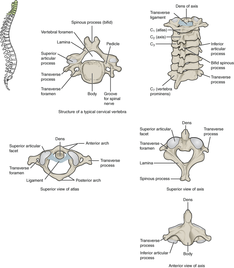
what travels through the transverse foramen in cervical vertebrae?
vertebral artery
which part of the vertebra do the intervertebral discs lie between?
vertebral bodies
what parts of the thoracic vertebrae do the ribs attach to?
each rib articulates with 2 adjoining vertebrae, with the rib being numbered the same as the most caudal vertebrae. the ribs articulate with costal facets on the thoracic vertebrae → rib heads articulate with facets of the vertebral bodies and rib tubercfles articulate with the facets of the transverse processes
what runs in the costal groove
intercostal vein artery and nerve
Intervertebral joints
Secondary cartilaginous joints made up of caudal surface of one vertebral body, cranial surface of caudal vertebral body and an interposed disc. Disc provides degree of movement between each pair of vertebrae and also a degree of stability and resistance to abnormal movements
molecule predominating outer layer of intervertebral disc?
collagen
provides strength and resistance to tearing
is the disc the same consistency throughout?
no
disc has a tough outer layer (Annulus fibrosus) and soft semi-liquid central region (nucleus pulposus)
Function of annulus fibrosus
provides a tough outer layer resistant to tearing and tension (collagen component)
function of nucleus pulposus
semi-liquid nucleus pulposus provides a hydrostatic centre which resists compression and helps to even the force over the whole vertebral body, rather than focusing it on small corners of the vertebra.
which region of outer layer (annulus fibrosus) is the thinnest?
dorsal region
clinical relevance:
The dorsal region of the annulus fibrosus underlies the spinal cord and is the most prone to tearing, inflammation and extrusion of the nucleus pulposus. This is significant as the spinal cord is susceptible to compressive and concussive injury
supraspinous ligament
resist flexion of spine
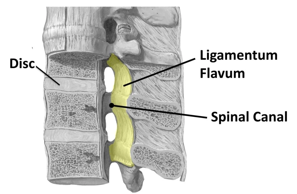
flaval (yellow) ligament
elastic ligament that helps return spine to extended / straight
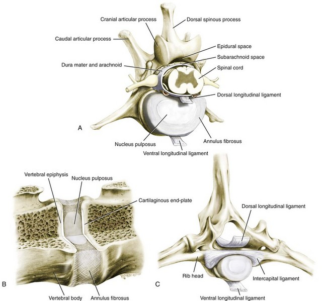
dorsal and ventral longitudinal ligament
dorsal longitudinal ligament
sits dorsally to vertebral body, resists flexion of spine
ventral longitudinal ligament
sits ventrally to vertebral body and resists extension of spine

intercapital ligament
runs between paired rib heads, restrains and attaches rib heads. clinically believed to reduce frequenxy of intervertebral disc compression in the thoracic spine (T2-T10) by increasing resistance to extrusion of nucleus pulposus.
why is the region T11-L3 the most commonly affected area for Intervertebral disc extrusion in dogs.
mobility and forces experienced between T11-L3 are the highest
greatest flexion and extension when moving
cranial to T10, the intercapital ligaments help provide some support.
function of epaxial muscles if they contract bilaterally
spine extension
epaxial = above vertebral column, so if contract will extend spine, not like ligaments that resist flexion if dorsal to spine
if epaxial muscles contract unilaterally
spine lateral bending towards side that contracts
external abdominal oblique a hypaxial muscle?
yes
hypaxial = below vertebral column
Innervation to epaxial muscles
segmental dorsal branches of the somatic/motor nerves. These nerves have somatic sensory components which supply the dorsal skin and muscles, but also rejoin the ventral branches to provide sensation and motor innervation to the body wall muscles (external abdominal oblique, external intercostal) and skin.
neck musculature
brachiocephalicus
unilateral contraction draws head and neck to side
bilateral contraction draws head and neck ventrally