Lec 5 - Nervous System I
1/51
There's no tags or description
Looks like no tags are added yet.
Name | Mastery | Learn | Test | Matching | Spaced |
|---|
No study sessions yet.
52 Terms
Organization and Components of the Nervous System
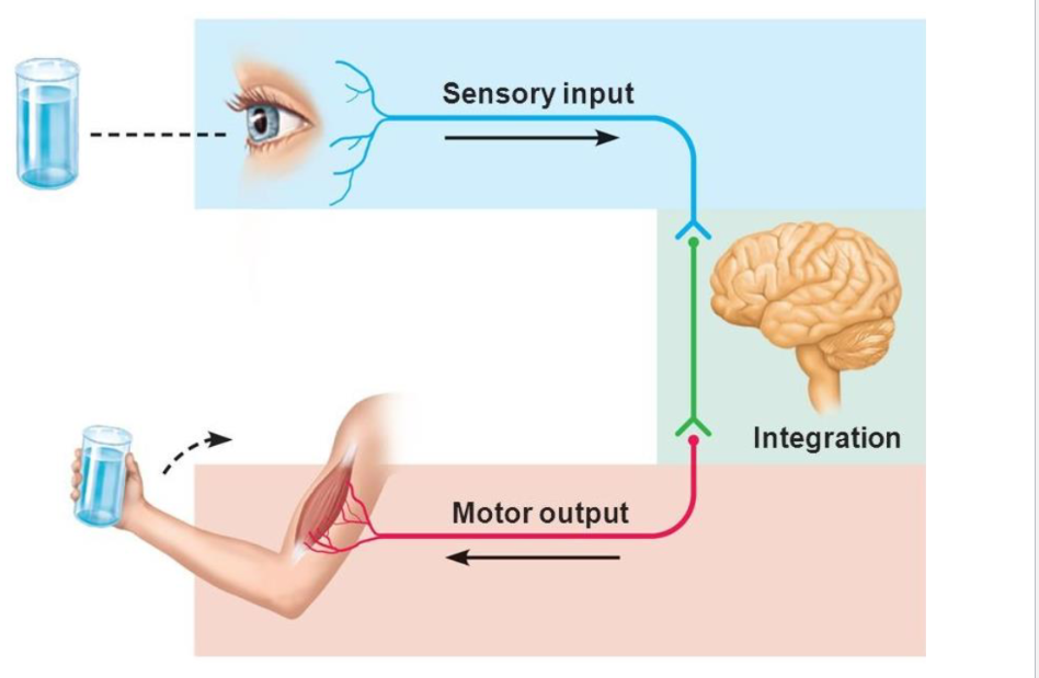
• Sensory Input
• Integration
• Motor Output
(it like negative feedback mechanism or homeostasis)
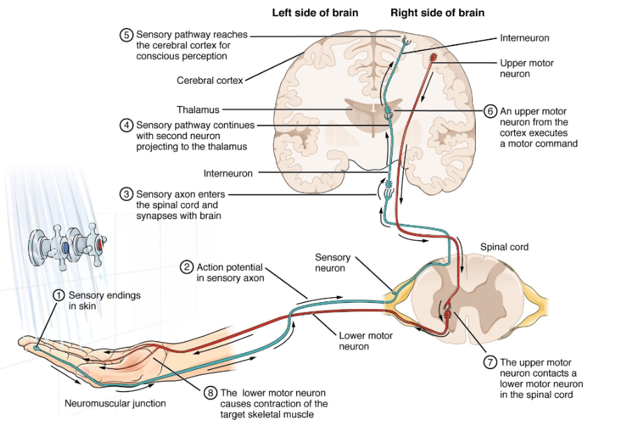
(thirsty is stimulus. Photoreceptor in your eyes is receptor. The sensory input is afferent neuron. It go to the interneuron in central neuvous system is control center. Motor output is efferent signal. The muscle is effector.)
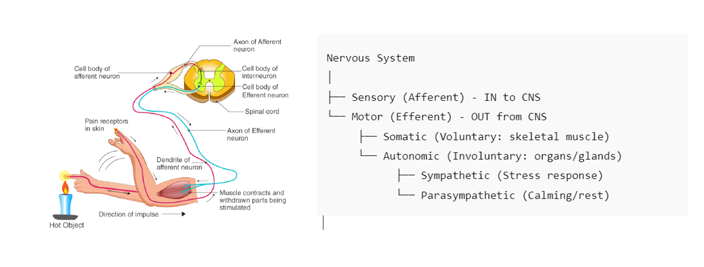
(if you touch something hot, you pull your hand away. This is also homeostatic or negative feedback mechanism)
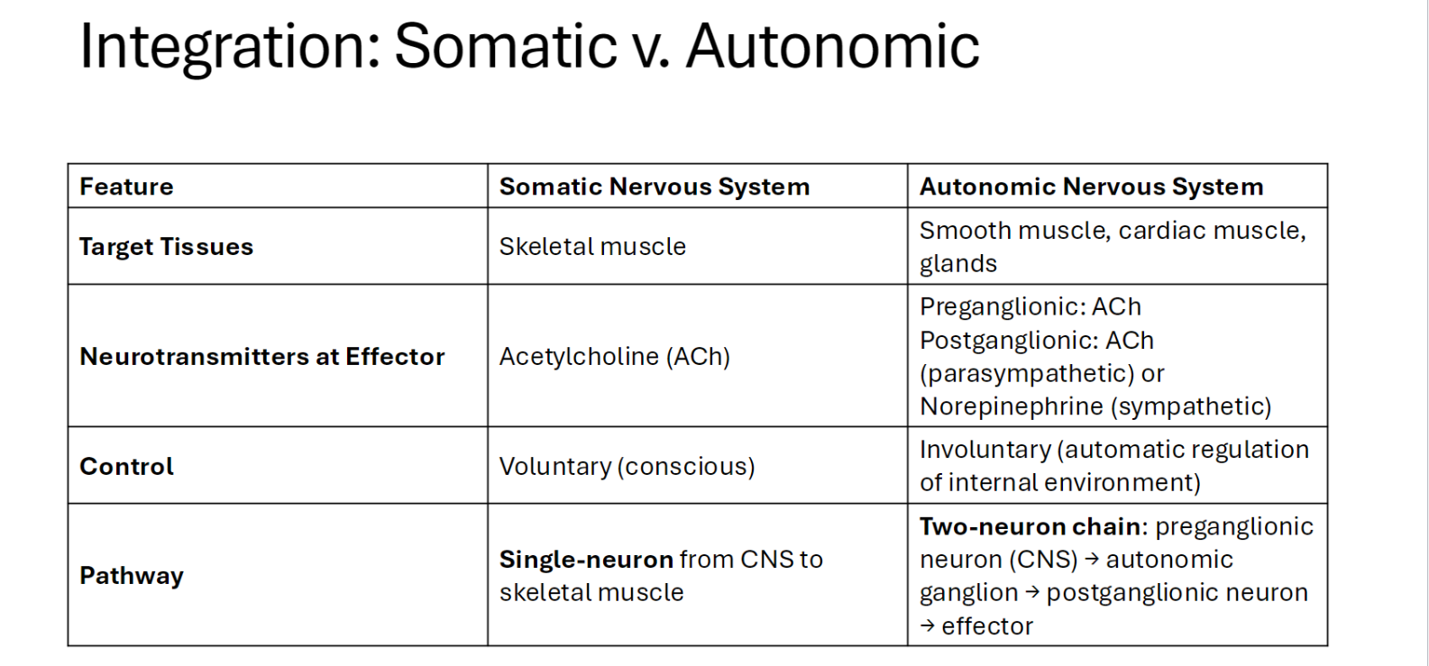
Functional Divisions of the Nervous System
(test will have question about different between the somatic and autonomic)
Sensory (Afferent)
(Carries information into the central nervous system (CNS).)
Motor (Efferent)
(Carries signals away from the CNS to muscles and glands.)
• Somatic (Controls voluntary movements or conscious thought. Example: Walking, writing, or speaking.)(also called somatic nervous system, somatic motor, somatic sensory)(2 lane highway)(not your organ, it recognizes pain or heat)
• Autonomic (Regulates involuntary or automatically functions systems. Example: Heart rate, digestion) (also call autonomic nervous system or autonomic motor)(1 way road)
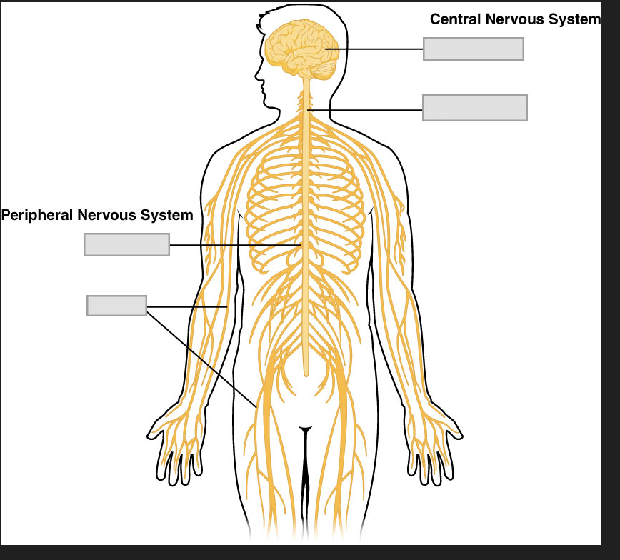
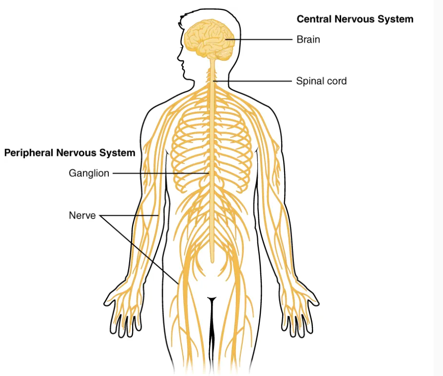
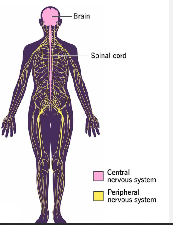
Structural Divisions of the Nervous System
Central Nervous System (CNS)
• Brain – in the cranial cavity
• Spinal Cord – in the vertebral column
Peripheral Nervous System (PNS)
• Spinal Nerves – originate from the spine (almost all the peripheral nervous system is the spinal nerves)
• Cranial Nerves – the rest of peripheral nervous system that don’t originate from the spine. 12 nerves in the PNS that originate from the brain or cranium)
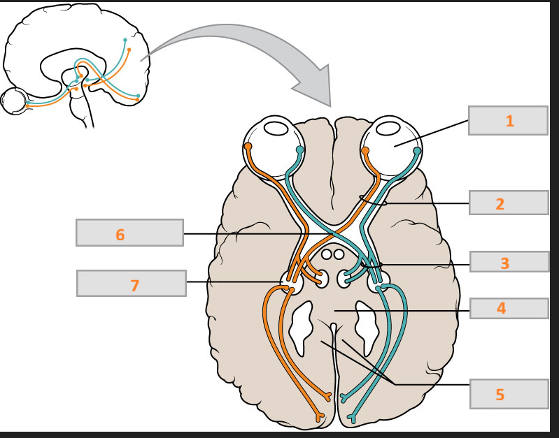
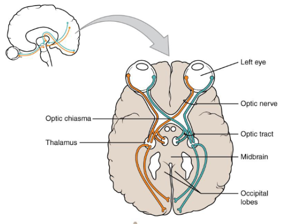
Every pathway crosses both (CNS and PNS)
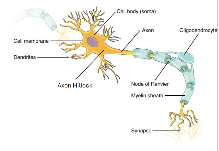
Neurons: The Signaling Cells
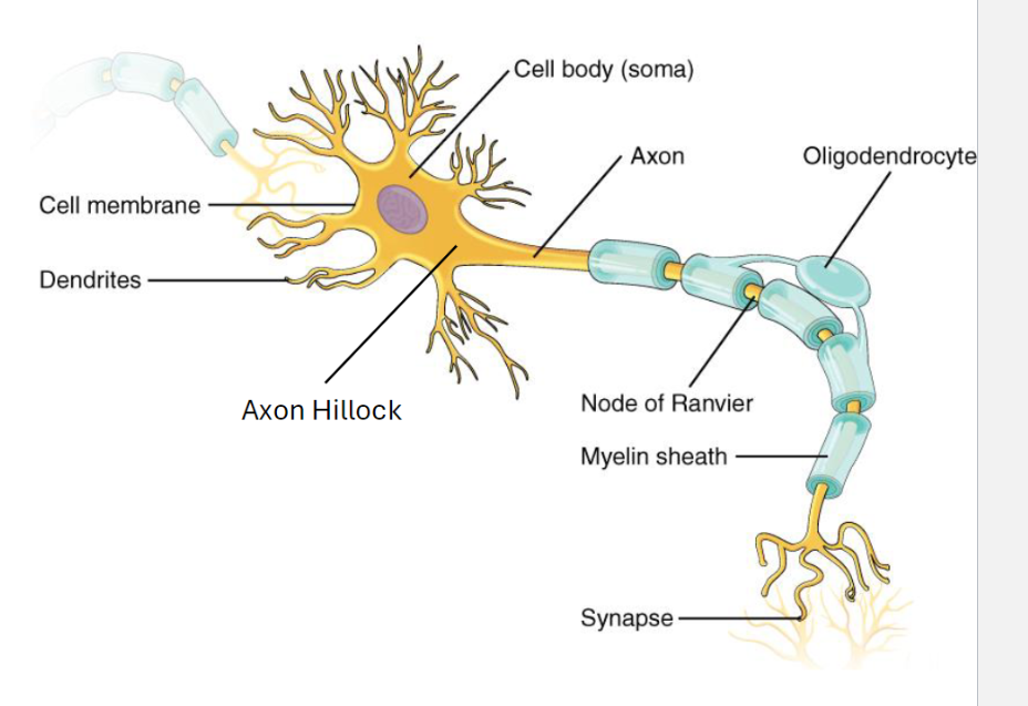
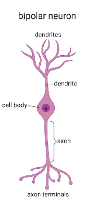
• Dendrites: these are highly branched processes that receive information from other neurons at the synapse.
• Soma or cell body: the main part of the cell or the neurons, and it has all the organelles and the nucleus.
• Axon: this is single projection from cell body that carries an impulse or action potential.
• Axon Hillock: This is a special region of the neuron, where the axon emerges from the cell body. (like trigger. If this is going to fire, it has to fire by the axon hillock.
• Synaptic Terminals: it is the end of our axon, and this is where we communicate with another target cell. They release neurotransmitters.
• Myelin Sheath: this is an insulating substance that wraps around the axon. It speeds up the action potential.
Nodes of Ranvier: they are the gaps between those.
(This is in the central nervous system)
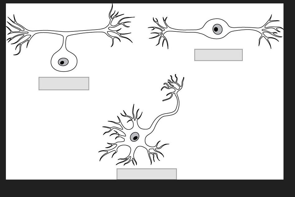
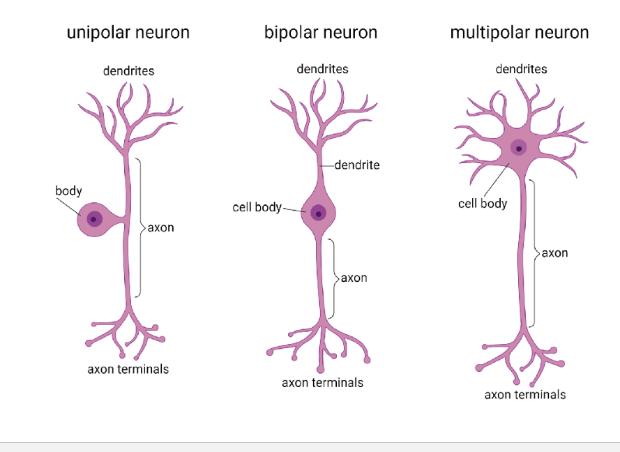
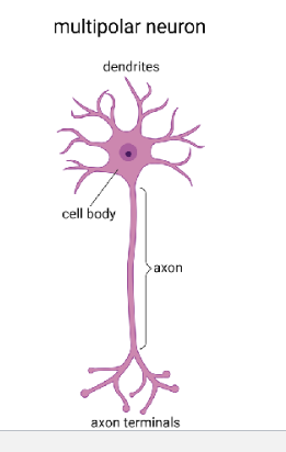
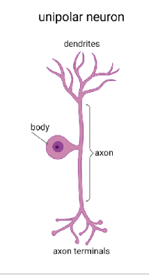
Neuron Classification: By Shape
• Multi-polar: this is the most of the nerves in the nervous system, especially for interneurons and motor neurons
• Bi-polar: neurons collect light in your eyes, pretty specialized
• Uni-polar: usually a sensory neuron. (where all the cell bodies from all the sensory neurons hang out together, called ganglia)
Neuron Classification: By Function
• Sensory
• Interneuron
• Motor
Multipolar
Key Role: Motor & integration (can send signal all places. It is integrating all the information and then deciding what it should do)
Classic Location: Motor neurons, interneurons
Unipolar
Key Role: Rapid sensory input
Classic Location: Dorsal root ganglia
Bipolar
Key Role: Special senses
Classic Location: Retina, olfactory epithelium, inner ear
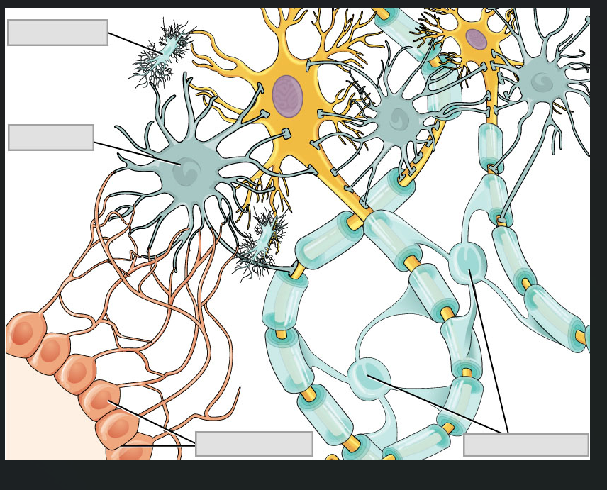
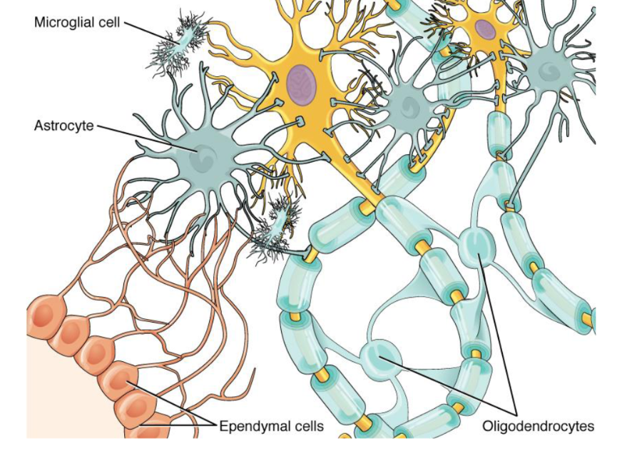
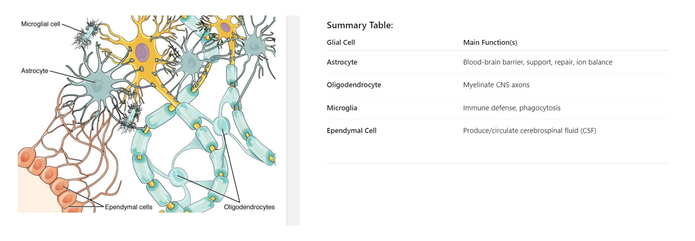
Neuroglia of the CNS
• Astrocyte – it is the star-shaped cell.
Main Functions:
Maintain the blood-brain barrier (control what enters/exits the brain from the blood).
Provide structural support to neurons.
Regulate ion and nutrient concentrations in the extracellular space.
Repair damaged nervous tissue.
Assist in neurotransmitter recycling.
• Oligodendrocyte – It insulates axon with myelin sheath. One oligodendrocyte can myelinate multiple axons.
• Microglia – Act as the immune cells of the CNS(brain). They protect against infection.
• Ependymal cell – create cerebrospinal fluid
(all these cells are only in central nervous system)
what cell myelinates and acts on in the central nervous system?
Oligodendrocytes
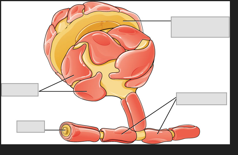
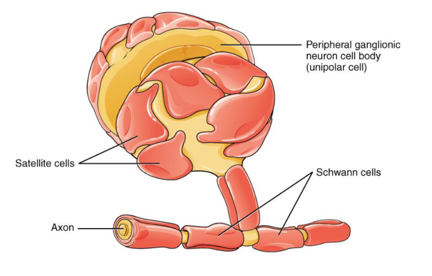
Neuroglia of the PNS
• Schwann Cells – these are myelinated axons in the PNS.Schwann cells myelinate only one axon segment.
• Satellite Cells – these are found in sensory and autonomic ganglia. Functionally similar to astrocytes. These are the support cells. They respond to damage. These are the cells that are maintaining that ion balance around the cell.
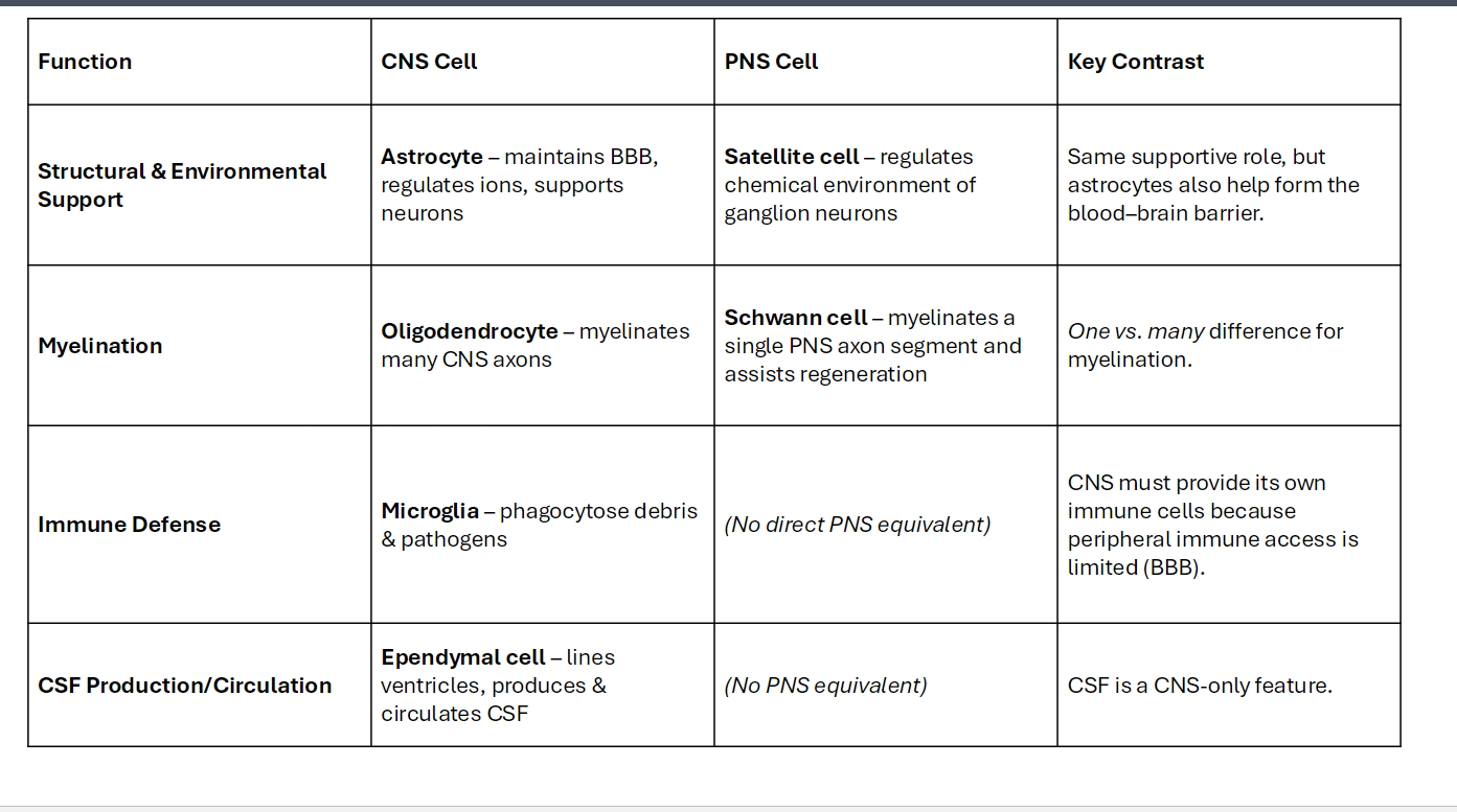
Function
Structural & Environmental Support
In CNS Cell
Astrocyte – maintains BBB, regulates ions, supports neurons
In PNS Cell
Satellite cell – regulates chemical environment of ganglion neurons
Key Contrast
Same supportive role, but astrocytes also help form the blood–brain barrier.
Function
Myelination
In CNS Cell
Oligodendrocyte – myelinates many CNS axons
In PNS Cell
Schwann cell – myelinates a single PNS axon segment and assists regeneration
Key Contrast
One vs. many difference for myelination.
Function
Immune Defense
In CNS Cell
Microglia – phagocytose debris & pathogens
In PNS Cell
(No direct PNS equivalent)
Key Contrast
CNS must provide its own immune cells because peripheral immune access is limited (BBB).
Function
CSF Production/Circulation
In CNS Cell
Ependymal cell – lines ventricles, produces & circulates CSF
In PNS Cell
(No PNS equivalent)
Key Contrast
CSF is a CNS-only feature.
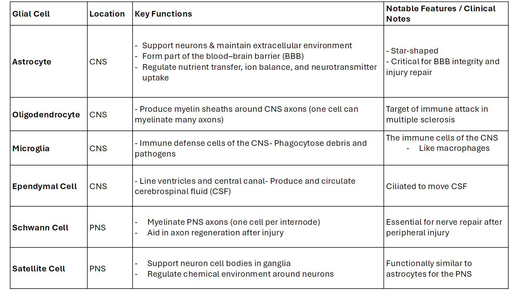
Glial Cell
Astrocyte
Cell Location: CNS
Key Functions:
- Support neurons & maintain extracellular environment
- Form part of the blood–brain barrier (BBB)
- Regulate nutrient transfer, ion balance, and neurotransmitter uptake
Notable Features / Clinical Notes
- Star-shaped
- Critical for BBB integrity and injury repair
Glial Cell
Oligodendrocyte
Cell Location: CNS
Key Functions:
- Produce myelin sheaths around CNS axons (one cell can myelinate many axons)
Notable Features / Clinical Notes:
Target of immune attack in multiple sclerosis
Glial Cell
Microglia
Cell Location: CNS
Key Functions:
- Immune defense cells of the CNS
- Phagocytose debris and pathogens
Notable Features / Clinical Notes:
The immune cells of the CNS
- Like macrophages
Glial Cell
Ependymal Cell
Cell Location: CNS
Key Functions:
- Line ventricles and central canal- Produce and circulate cerebrospinal fluid (CSF)
Notable Features / Clinical Notes:
Ciliated to move CSF
Glial Cell
Schwann Cell
Cell Location: PNS
Key Functions:
- Myelinate PNS axons (one cell per internode)
- Aid in axon regeneration after injury
Notable Features / Clinical Notes:
Essential for nerve repair after peripheral injury
Glial Cell
Satellite Cell
Cell Location: PNS
Key Functions:
- Support neuron cell bodies in ganglia
- Regulate chemical environment around neurons
Notable Features / Clinical Notes:
Functionally similar to astrocytes for the PNS
smooth muscle is involuntary. Skeletal muscle is voluntary.
Somatic nervous system always acetylcholine. Acetylcholine is going to be its neurotransmitter.
Nerve v. Tract
• Nerve – bundle of axons in the PNS (peripheral nervous system). It is going to transmit sensory and motor information.
• Tract – bundle of axons in the central nervous system
Ganglia v. Nucleus
Ganglia – collection of cell bodies in the PNS.
Nucleus – collection of cell bodies in the CNS.
Gray Matter and White Matter
Gray Matter –
non myelinated area
you have cell bodies.
you have dendrites.
you have unmyelinated axons.
• White Matter –
myelinated
myelin is a fatty material - white color
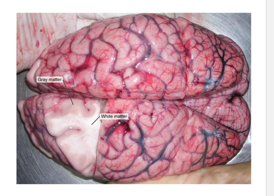
Where would you expect to find tracts?
In the White Matte because this where you would find myelinated.
Where would you expect to find nuclei?
In the gray matter because nuclei are collection of cell bodies, non-myelinated gray matter.
Multiple Sclerosis (MS)
(it is autoimmune disease in CNS. Immune system targets myelin.(antibodies))
Symptoms:
• Weakness
• Vision problems
• Loss of coordination
Autoimmune disorder leading to demyelination in the CNS
The importance of Myelin
Multiple sclerosis damages which glial cell?
Would you expect to find damage in gray matter or white matter? Why?
That is oligodendrocyte because it damages myelin sheath and the oligodendrocytes build the myelin sheath. It is in CNS.
you expect to find damage in white matter because white matter is myelin (ex: bunch of scarring in the white matter is the result of sclerosis damages)
(read to know don’t need to study)
Case Study
• Case: A 38-year-old construction worker falls and suffers a laceration that severs the left brachial plexus (neurons in the upper left arm).
Prompt:
• Describe which division(s) of the nervous system are affected.
• Predict the sensory and motor deficits he will experience in his left upper limb.
• Explain how this injury could impact somatic versus autonomic functions.
Describe which division(s) of the nervous system are affected.
The brachial plexus is part of the Peripheral Nervous System (PNS), specifically the somatic nervous system, which controls voluntary motor and sensory functions in the limbs.
Predict the sensory and motor deficits he will experience in his left upper limb.
Motor Deficits:
Complete or near-complete paralysis of the left upper limb.
Loss of movement in:
Shoulder
Arm and forearm muscles
Hand muscles
The limb may appear flaccid and atrophied over time.
Sensory Deficits:
Complete loss of sensation in the skin of the left upper limb
Explain how this injury could impact somatic versus autonomic functions.
Somatic Nervous System (SNS):
Severely impacted:
Complete loss of voluntary movement.
No conscious sensation in the left arm due to the interruption of sensory and motor pathways.
Profound disability in performing daily tasks
Autonomic Nervous System (ANS):
Loss of sympathetic innervation to skin and blood vessels in the left arm
Which division of the nervous system carries motor commands to skeletal muscles?
Somatic nervous system
The _______ nervous system consists of the brain and spinal cord.
Central nervous system
Neurons that carry information from sensory receptors towards the CNS are:
sensory neurons
Which neuroglia cell produces myelin in the CNS?
Oligodendrocytes
The gaps in a myelinated axon where action potentials are generated are called ___________.
Nodes of Ranvier
What type of glial cell is responsible for filtering blood to produce CSF at the choroid plexus?
ependymal cell
astrocyte
oligodendrocyte
Schwann cell
ependymal cell
Which part of a neuron transmits an electrical signal to a target cell?
dendrites
soma
cell body
axon
axon
Which term describes a bundle of axons in the peripheral nervous system?
nucleus
ganglion
tract
nerve
nerve
What type of glial cell provides myelin for the axons in a tract?
oligodendrocyte
astrocyte
Schwann cell
satellite cell
oligodendrocyte
Which part of a neuron contains the nucleus?
dendrite
soma
axon
synaptic end bulb
soma
What type of glial cell is the resident macrophage behind the blood-brain barrier?
microglia
astrocyte
Schwann cell
satellite cell
astrocyte
which cell type myelinatesaxon in the PNS?
satellite cells
myelin cells
schwann cells
oligodendrocytes
schwann cells