Radiology PA: Introduction to Chest Radiology
 Call Kai
 Learn
Learn Practice Test
 Spaced Repetition
 Match
Match Flashcards
 Knowt Play
1/110

There's no tags or description
Looks like no tags are added yet.
Name | Mastery | Learn | Test | Matching | Spaced | Call with Kai | Chat |
|---|
No analytics yet
Send a link to your students to track their progress
111 Terms
indication for this imaging technique include: pneumonia, pulmonary edema, effusions, pneumothorax, cardiomegaly, rib fractures, and lung nodules (screening)
x-ray
what imaging type is almost ALWAYS the first line modality for chest imaging
x-ray
the indications for this imaging type include: pulmonary embolism, coronary CT, lung mass, and trauma
CT
the indications for this imaging type include: assessing the blood flow in heart, pericardial effusion, an echocardiogram, point of care (POC) imaging (supplements physical exam and definitive imaging), eFAST, cardiac, PNA, and pleural effusion
US
what imaging type provides the MOST LIMITED evaluation of the lungs?
US
what imaging type is used when masses in the mediastinum are suspected?
MRI
What is the preferred view for an abled bodied pateint for a chest x-ray?
PA
what x-ray view is used to help visualize the retrocardiac space and lower lung lobes?
lateral
what x-ray view is used to visualize the lung apex to look for masses and TB?
lordotic
what x-ray view is used to identify a pleural effusion or pneumothorax?
lateral decubitus
to view possible EFFUSION on the lateral decubitus view, the effusion side of interest should be ________
down
to view possible PNEUMOTHORAX on the lateral decubitus view, the pneumothorax side of interest should be _____
up
to determine if cardiomegaly is present is a patient, the cardio-thoracic ratio needs to be grater than __%
50
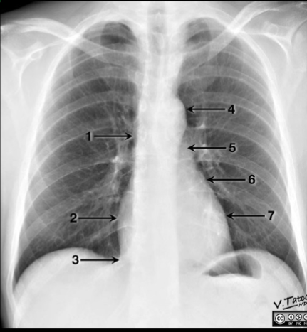
number 1 on the image is referring to what structure?
superior vena cava
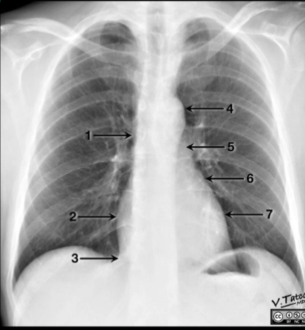
number 2 on the image is referring to what structure?
right atrium
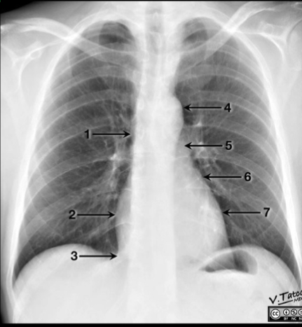
number 3 on the image is referring to what structure?
inferior vena cava
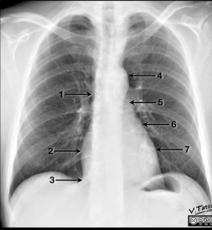
number 4 on the image is referring to what structure?
aortic arch
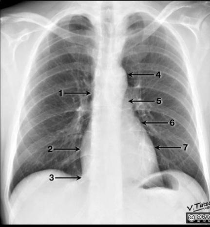
number 5 on the image is referring to what structure?
pulmonary trunk
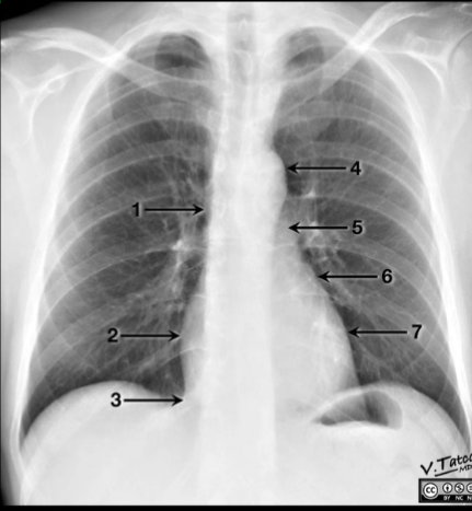
number 6 on the image is referring to what structure?
left artial appendage
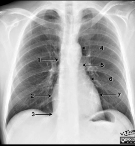
number 7 on the image is referring to what structure?
left ventricle
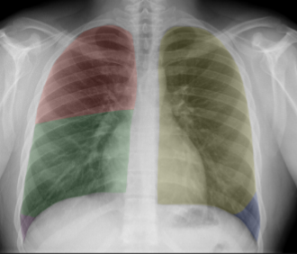
the area highlighted RED indicates:
the right upper lobe
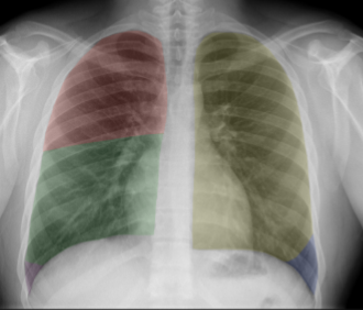
the area highlighted GREEN indicates:
right middle lobe
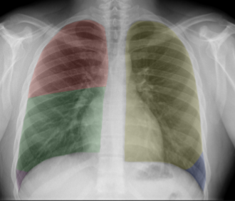
the area highlighted PURPLE indicates:
right lower lobe
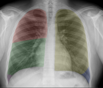
the area highlighted YELLOW indicates:
left upper lobe
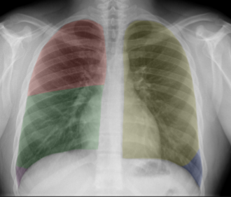
the area highlighted BLUE indicates:
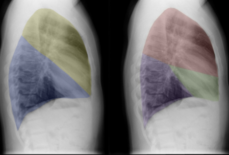
on the LATERAL view, the area highlighted RED indicates:
right upper lobe
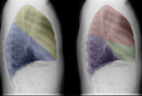
on the LATERAL view, the area highlighted GREEN indicates:
right middle lobe
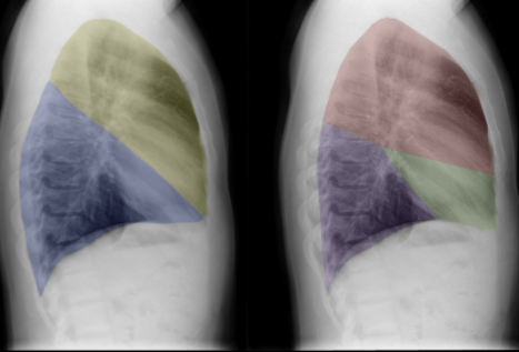
on the LATERAL view, the area highlighted PURPLE indicates:
right lower lobe
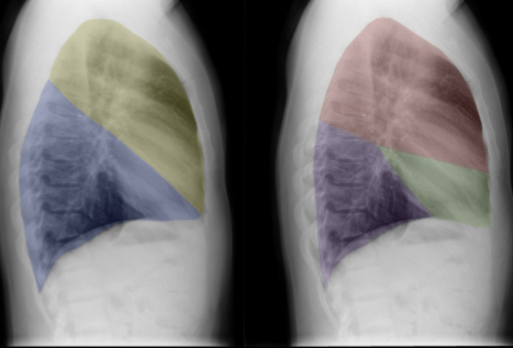
on the LATERAL view, the area highlighted YELLOW indicates:
left upper lobe
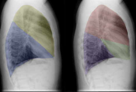
on the LATERAL view, the area highlighted BLUE indicates:
left lower lobe
to inspect x-ray image QUALITY, the acronym RIP is used and it stands for:
rotation, inspiration, and penetration
to review perform a SYSTEMATIC review of an xray, the acronym ATMLL is used and stands for:
abdomen, thorax, mediastinum, lungs, and line/tubes
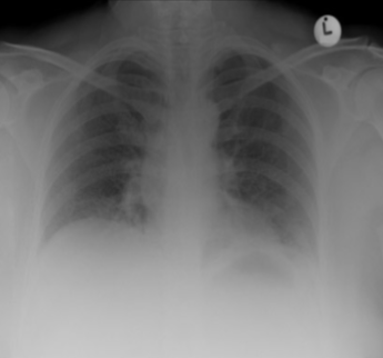
the included image is an example of _______ interferring with the x-ray as the ANTERIOR portion of 5-7 ribs are NOT visable at the midclavicular line
inspiration
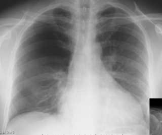
the included image has an example of a thoracic abnormality; what is it?
fractured clavicle
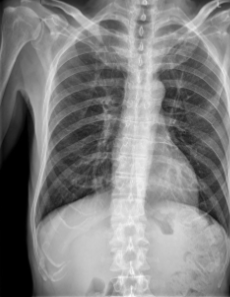
the included image has an example of a thoracic abnormality; what is it?
rib fracture on left side of image
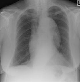
the included image has an example of a thoracic abnormality; what is it?
air located subcutaneous tissue above right clavicle
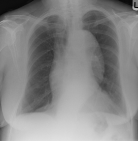
the included image has an example of a mediastinal abnormality; what is it?
widening of the aorta
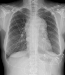
the included image has an example of a mediastinal abnormality; what is it?
mediastinal mass
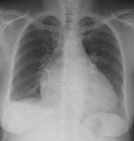
the included image has an example of a mediastinal abnormality; what is it?
cardiomegaly
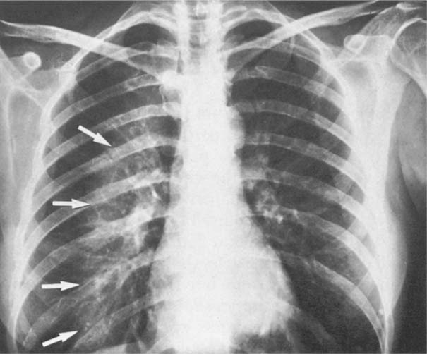
the included image has an example of infiltrate; what is it?
alveolar
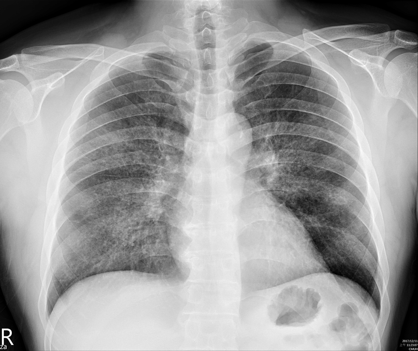
the included image has an example of infiltrate; what is it?
interstitial
what sign or indicator is noted when the border of the heart, aorta, or diaphragm is obliterated or obscured?
silhouette sign
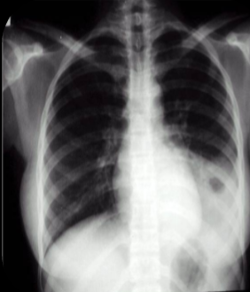
where is the infiltrate in this image?
left lower lung
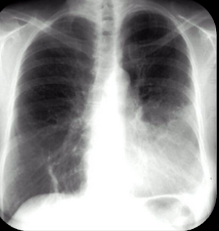
where is the infiltrate in this image?
lingula of left lung
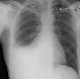
this x-ray has an example of a sign of pleural effusion: what is it?
meniscus sign on right side
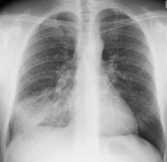
is the attached image an example of effusion or infiltrate?
infiltrate
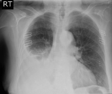
is the attached image an example of effusion or infiltrate?
effusion
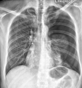
what is the abnormality is noted on the patient’s left side in this image?
pneumothorax
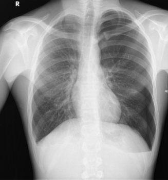
what is the abnormality is noted on the patient’s left side in this image?
pneumothorax
Is an inspiration view or expiration view more benficial to see a subtle pneumothorax?
expiration
what is the name of the horizontal fine white lines (which are found along the periphery of the lung and aim straight into the pleura) noted on a chest x-ray due to fluid build-up in the lobar septa?
Kerly B lines
what Kerly B lines, hilar vascular congestion, cephalization of vessels, and cardiomegaly all signs of if viewed on a chest x-ray?
congestive heart failure
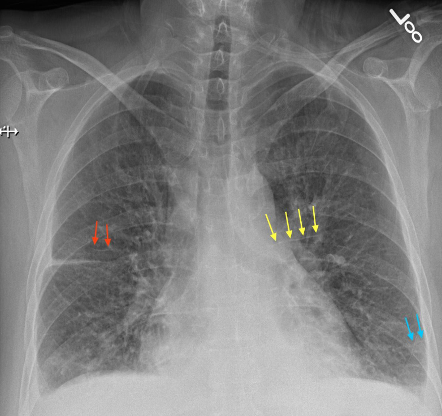
what abnormality is highlighted by the blue lines in this image
Kerly B lines
if trauma is the mechanism of injury, what imaging type should be used?
CT
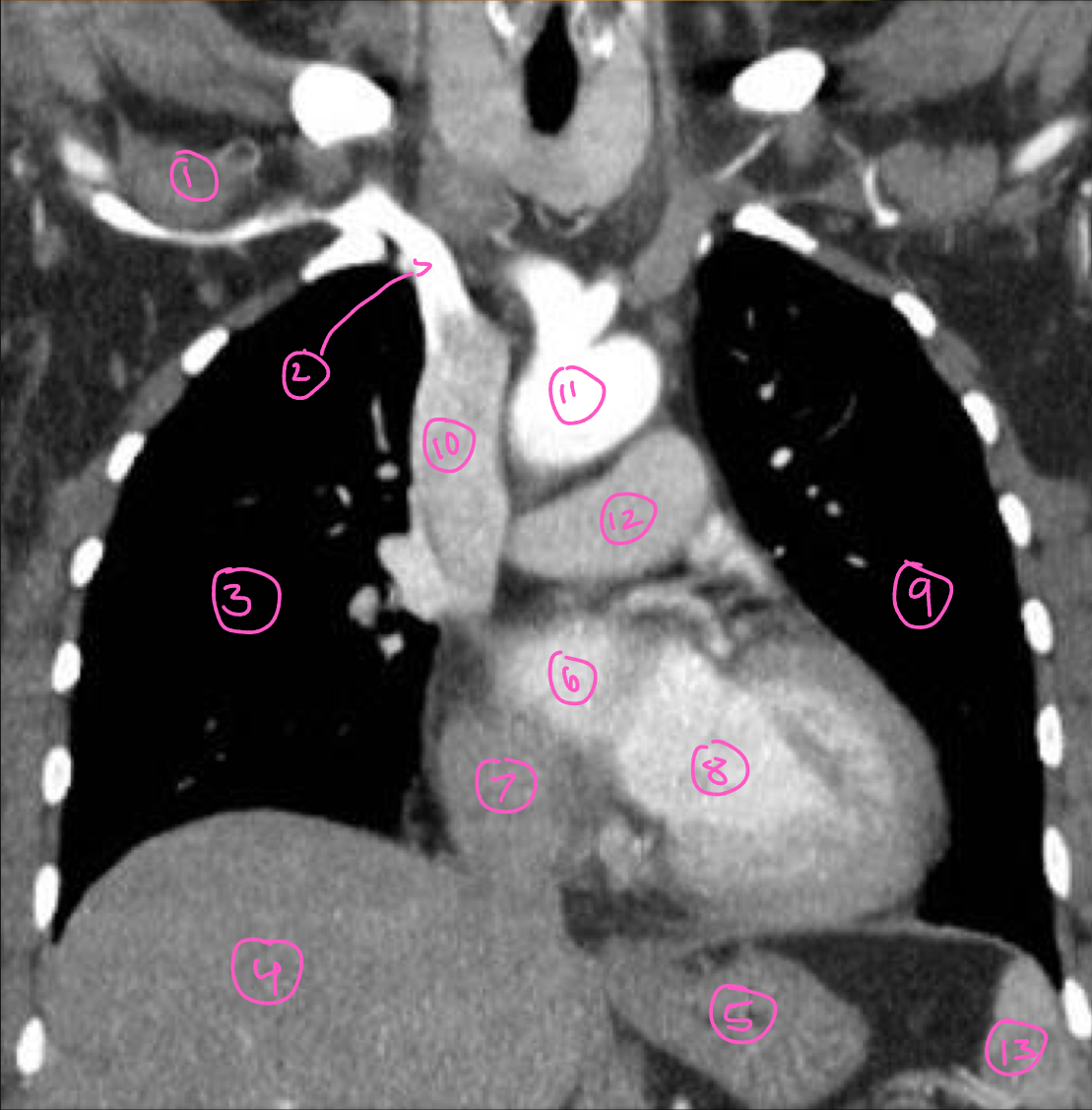
The structure labeled 1 in this image is:
right subclavian vein
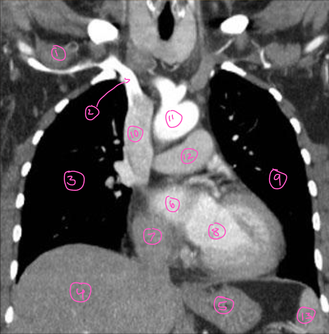
The structure labeled 2 in this image is:
right brachiocephalic vein
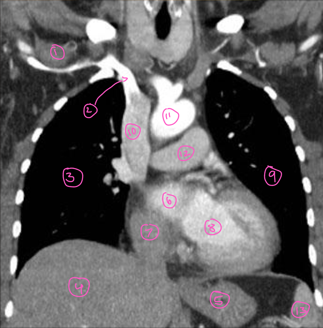
The structure labeled 3 in this image is:
right lung
The structure labeled 4 in this image is:
liver
The structure labeled 5 in this image is:
stomach
The structure labeled 6 in this image is:
left atrium
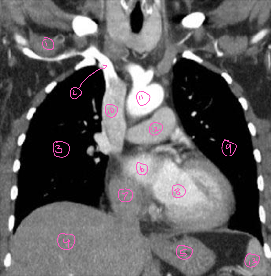
The structure labeled 7 in this image is:
right atrium
The structure labeled 8 in this image is:
left ventricle
The structure labeled 9 in this image is:
left lung
The structure labeled 10 in this image is:
superior vena cava
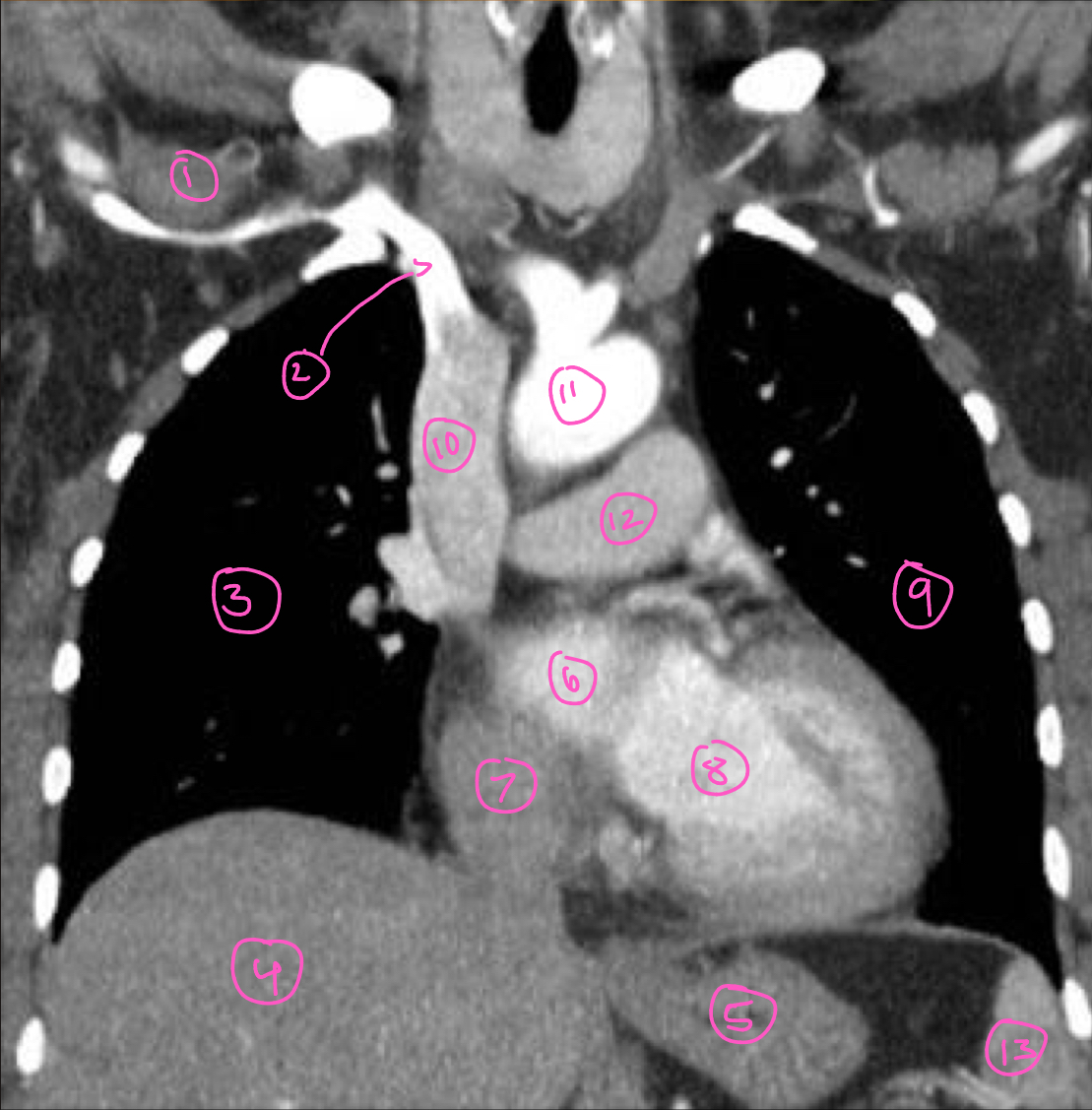
The structure labeled 11 in this image is:
aorta
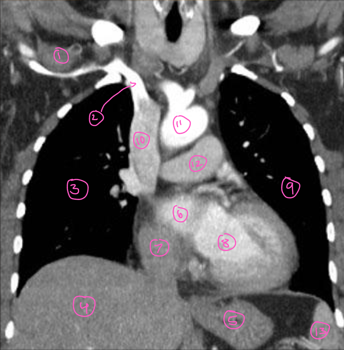
The structure labeled 12 in this image is:
pulmonary trunk
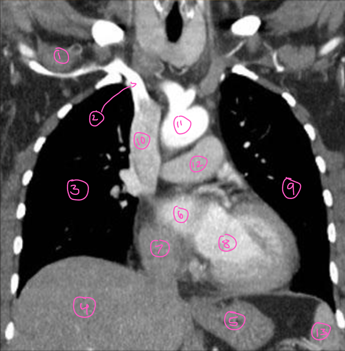
The structure labeled 13 in this image is:
spleen
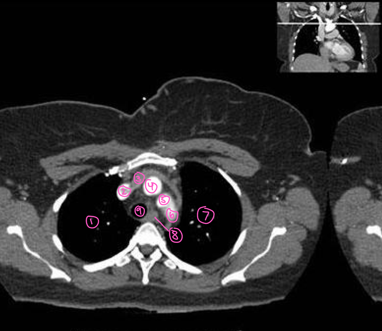
The structure labeled 1 in this image is:
right lung
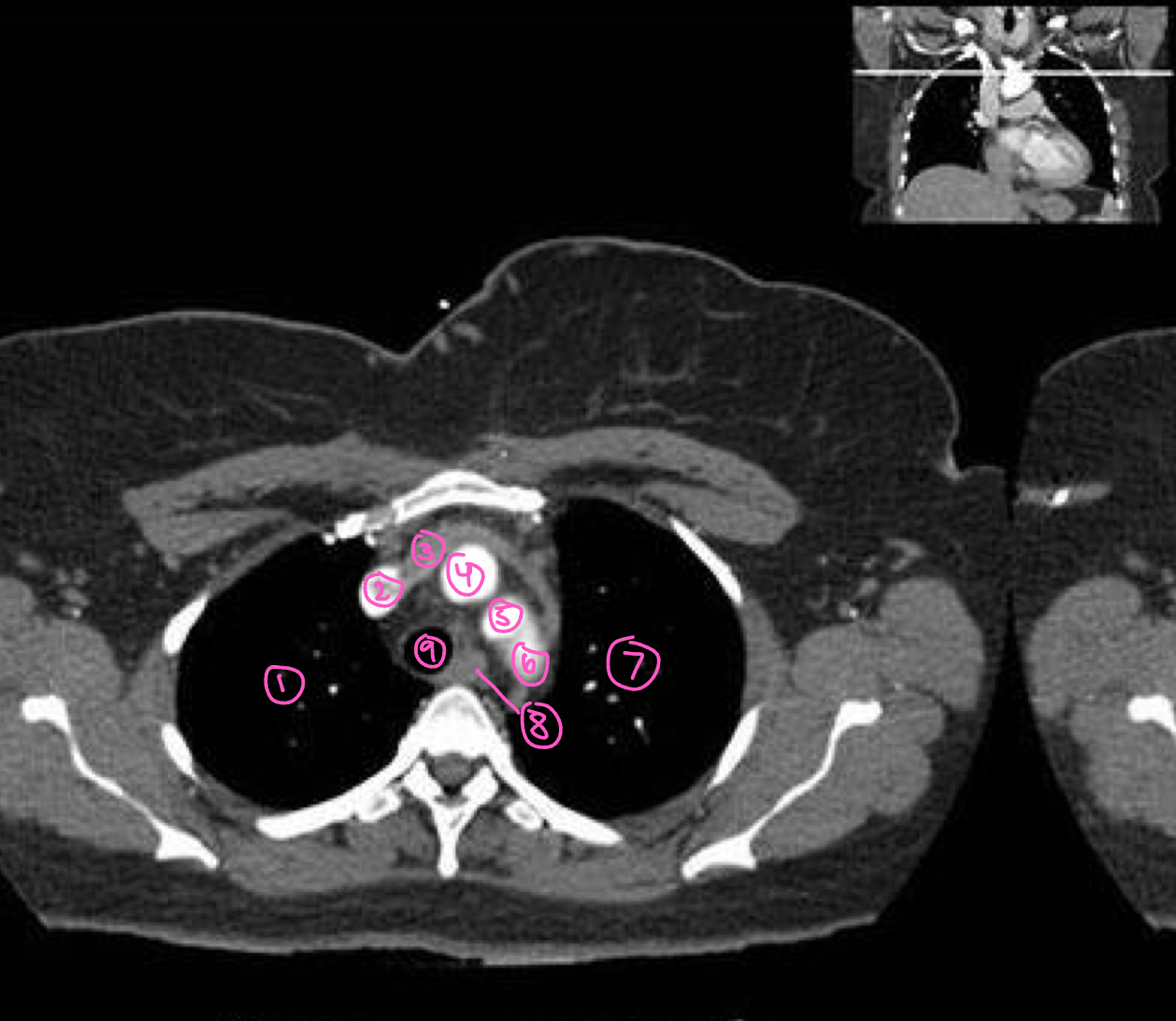
The structure labeled 2 in this image is:
superior vena cava
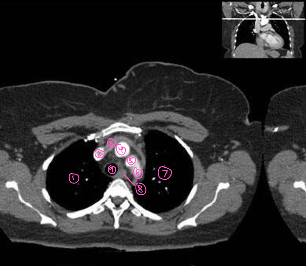
The structure labeled 3 in this image is:
left brachiocephalic vein
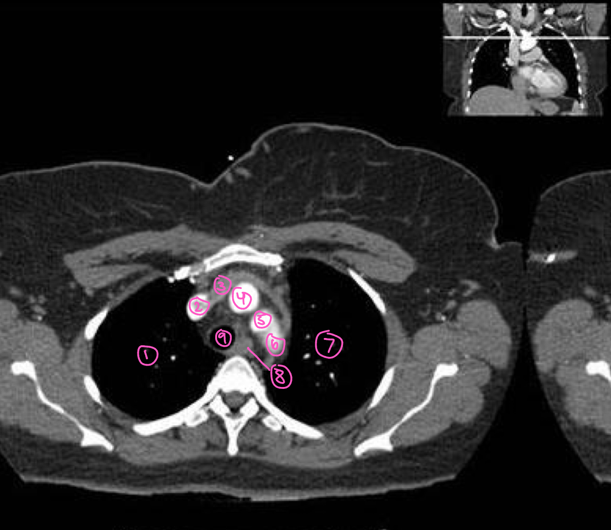
The structure labeled 4 in this image is:
right brachiocephalic artery
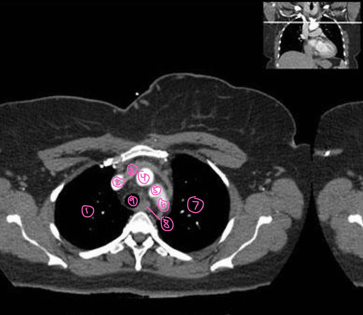
The structure labeled 5 in this image is:
left common carotid artery
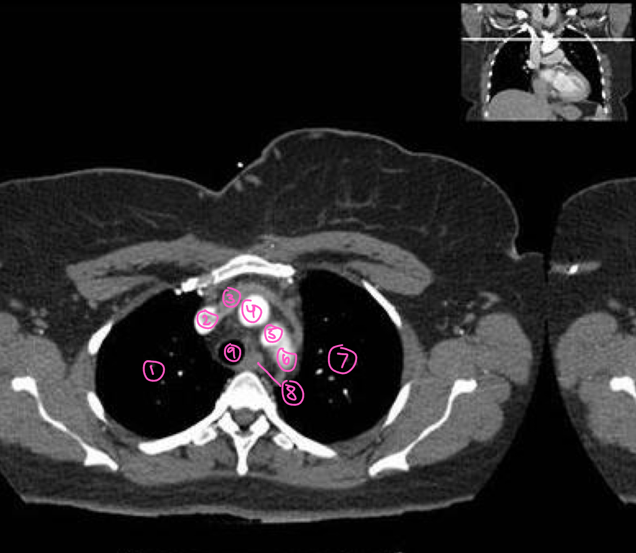
The structure labeled 6 in this image is:
left subclavian artery
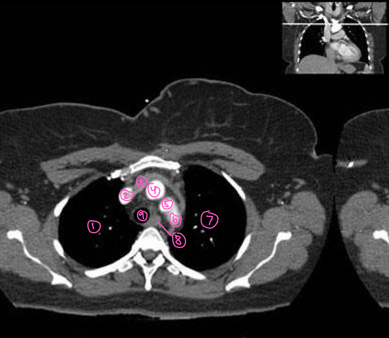
The structure labeled 7 in this image is:
left lung
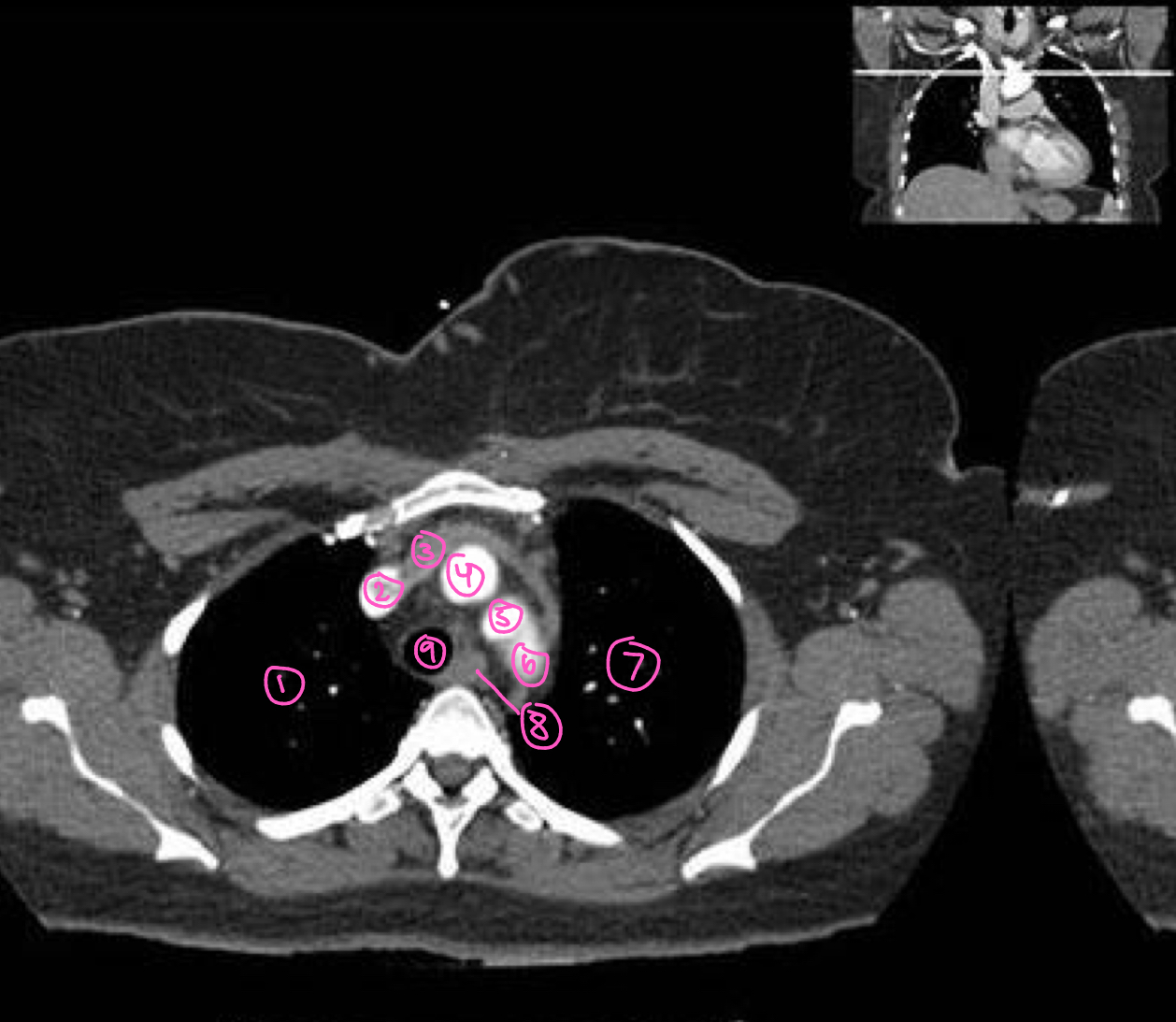
The structure labeled 8 in this image is:
esophagus
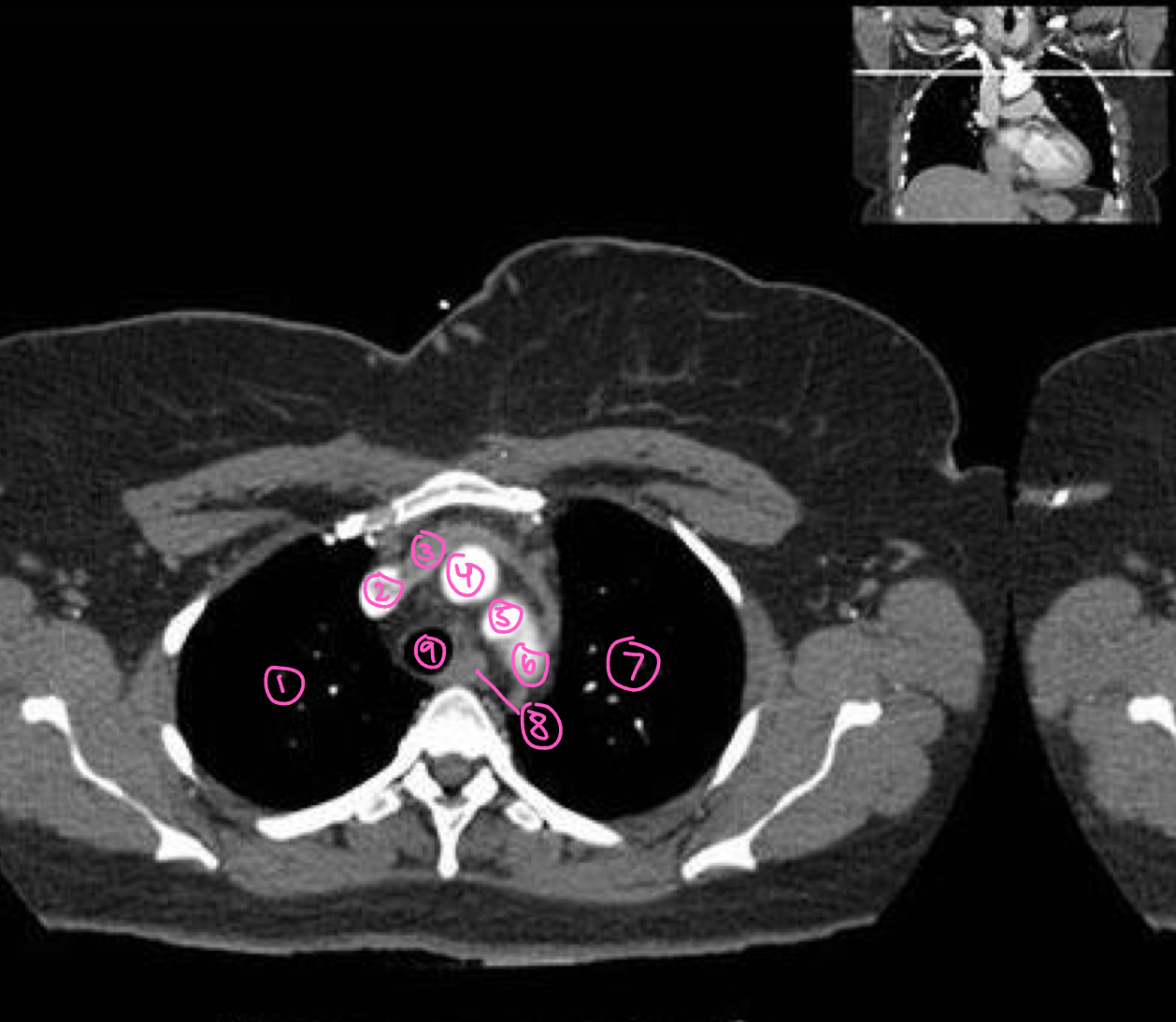
The structure labeled 9 in this image is:
trachea
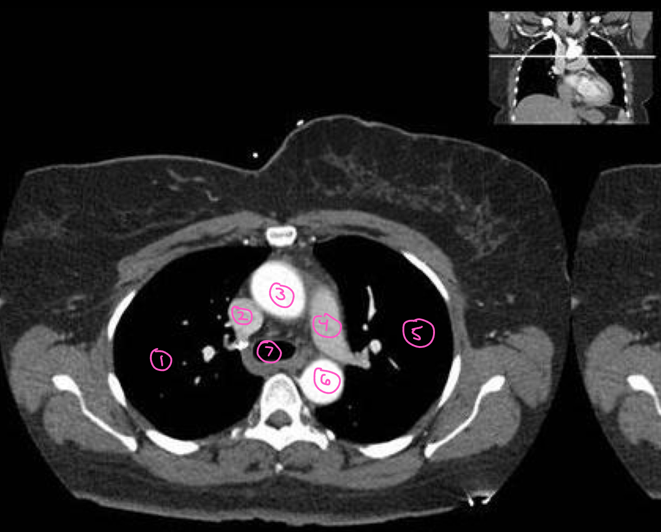
The structure labeled 1 in this image is:
right lung
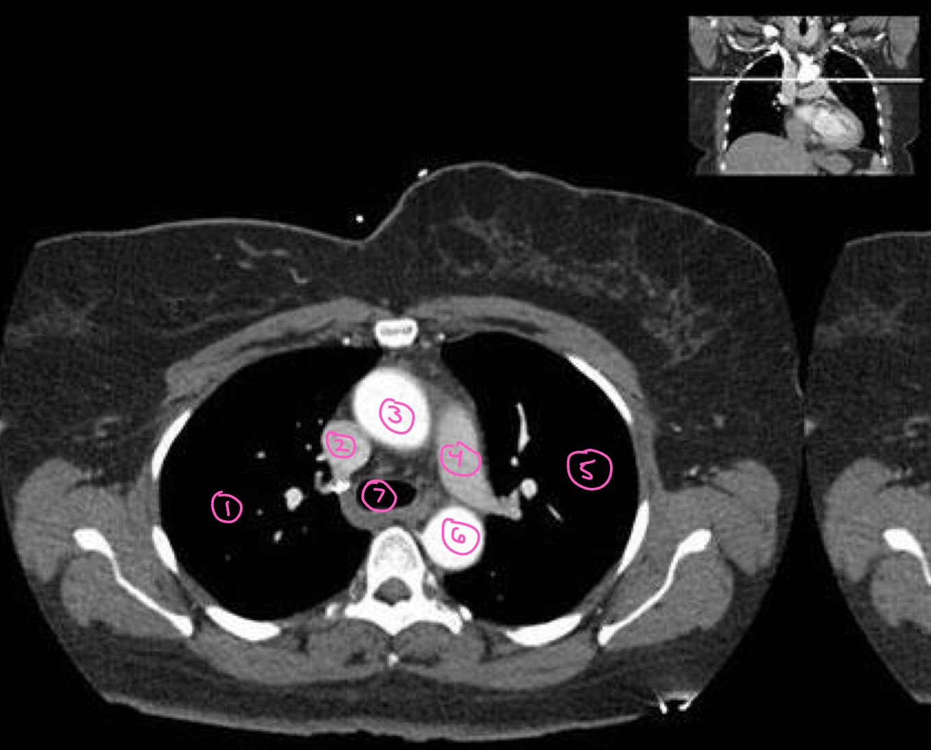
The structure labeled 2 in this image is:
superior vena cava
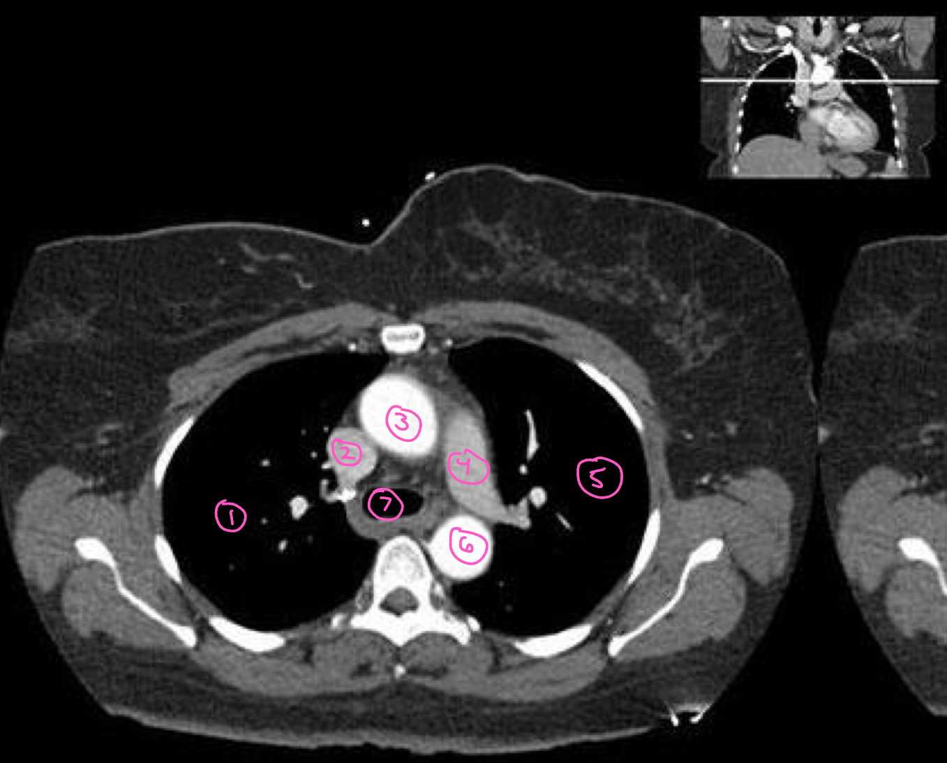
The structure labeled 3 in this image is:
ascending aorta
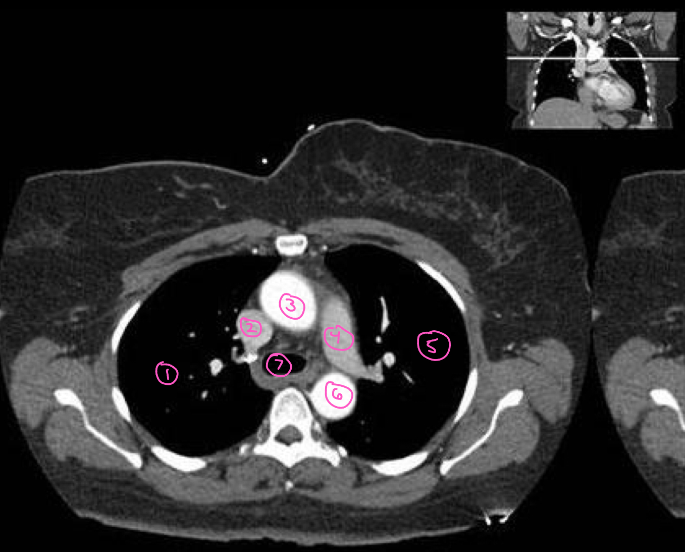
The structure labeled 4 in this image is:
left pulmonary artery
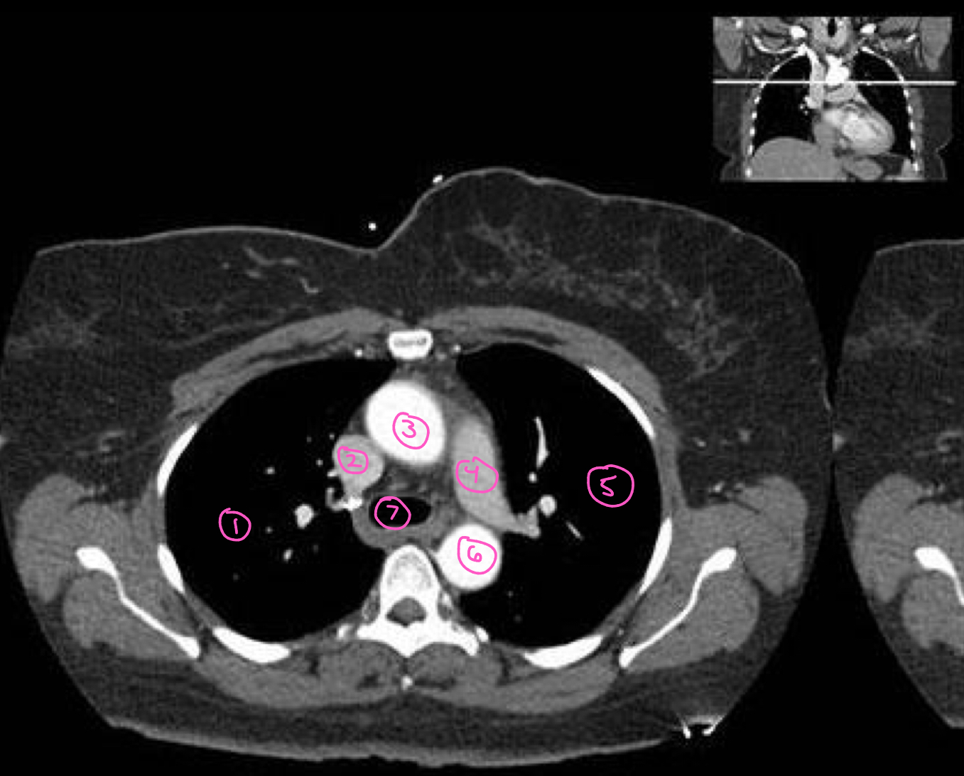
The structure labeled 5 in this image is:
left lung
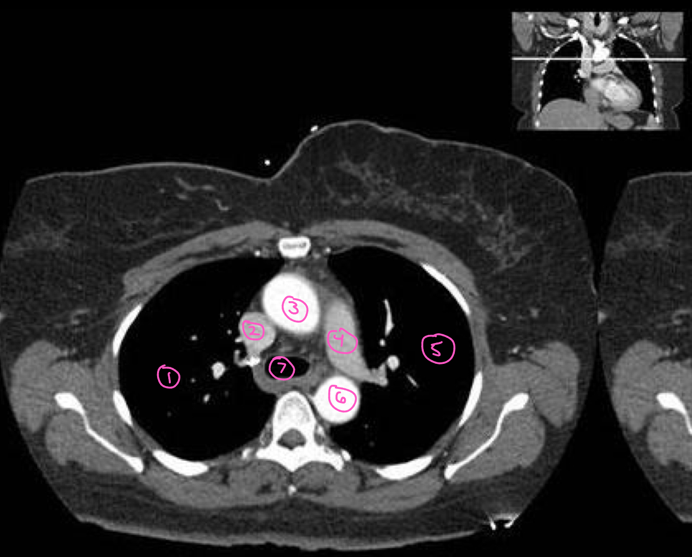
The structure labeled 6 in this image is:
descending aorta
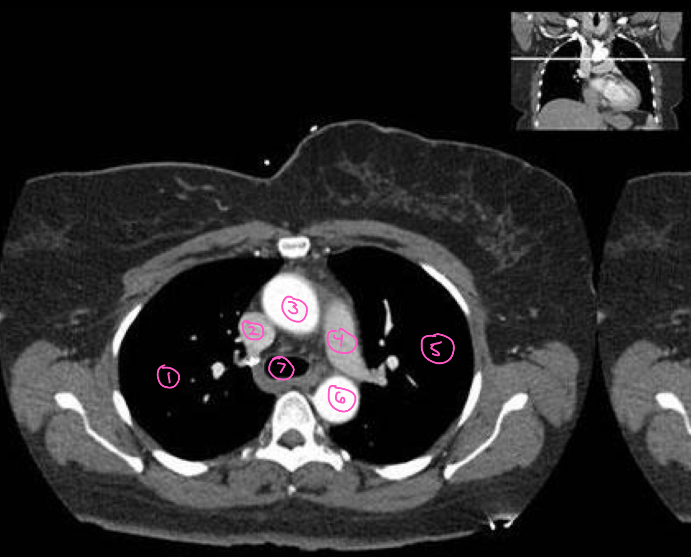
The structure labeled 7 in this image is:
trachea
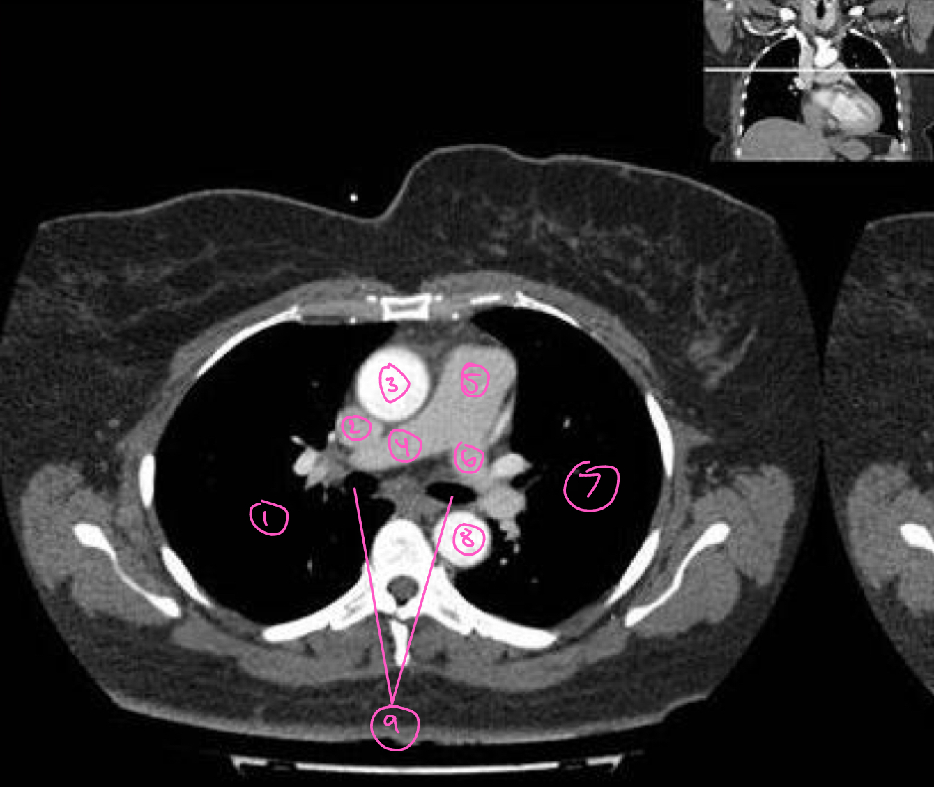
The structure labeled 1 in this image is:
right lung
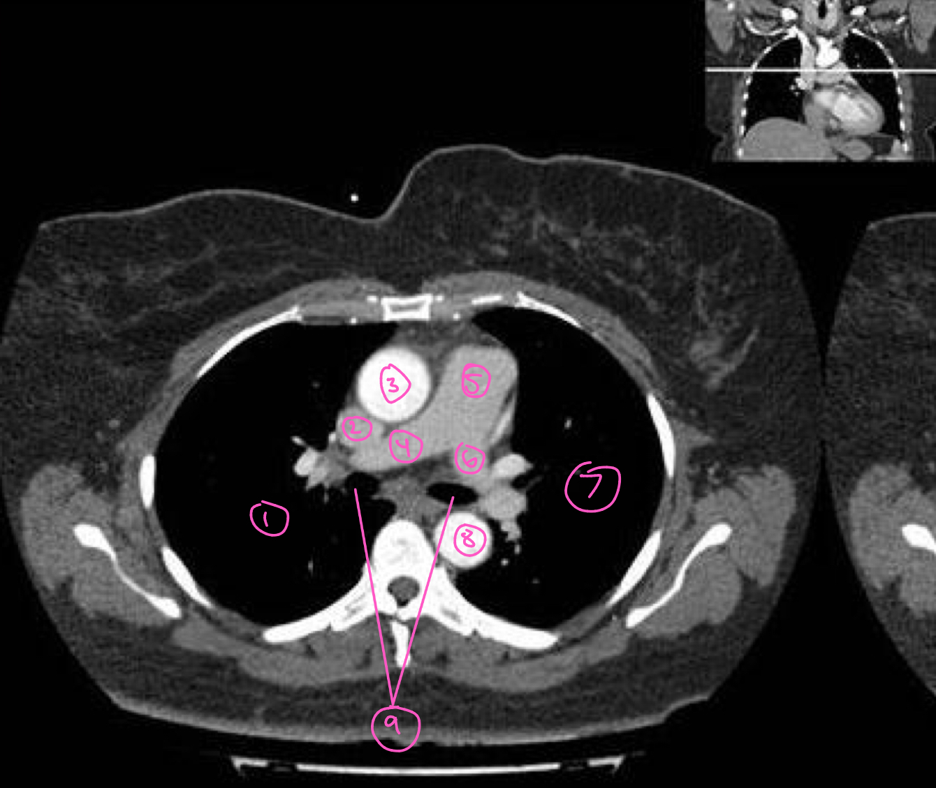
The structure labeled 2 in this image is:
superior vena cava
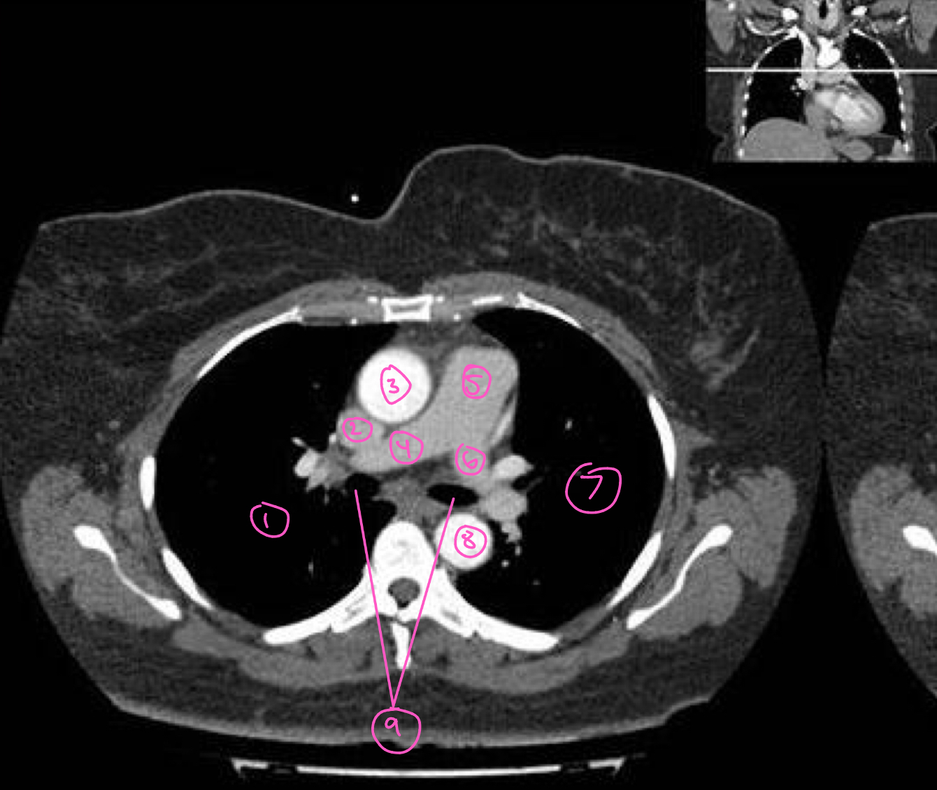
The structure labeled 3 in this image is:
ascending aorta
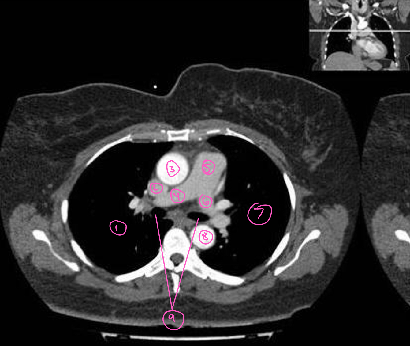
The structure labeled 4 in this image is:
right pulmonary artery
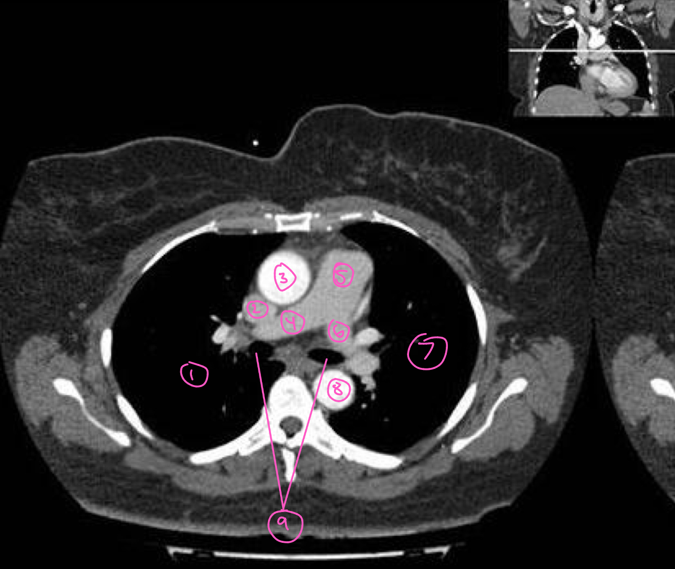
The structure labeled 5 in this image is:
pulmonary trunk
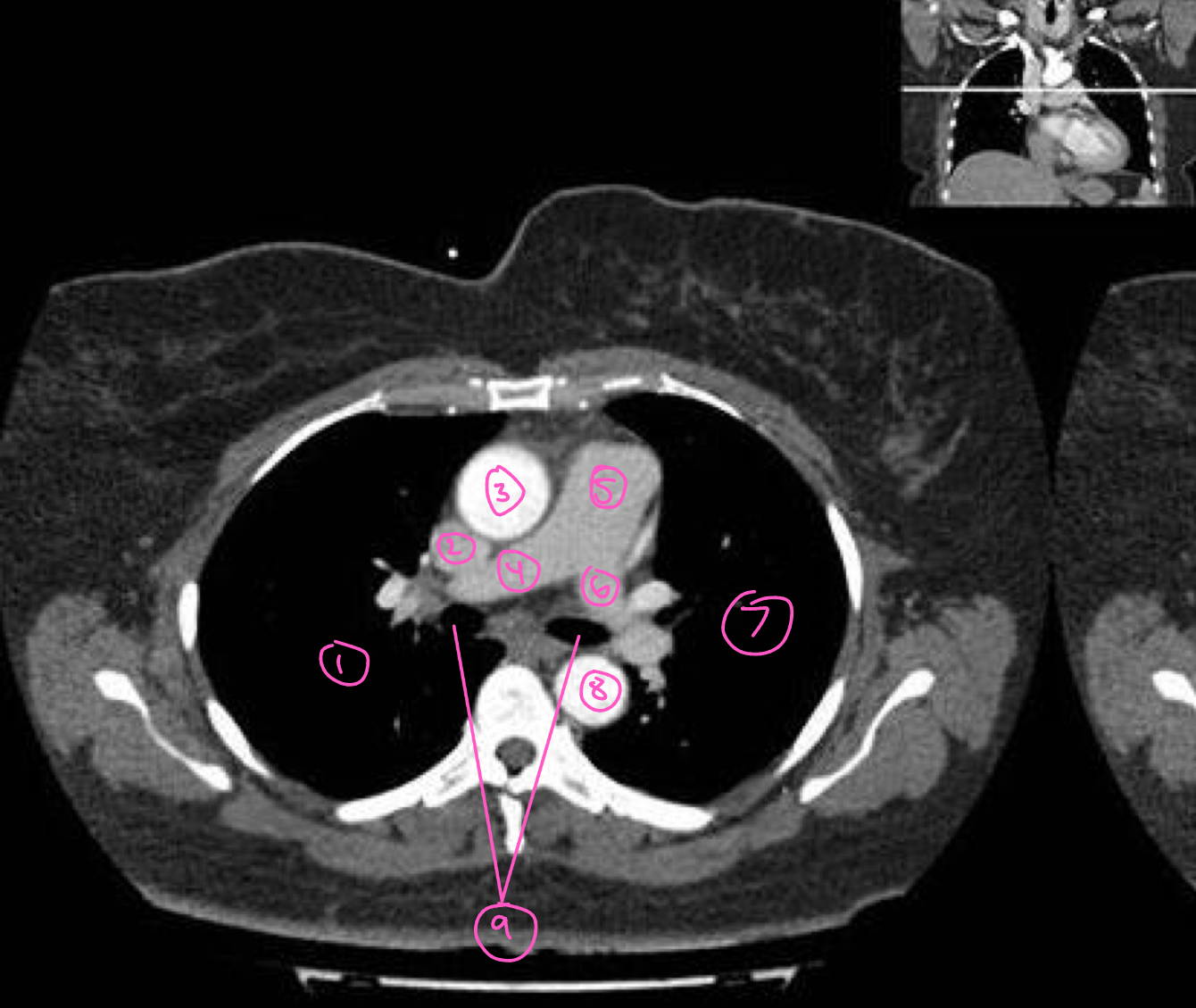
The structure labeled 6 in this image is:
left pulmonary artery
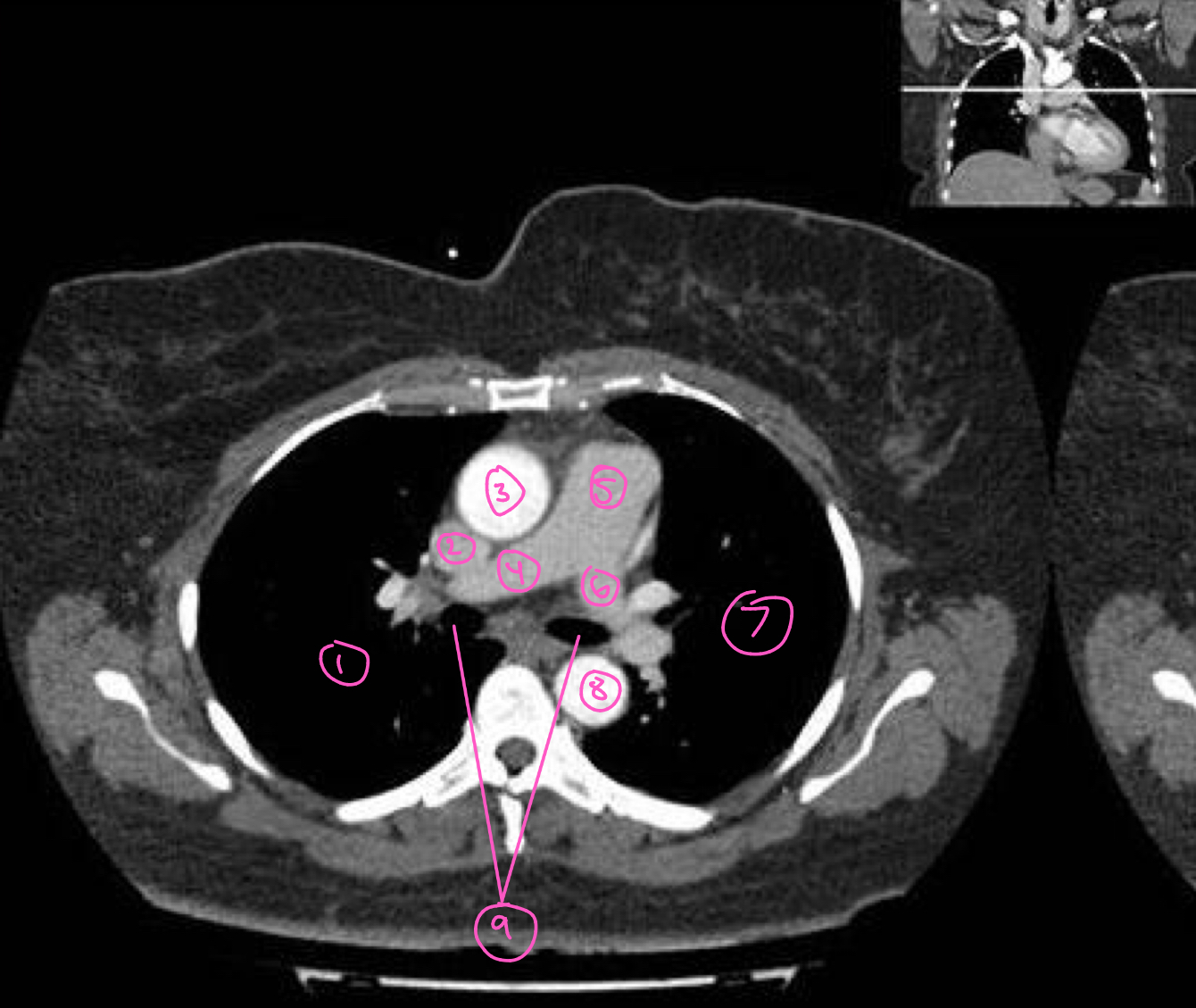
The structure labeled 7 in this image is:
left lung
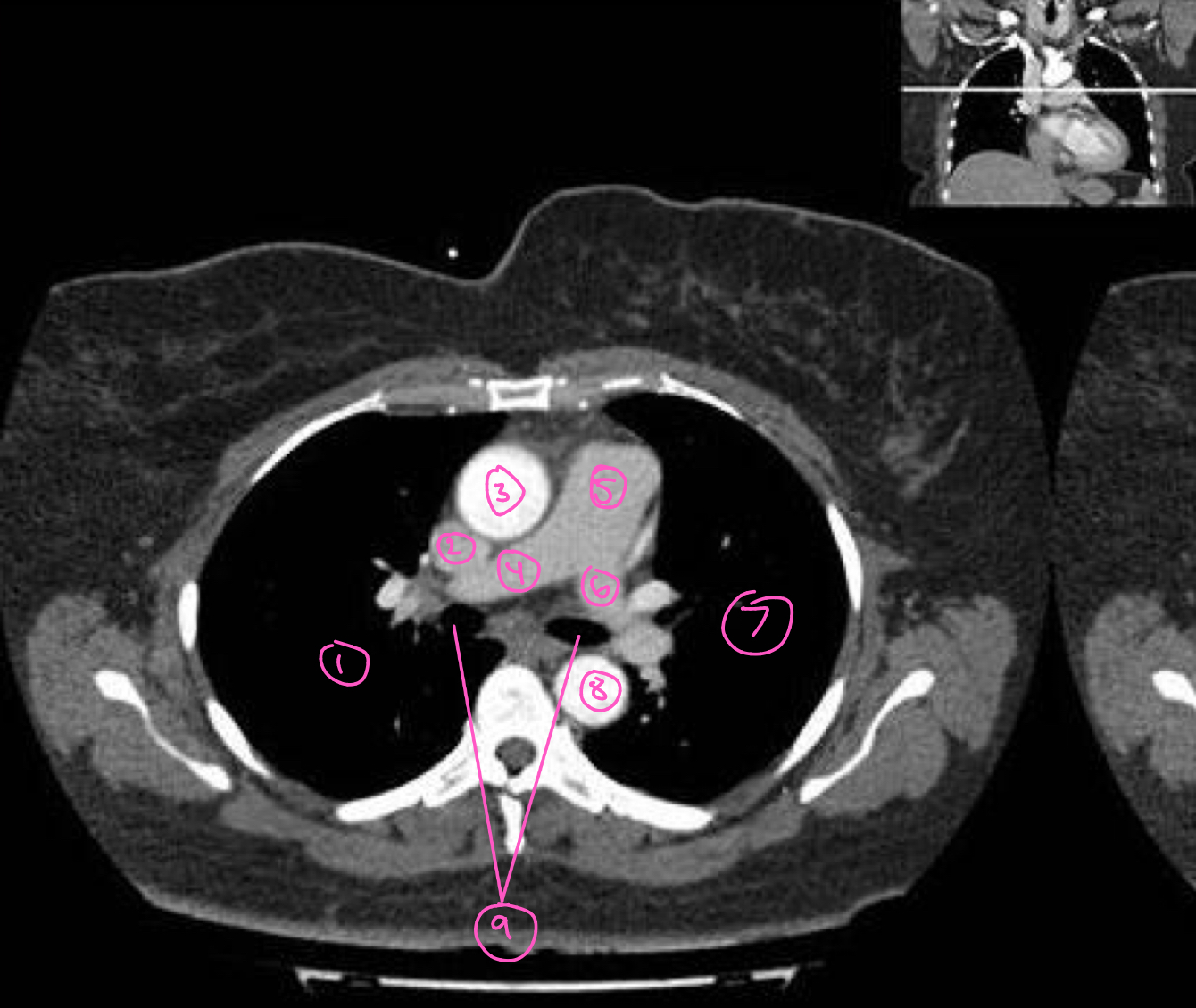
The structure labeled 8 in this image is:
descending aorta
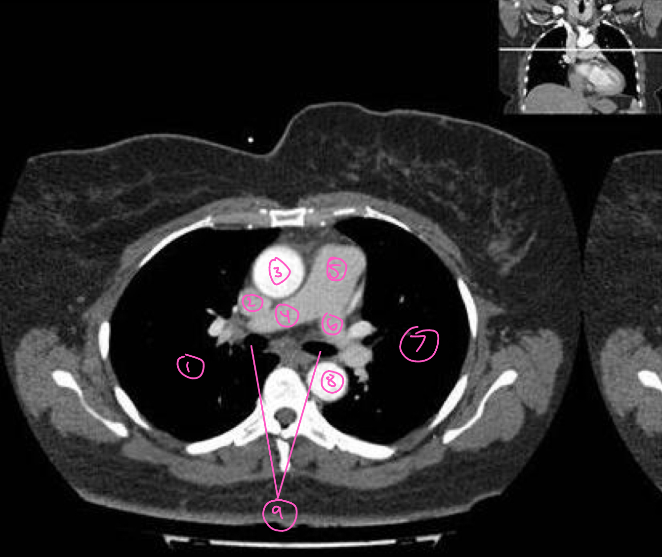
The structure labeled 9 in this image is:
main bronchi
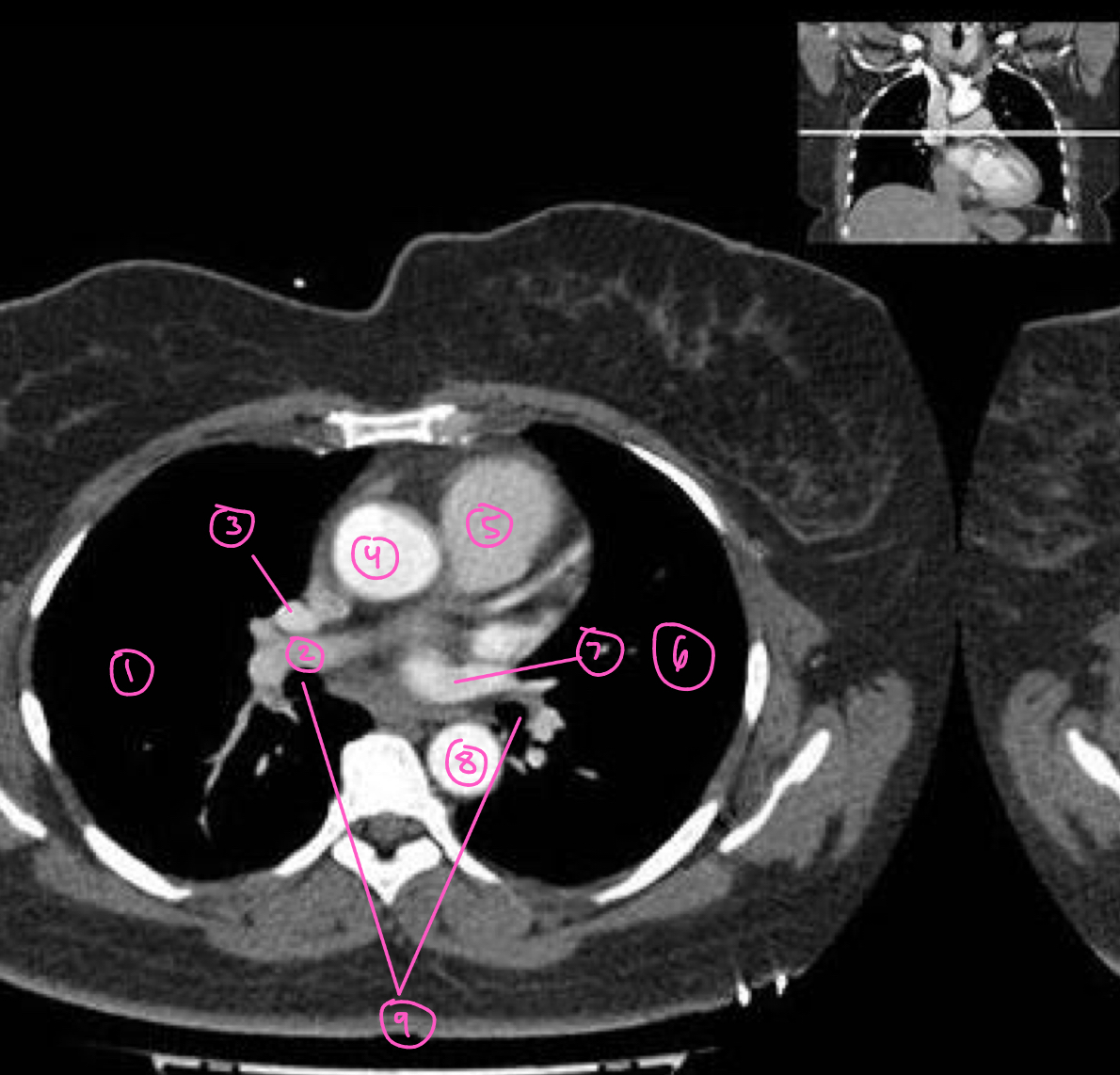
The structure labeled 1 in this image is:
right lung
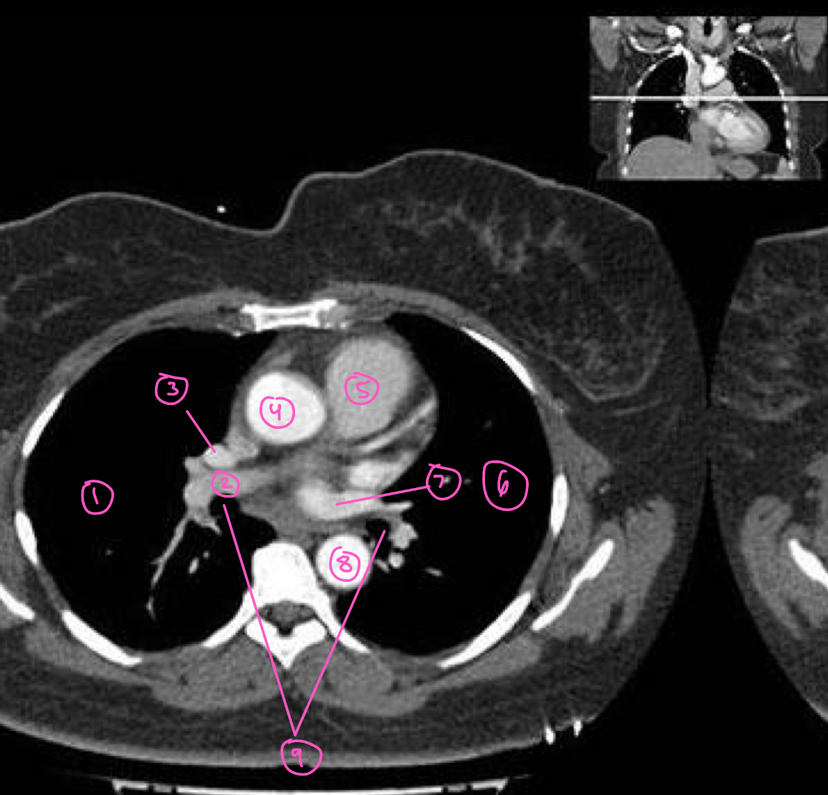
The structure labeled 2 in this image is:
right pulmonary artery
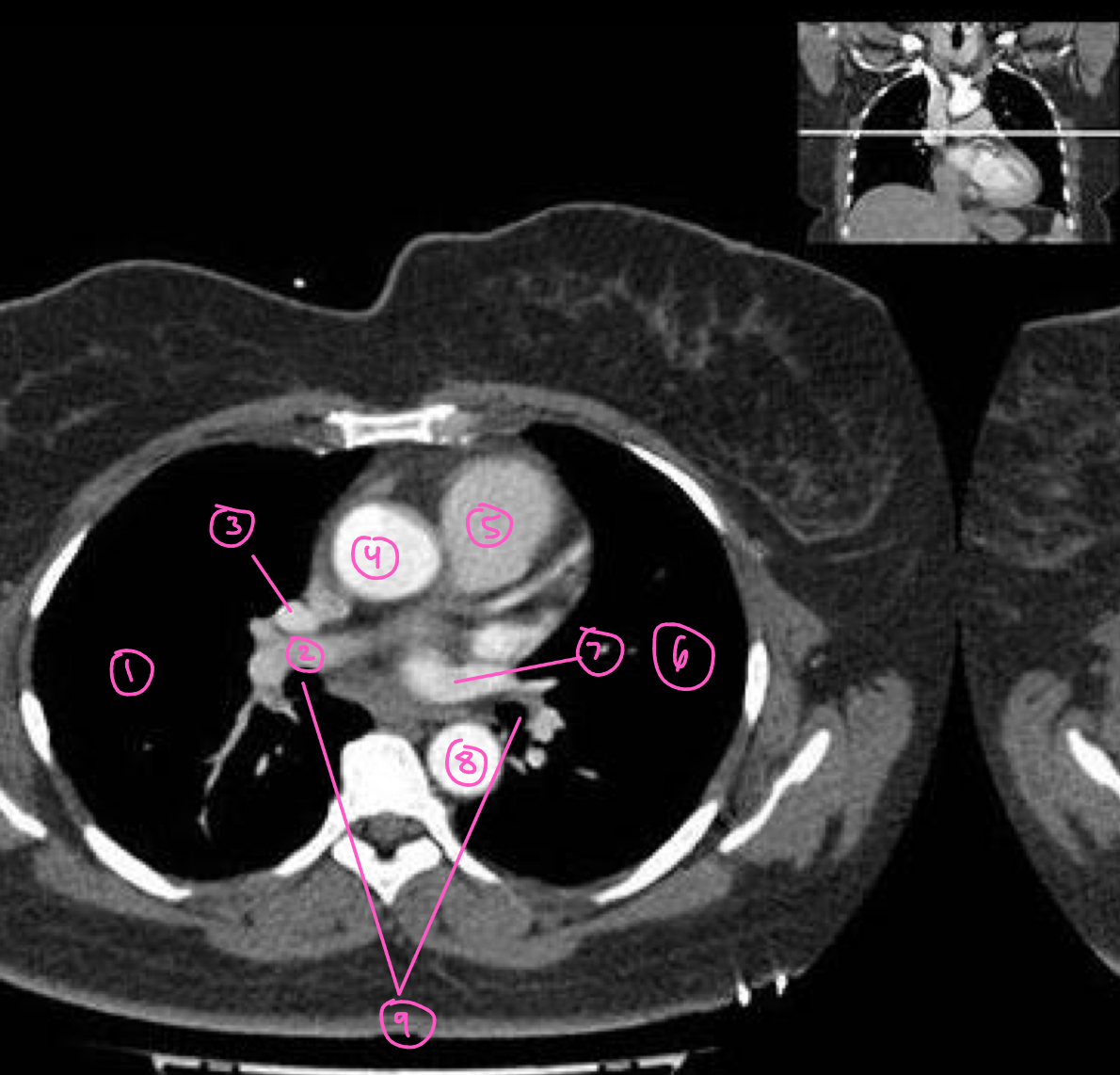
The structure labeled 3 in this image is:
superior vena cava
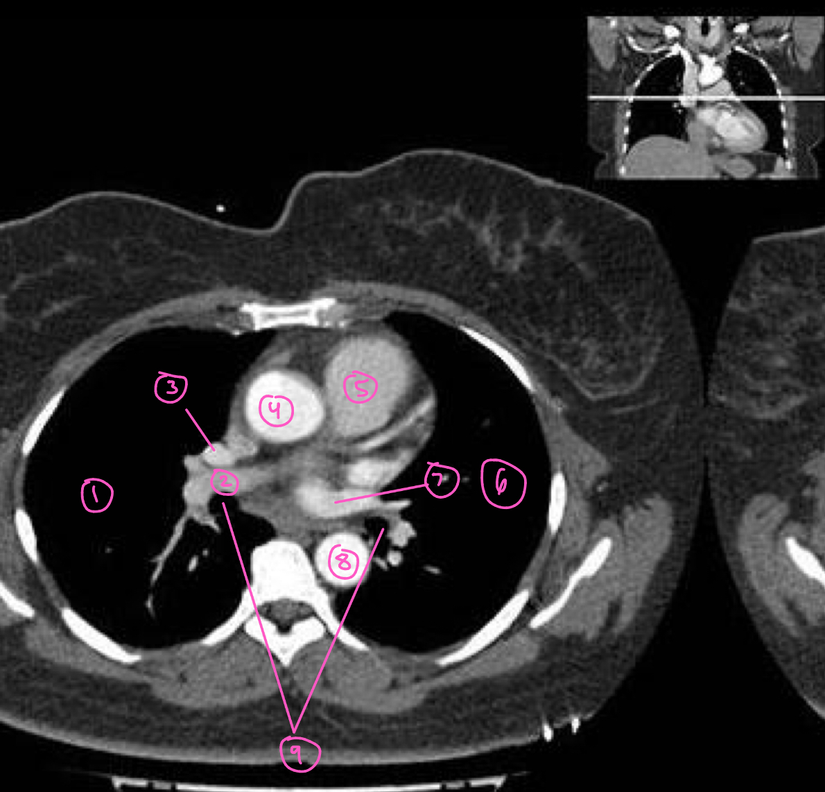
The structure labeled 4 in this image is:
ascending aorta
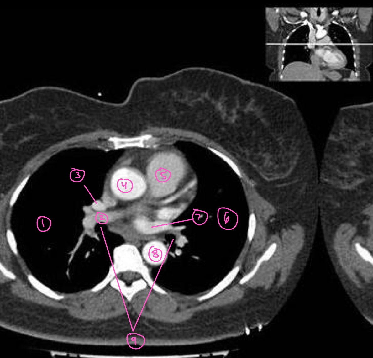
The structure labeled 5 in this image is:
right ventricle
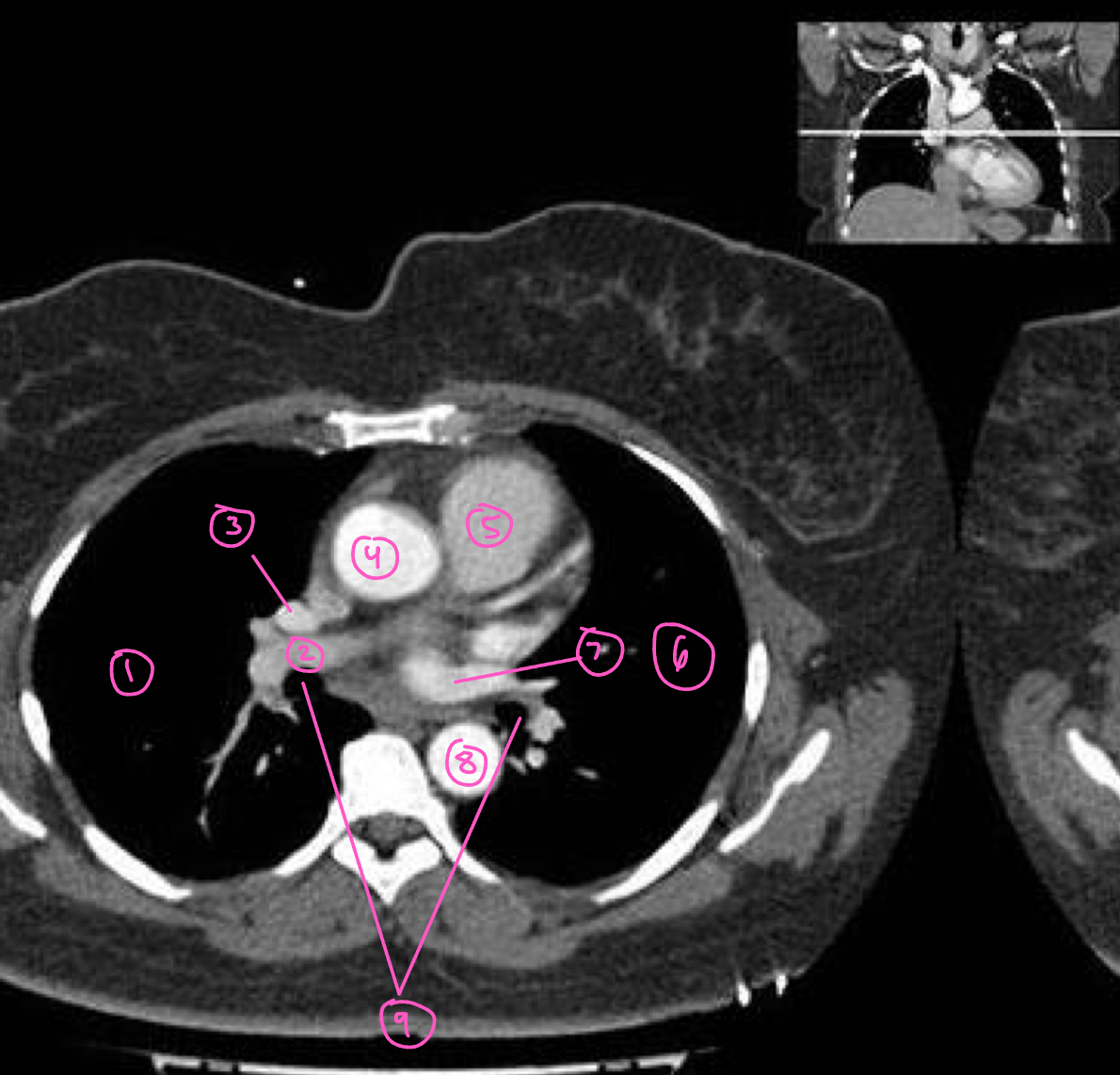
The structure labeled 6 in this image is:
left lung
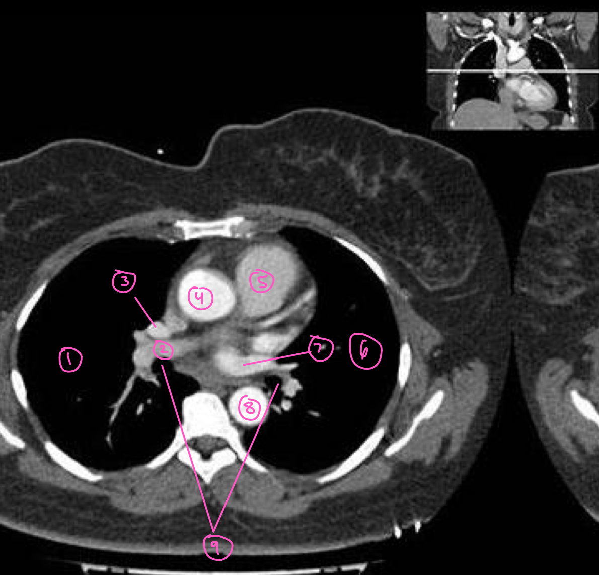
The structure labeled 7 in this image is:
left pulmonary artery
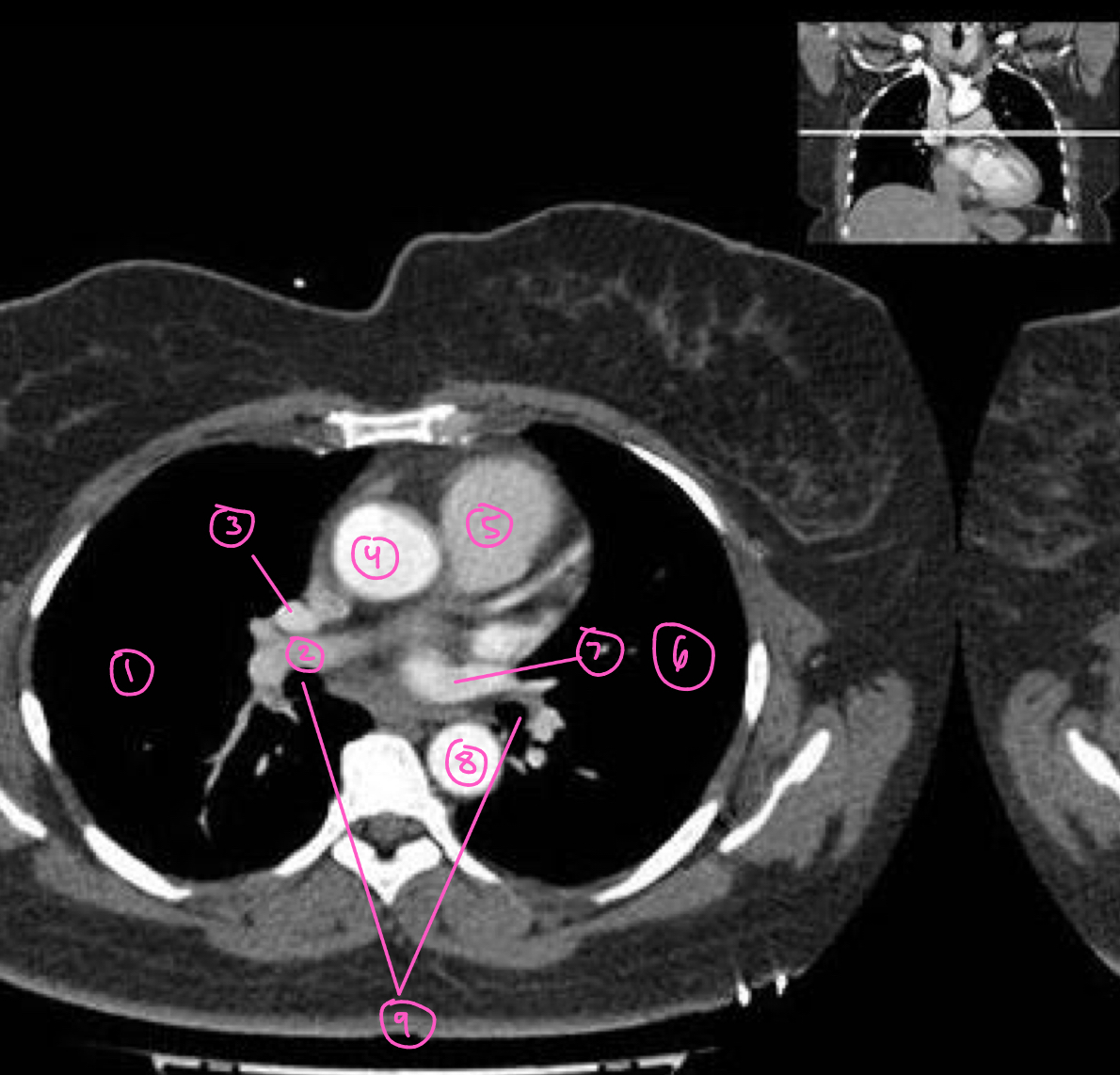
The structure labeled 8 in this image is:
descending aorta