Kinesiology Exam 1
 Call Kai
 Learn
Learn Practice Test
 Spaced Repetition
 Match
Match Flashcards
 Knowt Play
1/118

There's no tags or description
Looks like no tags are added yet.
Name | Mastery | Learn | Test | Matching | Spaced | Call with Kai | Chat |
|---|
No analytics yet
Send a link to your students to track their progress
119 Terms
Osteokinematics
Movement of bones around a joint axis
Arthrokinematics
The motions of joints in the body
Cephalic
Closer to head
Caudal
Closer to feet
Ventral
Front
Dorsal
Back
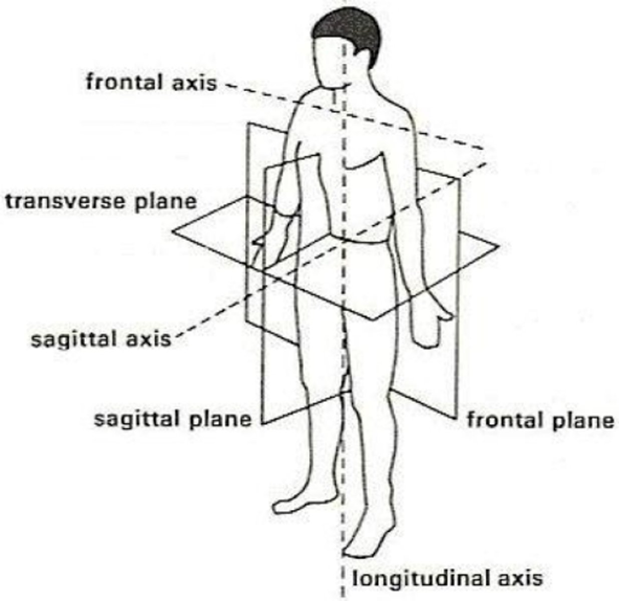
Sagittal plane
Divide body into right & left sections
Frontal/Coronal plane
Divide body into front & back sections
Horizontal/Transverse
Divide body into upper and lower sections
Axis of rotation is always perpendicular to the plane of motion
Sagittal plane-- Frontal axis
Frontal plane-- Sagittal axis
Transverse plane-- vertical axis
Linear motion
All parts of body move in same direction at same speed
Rotation (Angular) motion
Rotate on pivot point
Body parts move different distances at different angles depending on pivot
General motion
Combination of linear and angular motion
Human movement is mostly general
Anatomical position
Most of the joints are in neutral position
Least amount of tension for many structures
Motions of sagittal plane
Flexion & extension
DF/PF
Motions of frontal plane
Abduction & Adduction
Lateral flexion
ex) Ulnar/Radial deviation
eversion.inversion
Motions of horizontal plane
Internal & external rotation
ex) axial rotation
Pronation (foot)
Eversion + ABDuction + Dorsiflexion
Supination (foot)
Inversion + ADDuction + plantar flexion
Dorsal surface of foot
Superior (Top)
Plantar surface of foot
Inferior (Bottom)
Linear motion has no axis of rotation
Scapular depression/elevation - No axis of rotation
Motion: Walking
Plane of motion:
Axis of rotation:
Best view:
Motion: Walking
Plane of motion: Sagittal plane
Axis of rotation: Frontal axis
Best view: Side View
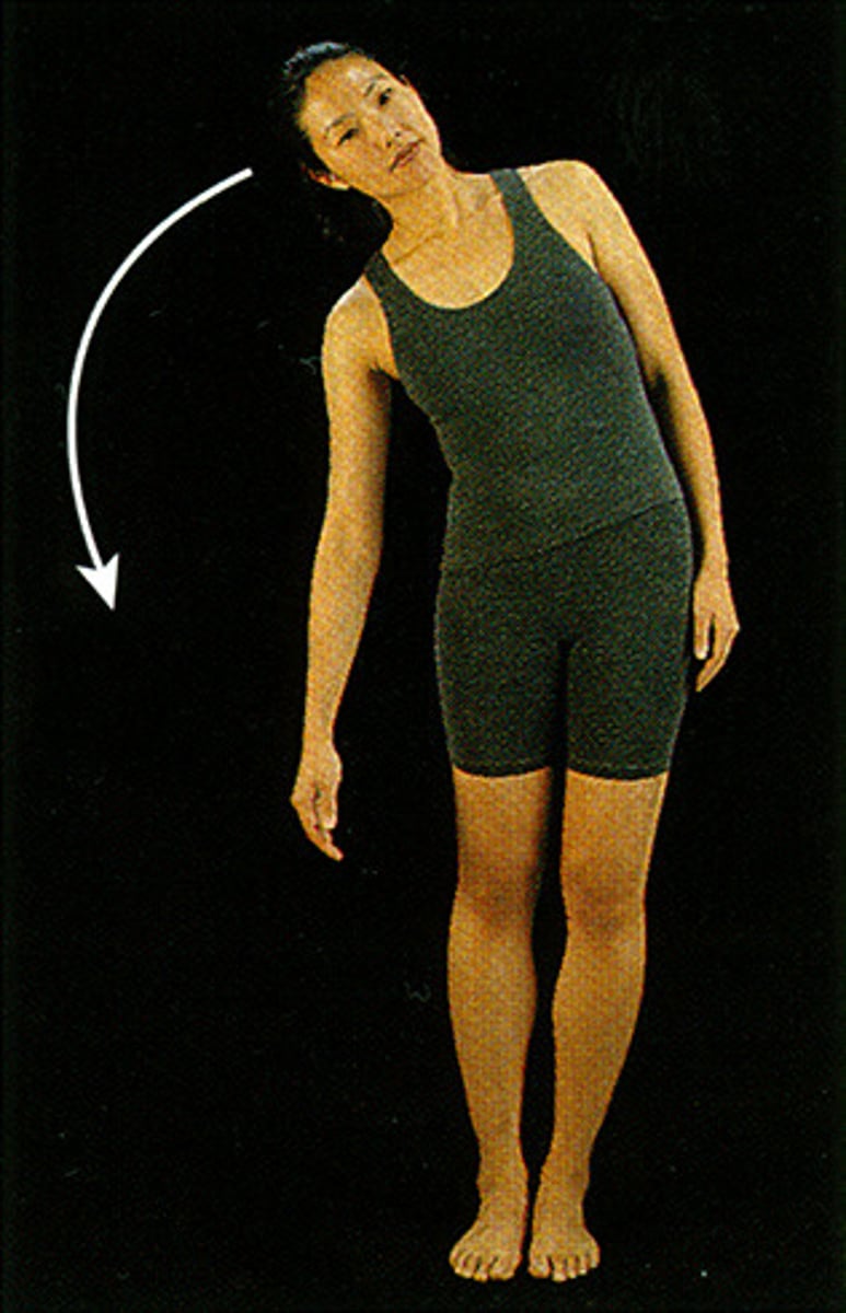
Motion: Lateral trunk flexion
Plane of motion:
Axis of rotation:
Best view:
Motion: Lateral side bend
Plane of motion: Frontal plane
Axis of rotation: Sagittal axis
Best view: Anterior/posterior
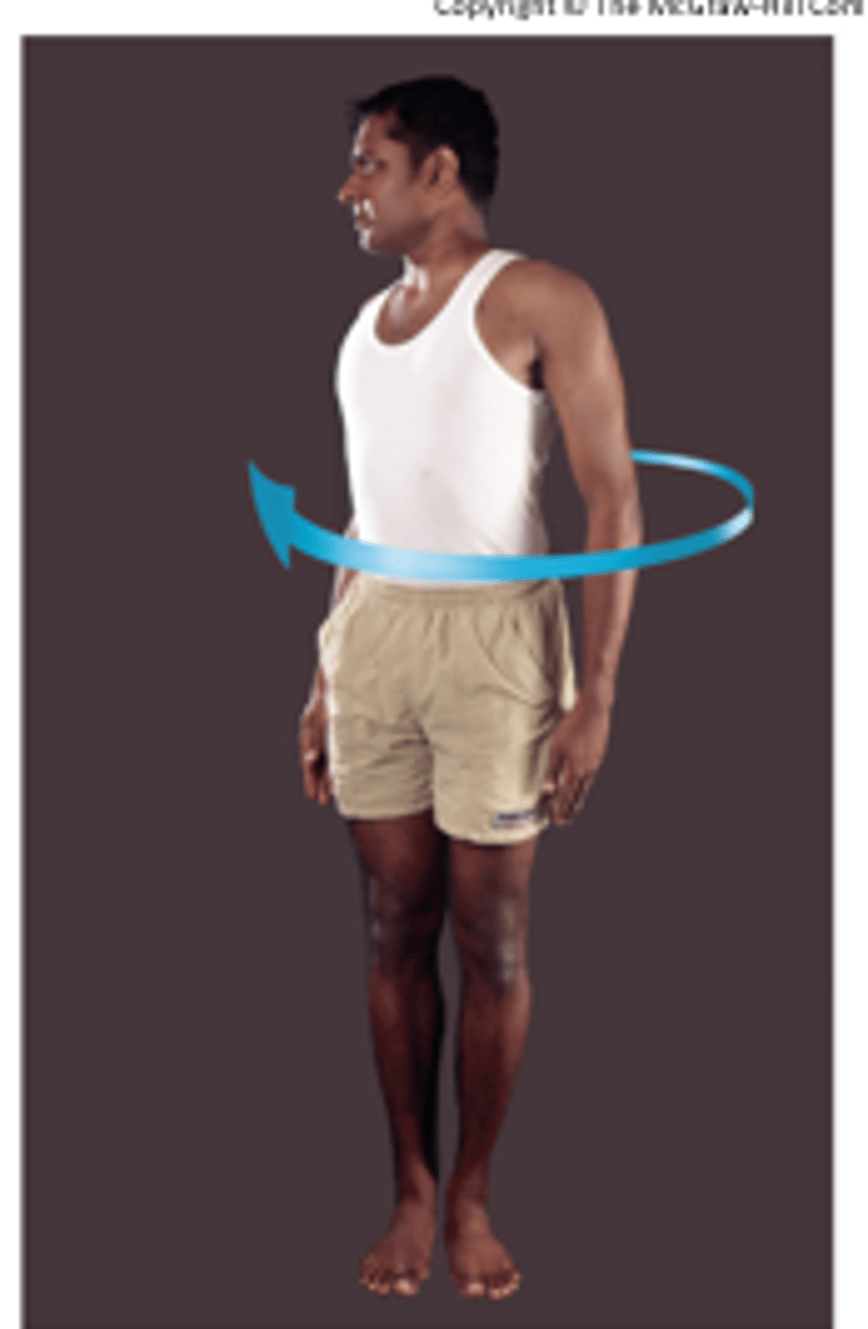
Motion: Lateral trunk rotation
Plane of motion:
Axis of rotation:
Best view:
Motion: Lateral side bend
Plane of motion: Transverse
Axis of rotation: Vertical axis
Best view: Top
Open chain
Proximal segment fixed
Distal segment free
Closed chain
Proximal segment free
Distal segment fixed
Closed chain movements
Push ups
pull ups
squats
deadlifts
lunges
dips
Open chain movements
Bicep curls
Hamstring curls
Leg extensions
Open chain:
Hip flexion
Right hip IR
Knee flexion
Ankle DF
Shoulder extension
Closed chain:
Trunk flexion
Trunk rotate to R hip
Squat
Tibia lean forward
"Muscle up"
Epimysium
Muscle body is surrounded by
Fascicle
Several bundles within muscle
Perimysium
Covers fasicles
Muscle fibers
Bundles together to form fasicle
Endomysium
Surrounds induvidual muscle fiber
Myofibrils
Bundled together to form muscle fibers
Myofilaments
Bundled together to make up myofibril
Components of myofilaments
actin
myosin
titin (passive)
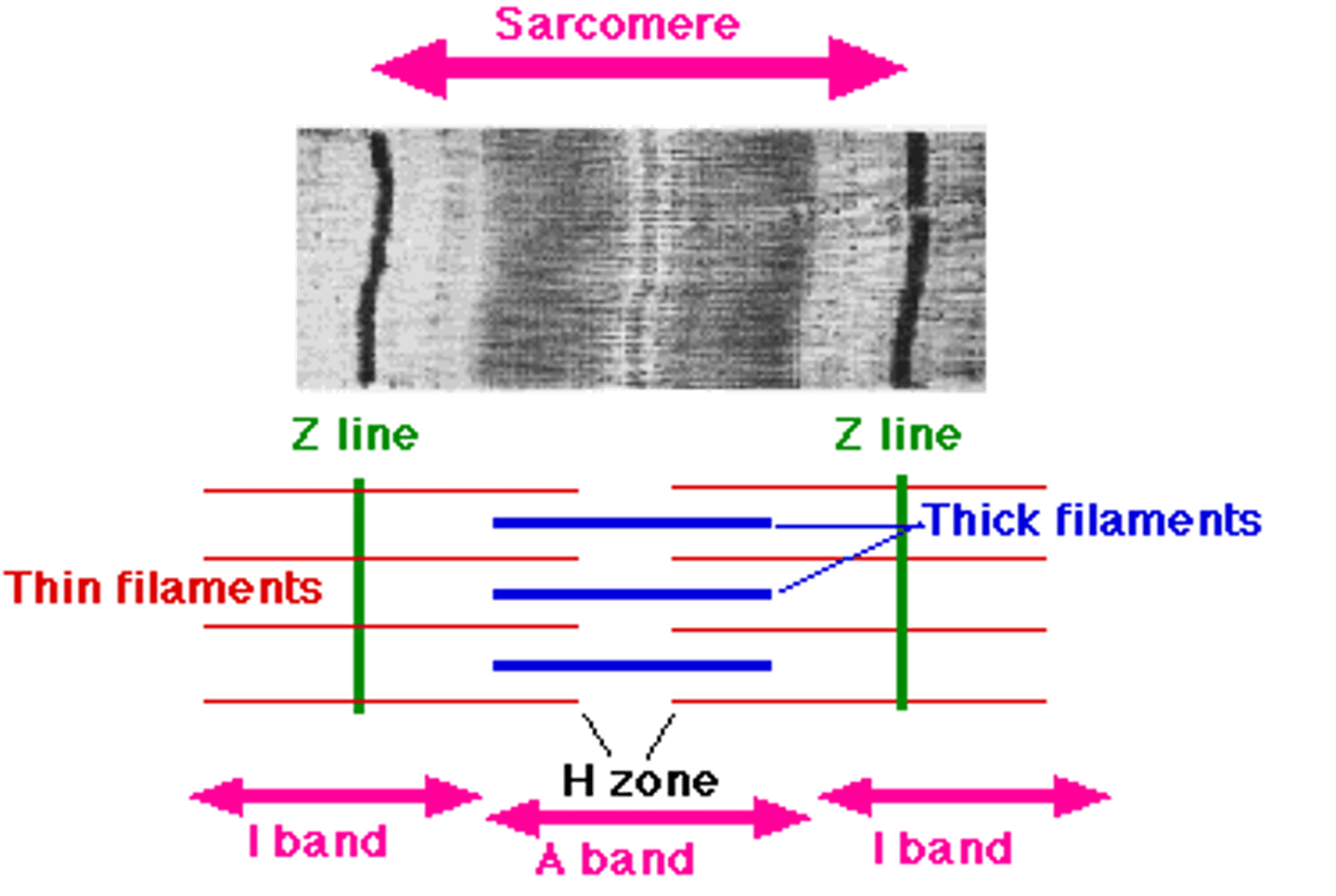
Sarcomere
Contractile unit of muscle
Z line to z line defines sarcomere
Myosin head pulls actin fibers together
Fast glycolytic fibers
Fast twitch
Type II
Large motor units
High innervation ratio
Easily fatigued
Slow oxidative fibers
Small motor unit
Low innervation ratio
Fatigue resistant
Type I
Muscle morphology
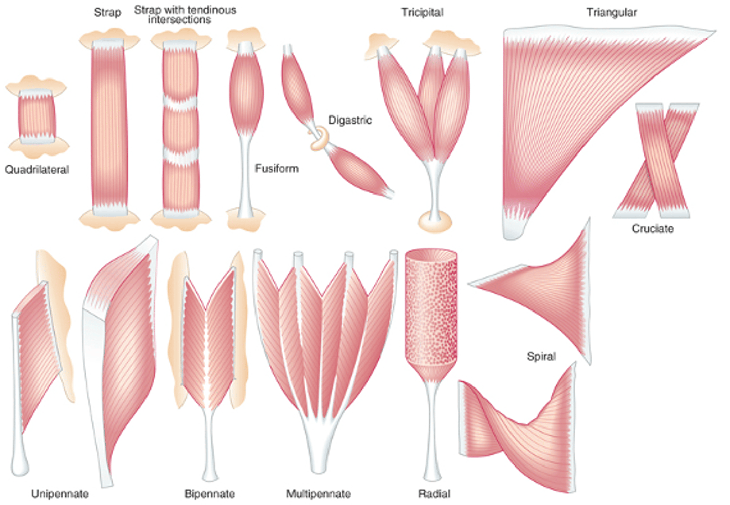
Fusiform
Muscle fibers parrellel to each other and the tendon
Pennate
Muscle fibers attach to tendon at angle
Pennation angle
Angle of orientation between muscle fibers and tendon
affects the force production capabilities as well as the range of motion of a muscle
Pennation angle - 0 degree = 100% force transmission
Pennation angle - 30 degrees = 87% force transmission
Cross-sectional area
Amount of muscle available to generate active force
Titin
A series elastic component protein responsible for allowing the sarcomere to stretch and recoil
Attach myosin to tendon
Titin & Tendon
Series elastic components
Muscle contractions starts with
Myosin pulling actin
Cross bridge
Z-line shortens
Sarcomere shortens
Pulling on titin
Elongation of muscle tendon during contraction
Parellel elastic components
Wraps around muscle fibers
Not stretched in contraction
Does not have active tension during contraction
Series elastic component
Will be stretched during contraction
Titin
Tendons
Contractile (active) components
Actin
Myosin
Non-contractile (passive) components
Series elastic :
Tendon & Titin
Parallel elastic:
Extracellular connective tissue
(epimysium, perimysium, endomysium)
Structural proteins
Internal forces
Active forces
Active movement
External forces
Passive forces
-Stretch of connective tissue
Passive movement
Passive forces
-Pull of gravity
-Push from person
80-120% length of muscle
Optimal range for contraction
100% generates most force
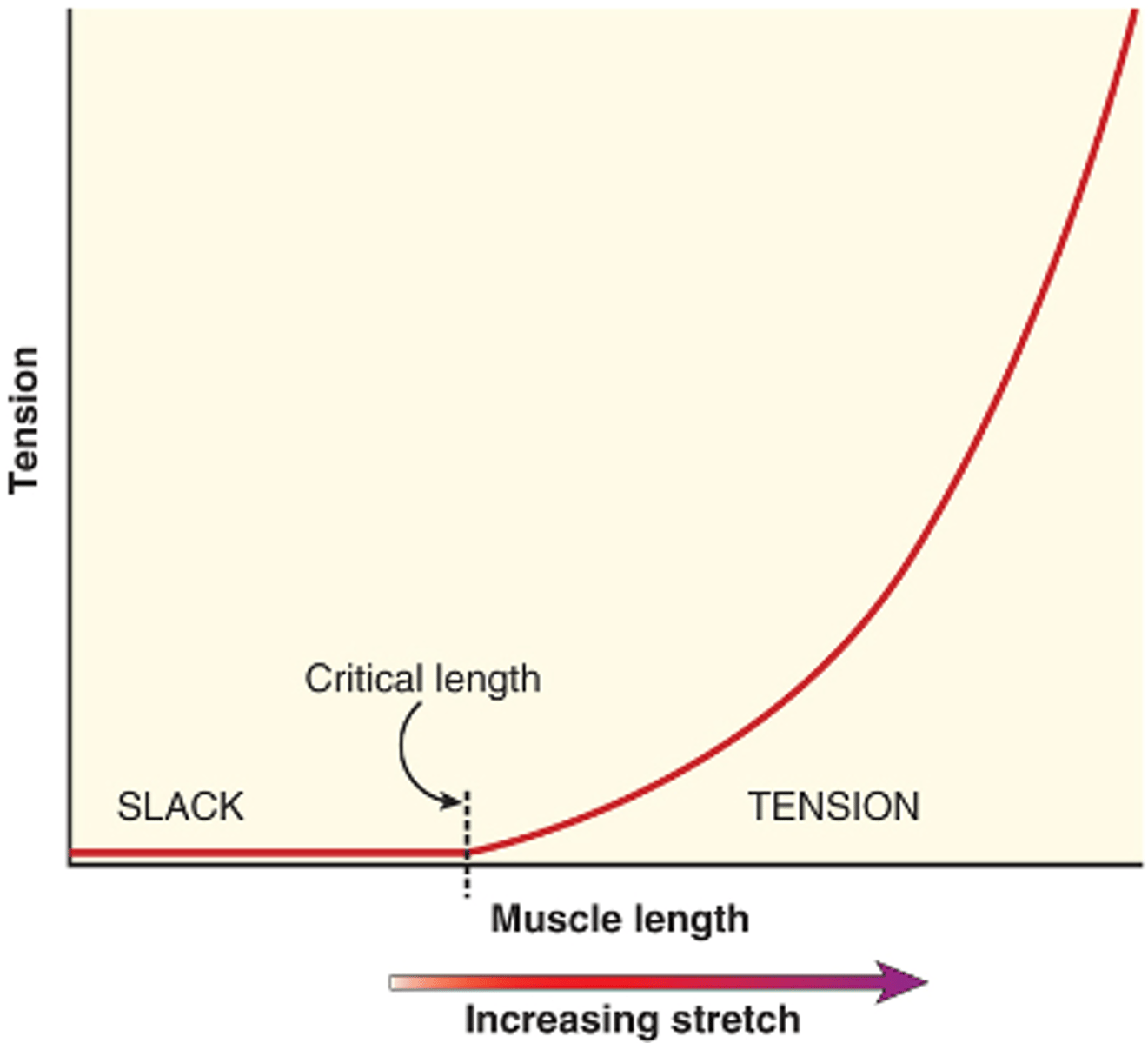
Passive-length tension
as a muscle is progressively stretched, the tissue is slack during the muscle's initial shortened length until it reaches a critical length at which it begins to generate passive tension.
beyond this critical length, the tension builds as an exponential function
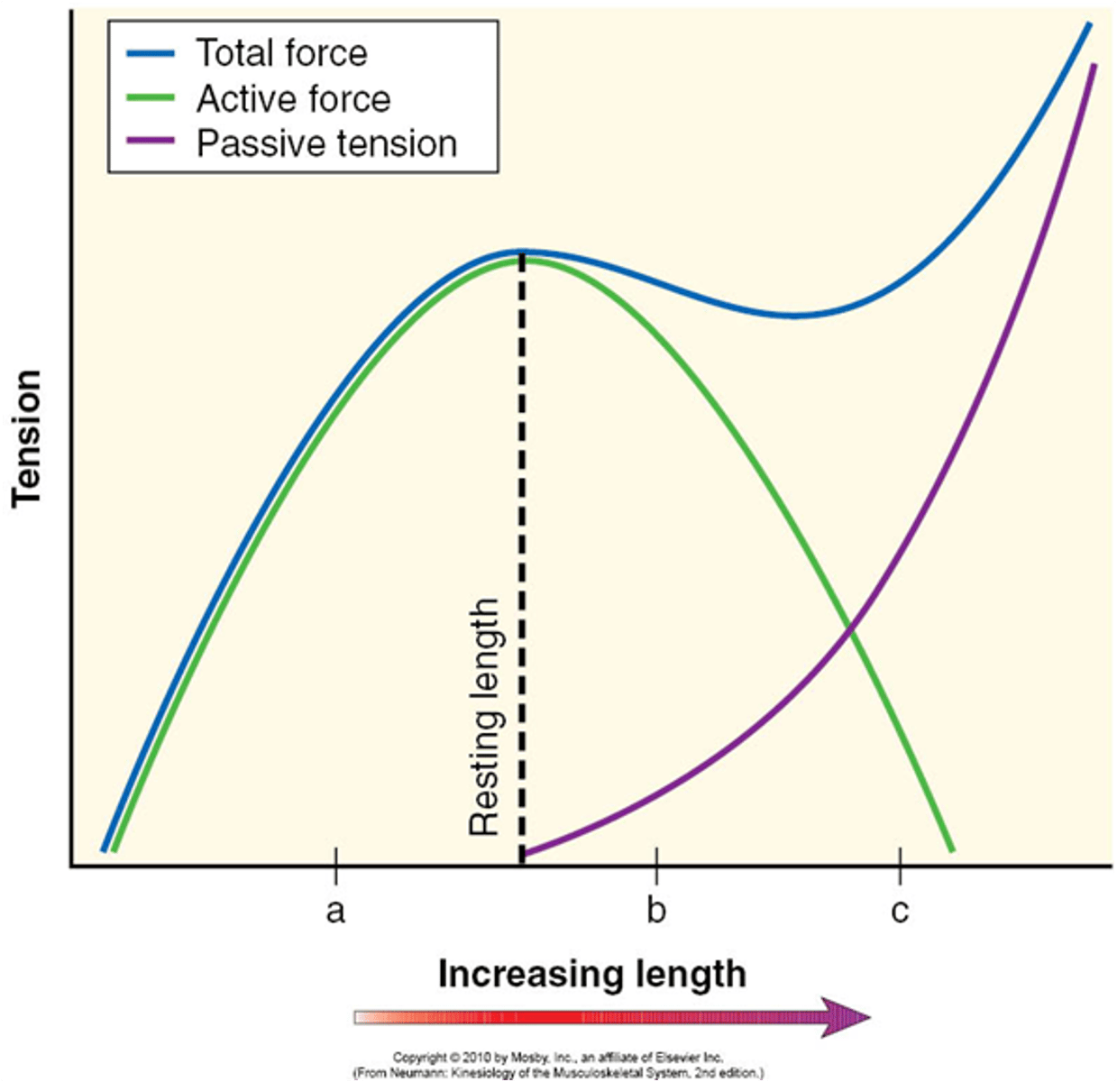
Total-length tension curve
Below resting length, active force is the only contributor to tension
After resting length, passive force contributes to tension
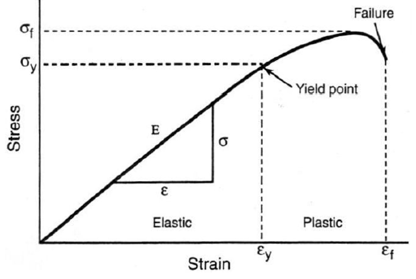
Elastic region and plastic region
Stress-strain relationship
Elastic region - physiological range
Tension increases linearly
Yield point separates elastic and plastic regions
Plastic region - Approaches failure point
Greater slope = greater stiffness
Strain %
deformation / original length
To increase range of motion, body part must stretched in...
Pastic region
Agonist
Directly related to the initiation and execution of a particular movement
Antagonist
Have the opposite action of a particular agonist
Synergists
All the muscles involved in the execution of a particular movement
No change in muscle length
No work
Shortening of muscle length
Concentric contraction
Positive work
Agonist is activated
Move in direction of internal force
Lengthening of muscle length
Eccentric contraction
Negative work
Antagonist activated
Move in direction of external force
Concentric elbox extension
Agonist - Elbow extensors
Antagonist - Elbow flexors
Activated muscles - Elbow flexors
Force output by contraction
1) Eccentric
2) Isometric
3) Concentric
Faster eccentric > slower eccentric
Faster concentric < slower concentric
Active insufficiency
A muscle crosses two or more joints is unable to produce effective tension at all joints at the same time
Failure to produce force when slack
active insufficiency examples
Flexor Digitorum Superficialis
•Crossing wrist and joints in the hand anteriorly
•Shortened in wrist flexion
•Shortened in finger flexion
Unable to form a tight fist with wrist fully flexed
active insufficiency examples
Crossing knee and ankle posteriorly
Shortened in knee flexion
Shortened in plantar flexion
Unable to produce sufficient plantar flexion with knee flexed
Also unable to produce sufficient knee flexion with dorsiflexion
passive insufficiency
A muscle crosses two (or more joints) is unable to stretch enough to allow full range of motion at both (or all) joints at the same time
Range of motion is limited by muscle length of a two-joint muscle
passive insufficiency examples
Finger flexors cross wrist and joints in the hand anteriorly
Stretched during wrist extension
Stretched during finger extension
Wrist extension is limited by finger extension
Finger extension is limited by wrist extension
Series elastic fibers
Tight in both concentric and eccentric contractions
Parallel elastic fibers
Only tight in eccentric contractions
Vector
Magnitude
Spatial orientation
Direction
Point of application
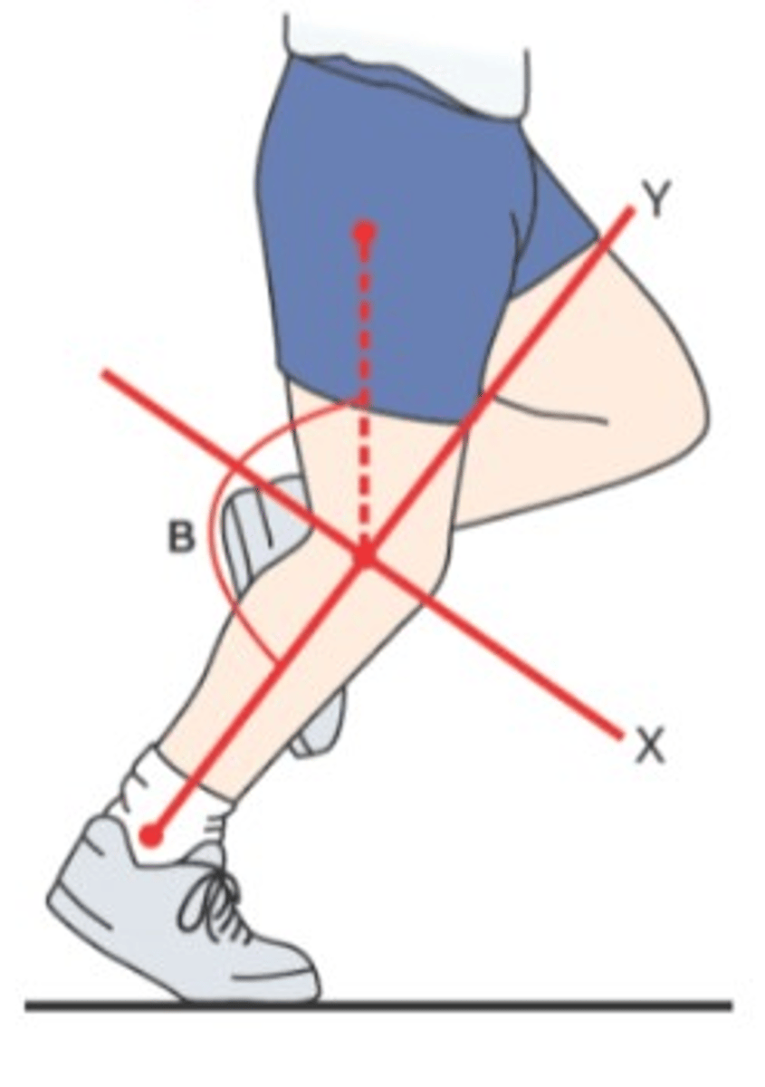
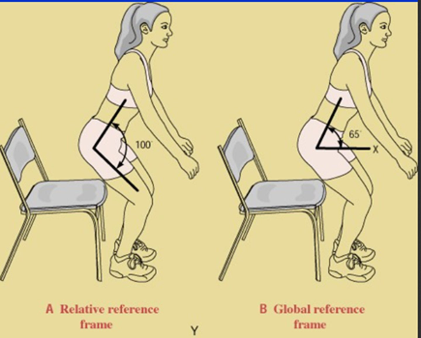
Relative reference frame
Specific joint is being considered
XY axis can be tilted
Global reference frame
XY axis not tilted
Free body diagram steps
•Step I: Identify and isolate the free body under consideration
•Step II: Establish a coordinate reference frame
•Step III: Draw the internal (muscular) and external forces that act on the system
•Step IV: Draw the joint reaction force
•Step V: Write the governing equations of motion
Internal forces in free body diagram
Muscle forces within diagram or muscle itself
External forces in free body diagram
Forces acting on the body
Including weight because it is a component of gravity
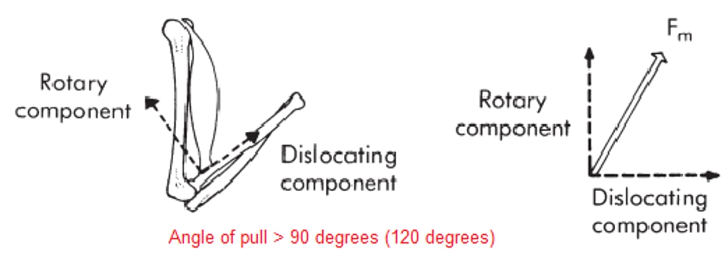
Dislocating component
Move away from the joint
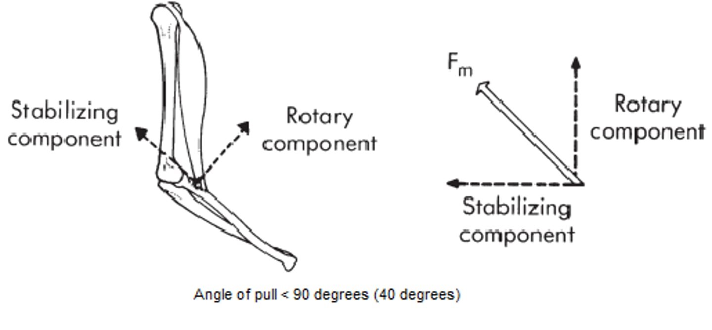
Stabilizing component
Move into the joint
Joint-stabilizing force
Counteracts the muscle force
Load
Tension- Pul apart
Compression- Push together
Bending- Tension on convex side, compression on concave side
Shear- Displaced in opposite directions on top of each other
Torsion- Twist
Force
A push or a pull
Characterized by magnitude, direction, and point of application
F = ma
Unit is the Newton (N)
Torque
The rotary effect of a force
Torque = Force (N) × Moment arm (m)
T = Fd
Unit: Nm
Moment arm: perpendicular distance from the force's line of action to the axis of rotation
When drawing moment arm
Trying to connect axis and rotation to force line
When force passes over moment arm
No torque is produced
Isometric
No change of muscle length
No work
Internal Torque = External Torque
Concentric
Shortening of muscle length
Positive work
Internal Torque > External Torque
Direction of motion is the same with the direction of internal torque
Agonist is activated
Eccentric
Lengthening of muscle length
Negative work
Internal Torque < External Torque
Direction of motion is opposite to the direction of internal torque
Antagonist is activated
Torque generates angular acceleration (α)
Linear - F=ma
Angular - T=lα
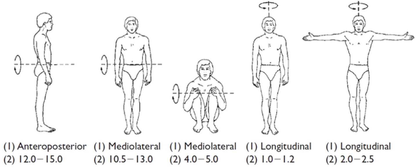
Mass moment of inertia (I)
The inertial property for rotating bodies represents resistance to angular acceleration based on both mass and the distance the mass is distributed from the axis of rotation
I = mk^2
I= moment of inertia
m= mass
k= radius of gyration
Radius of gyration (k)
The distance from the axis of rotation to a point where the total mass of the body is supposed to be concentrated
Law of inertia
A body will maintain a state of rest or constant velocity unless acted on by an external force that changes the state
Static equilibrium - Not moving
dynamic equilibrium -Same direction & velocity
Net force/net torque is zero
Law of acceleration
F = ma