Physiology 2130 Unit 2: Excitable Cells and Synaptic Transmission
1/119
There's no tags or description
Looks like no tags are added yet.
Name | Mastery | Learn | Test | Matching | Spaced | Call with Kai |
|---|
No analytics yet
Send a link to your students to track their progress
120 Terms
excitable cell
cell use its resting membrane potential (RMP) to generate action poteitnal
action potential = electrochemical impulse
excitable cell VS non-excitable cell
non-excitable cell = X generate action potentials
EX → excitable cells = neurons, muscles, some endocrine
EX → rest of body non excitable
What is an action potential?
rapid electrical signal generated when an excitable cell depolarizes beyond threshold
occur when open voltage gated ion channel
all or nothing response → same magnitude
How do excitable cells communicate through action potentials?
communicate by generating and propagating action potentials along the neuron
occur w depolarization events in cell if enough depolarization occurs & excitable cells fire to comm. w adjacent cells
What is depolarization?
process by which ions move in and out of the cell
GOAL = inside of the cell INC positive relative to the resting membrane potential
occur cell constant but only when beyond -55mV, AP trigger
what is ion movement is controlled by?
controlled by membrane proteins such as channels and pumps on membrane
chemically/ligand gated & voltage gated channels help movement of ions in & out of cell
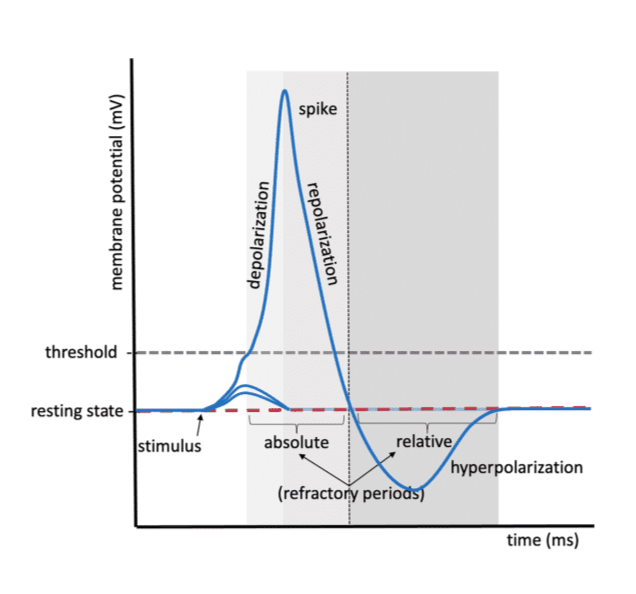
What are the 6 main components of an action potential?
stimulus & RMP
trigger VV depolarization events in excitable cell → inside cell INC +
RMP ~ −70 mV
K⁺ leak channels leaky & X fully closed at rest
voltage gated Na channel & chem gated K channel closed
Threshold
stimulus reaches approximately −55 mV
AP triggered
Depolarization
Voltage-gated Na⁺ channels open
Na⁺ enters cell → inside INC (+) → INC K move out of cell to counteract
membrane potential -70mV to -30mV
leaky K channel open
chem gated K channel closed → need chem bind to open
Repolarization
Voltage-gated K⁺ channels open
K⁺ leaves cell
voltage gated Na⁺ channels close
Hyperpolarization
cell INC (-) than RMP = DEC chance AP occur
Relative refractory period occurs → harder to do AP
chem gated K+ channel open → INC K+ leave cell
voltage gated Na+ channel closed → bc open voltage goated K+ channel
all channels move K+ out of cell
Return to resting membrane potential & resting state
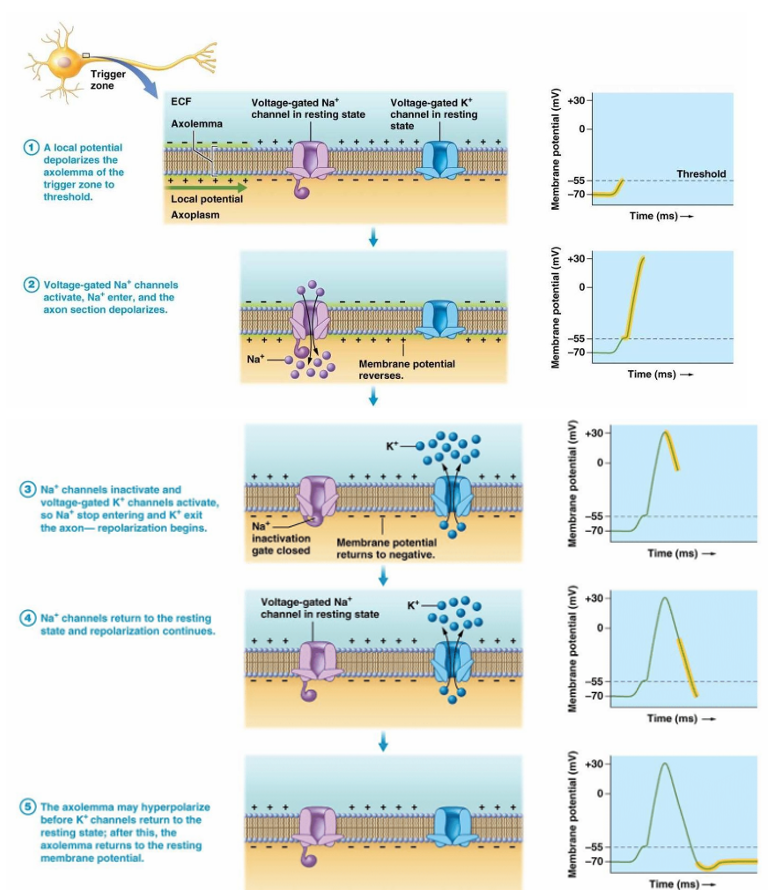
example of Na+ & K+ channel AP
local potential depolarizes trigger zone’s axolemma to threshold of -55mV
voltage gated Na+ channel active → Na+ enters, axon section depolarizes
Na+ channel inactive & voltage gated K+ channel actives
Na+ X enter
K+ excite axon → repolarize
Na+ channel returns resting state, repolarize cont.
axolemma can hyperpolarize before K+ channel becomes resting state
then return RMP
During hyperpolarization, channels are more selectively permeable to K than leak channels. True / False
True
What happens to the channels during the resting potential and what are they more permeable to
The leak channels open at rest, 20-25 times more permeable to K+ than to N+
What happens to the channels during the depolarization
The channels are selectively permeable to N+ than to K+
Failed initiations
The depolarization events below threshold
Why are voltage-gated Na+ and K+ channels called "voltage-gated"?
b/c it is a change in voltage that triggers their opening
relative VS absolute refractory period
refractory period = time frame after neuron makes AP & X able to fire
absolute = X stimuli makes AP
relative = another AP possible but need INC strong stimuli
inside cell = INC (-) & harder to reach threshold
what forms the absolute refractory period of the AP?
Depolarization and repolarization phases
Na channel inactive & X reopen until membrane repolarized enough
During this time, no AP can be elicited → ensure 1 direction AP travel
An AP can be generated during the relative refractory period T/F?
T, but a larger intensity stimulus would be required to produce an AP because the membrane is hyperpolarized
refractory period = cell is more negative, reaching approximately -90 mV
now more difficult to reach the threshold of -55M = INC stimulus needed to reach the threshold bc of how (-) cell is
Because of the absolute refractory period during which time the Na+ voltage-gated channels are closed, two APs cannot be fired one on top of the other. True/ False?
True
how do the channels change during a AP?
Na+ channels active = INC in membrane potential & start of AP
K+ channels help membrane repolarize
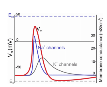
Why does the closing of the potassium channels cause the inside of the membrane to become more positive?
closing of potassium channels slows the outward flow of K⁺
cause the inside of the membrane to become less negative (or slightly more positive) before it fully returns to the resting potential
neurons
excitable cells
comm. w AP
structure
Soma (cell body)
Dendrites
Axon
Axon terminals
Myelin sheath
Schwann cells
Nodes of Ranvier
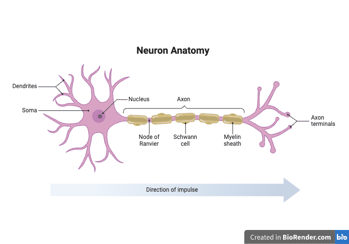
Soma
(cell body)
has nucleus & most organelles
Dendrites
branch-like projections from soma
get signals & info from other neurons → soma
direct AP → soma
Axon
projections of cell body
AP AWAY from soma
Axon terminals
ends of axon
Release neurotransmitters to communicate with next cell
Myelin sheath
Fatty acid & protein insulating layer surrounding the axon
speeds signal transmission
myelin
rapid move of APs thru saltatory conduction thru axon
prevent decay of AP when travel along axon
Schwann cells
Produce myelin and support neuron survival in the PNS
cell surround axon
Nodes of Ranvier
-Gaps in the myelin sheath rich in ion channels that aid rapid AP propagation
-Unmyelinated axon membrane
What is the direction in which an action potential propagates?
Dendrites → Soma → Axon → Axon terminals
DSAAT
saltatory conduction
occurs in myelinated neurons
AP jumps from one Node of Ranvier to the next instead of traveling continuously along every section of membrane
Advantages of saltatory conduction
INC transmission speed by 10-15 times compared with unmyelinated neurons
INC efficiency
rapid communication over long distances
conserve E because fewer ions cross the membrane
What is the all-or-nothing principle of an action potential?
if membrane depolarization reaches threshold (~−55 mV) → an action potential occurs
if threshold is not reached → no action potential occurs
action potentials always same amplitude
What determines the direction of the propagation of an action potential?
The direction is determined by the refractory periods, especially the absolute refractory period.
What happens during the absolute refractory period?
Voltage-gated Na⁺ channels become inactive
Another action potential cannot immediately occur in the area that just fired
Because the membrane behind the AP cannot fire again immediately, the signal moves forward only, preventing backward propagation.
Another AP cannot be elicited while the previous one is in the absolute refractory period. Why?
Because the ion channels are inactive during this time.
The relative refractory period (or hyperpolarization phase) makes the membrane more negative relative to the resting potential. T/F?
T, As a consequence, it is harder to reach threshold. The depolarization of the membrane will ONLY move in one direction
The AP only travels in one direction due to the absolute refractory period in only myelinated neurons. T/F?
False, both myelinated and unmyelinated neurons.
propagation of AP
propagate 1 direction in neuron
dendrite → soma → axon → axon terminals
neurotransmitter released from presynaptic neuron
bine to ion channel in postsynaptic cell & depolarize
depolarize = inside INC + than RMP & INC chance AP occur bc + inside closer to -55mV
signal propagates to soma
speed vary if axon myelinated or unmyelinated
how is AP propagation unidirectional?
bc refractory period & another AP X happen when previous one in absolute refractory period
ion channels inactive
only move in 1 direction towards axon terminals from soma
BUT during relative refractory period/hyperpolarization, membrane INC (-) can possible but INC force needed bc harder to reach threshold
what cells are in the brain?
neurons → info transmit & process for body
gilal cells → make enviro for neuron f(x)
What are glial cells?
neuroglia
support cells of the nervous system
provide the environment necessary for neurons to function properly
~ 90% of the brain
Unlike neurons, gilal cells do not primarily transmit electrical signals. T/F?
T
Glial roles include:
Support
Protection
Nutrient delivery
Insulation (myelin production)
Maintenance of neuronal environment
What are different types of neurons present in the brain?
for mammals
Bipolar neurons
Unipolar neurons
Multipolar neurons
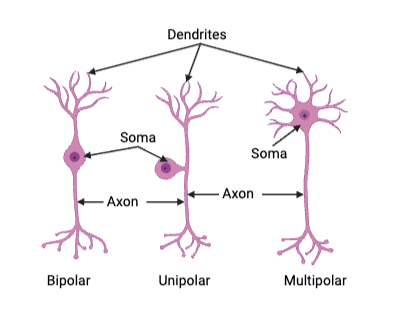
Bipolar neurons
1 axon + 1 dendrite w branches (2 processes extend from cell body)
mainly in specialized sensory structures such as the retina
Unipolar neurons
1 process extending from the cell body
straight connect axon & dendrite → soma separate on side
mainly sensory neurons in the PNS → send sinals to & from spinal cord
Multipolar neurons
1 axon with many dendrites
most common neuron type in the CNS & connect CNS w effector organs
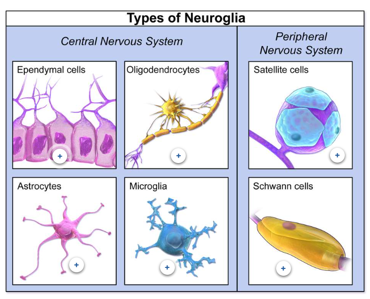
What are 6 examples of glial cells?
CNS →
Astrocytes
Oligodendrocytes
Ependymal cells
Microglia
PNS →
Schwann cells
Satellite cells
Astrocytes (Astrocytes are the most abundant cells in the brain.)
#1 in brain
star shape
Physical and nutritional support
Transport nutrient to neurons
Hold neurons in place
Remove debris
Digest dead neurons
Regulate extracellular environment
Promote synaptic connections
Participate in injury response
Schwann cells
gilal cells of PNS
surrond neruons & keep alive by cover w mylein
work for dvlp, maintain, f(x) & regen peripheral nerves
Oligodendrocytes
Produce myelin in CNS → layered phospholipid membrane support & insulate axon
1 cell can myelinate several axons
Ependymal cells
line ventricles of brain & spinal cord
Regulate ion and glucose movement
Help distribute hormones and signal molecules associated with the CNS
shape of cuboidal w cillia & microvilli used to circulate & make cerebrospinal fluid (CSF)
Microglia
immune defense cells → dynamic move to look invaders
emove damaged tissue and pathogens → engulf
small cells sparsely located
remove previously formed synapses X needed
Satellite cells
Support neurons in the PNS
Provide nutrients and structural support → bundle axons together & stop from touching
like astrocytes of CNS
Multiple Sclerosis (MS)
autoimmune disease → 2X W get
can stop natural flow AP & X transmission occur
progressive disease of CNS → X cure
chronic inflame response for myelin sheath & immune system attack them around axons
EFFECTS
myelin damage slows or blocks action potential transmission
Communication between neurons becomes impaired
Muscles may fail to receive signals
lead to weakness or paralysis → if nerve damaged connected to muscle & X contract
nervous system formation
central nervous system (CNS)
brain
spinal cord
peripheral nervous system (PHS)
somatomotor → voluntary w skeletal
autonomic → automatic w organs & control brain
nerves go from CNS to muscles & organs
central nervous system (CNS)
Brain + Spinal cord
Main function:
integrates and processes information
Coordinates responses and body functions
Peripheral Nervous System (PNS)
nerves connecting the CNS to the rest of the body
carry signals between organs and the CNS
(1) Somatomotor system
voluntary w skeletal
(2) Autonomic nervous system
automatic involuntary w organs & control brain
Compare the central and peripheral nervous system
Comparison: Both systems communicate through neurons and action potentials, but the CNS mainly processes information while the PNS transmits it.
what are the anatomical and functional structures of the brain?
2 cerebral hemispheres → L & R
contralateral control → L control R, R control L
control muscles, sensory info
gyri = bumps on brain
sulci = dips/valley
4 lobes
frontal
temporal
parietal
occipital
what is the use of gyri and sulci?
INC SA of brain
landmarks divide cerebral hemispheres into lobes (4)
Frontal lobe
planning & perception of stimuli
(1) Primary motor cortex → process input from skeletal muscles
(2) Premotor cortex → motor association area
work w/ prefrontal cortex → integrates info abt movement w other sensory input to make perception of stimuli
(3) Prefrontal cortex
Temporal lobe
olfaction
short term memory → mediate storage & recall
sound
get & process signals from auditory nerve & integrate w other sensory input
(1) primary auditory cortex
(2) auditory association area
Parietal lobe
touch and sensory integration
(1) primary somatosensory cortex → get input from major senses
EX →
(2) somatosensory association areas → intergrate sensory info w other association areas
Occipital lobe
vision & visual processing
(1) primary visual cortex → input from optic nerve
(2) visual association area → process visual info & integrate w other sensory information
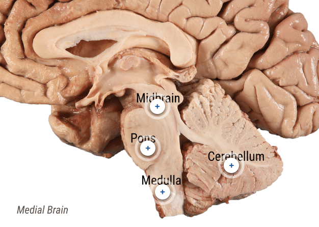
Cerebellum
posterior → under occipital lobe & above brain stem
ROLES →
process sensory info to
coordinate moves
#1 # of neurons in the brain → get input for many things
somatic receptors
receptors for equilibrium
balance and motor neurons from higher centers of the brain

Brain stem
controls some basic functions of body → heart rate & respiration
includes 9 cranial nerves
formation → midbrain, pons, medulla oblongata
medulla is continuous to the spinal cord
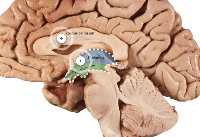
Corpus callosum
dense bundle nerve fibers
path & connect 2 cerebral hemispheres → help integrate sensory & motor info both sides & coordinate whole body movement & f(x)

diencephalon
(1) thalamus → get sensory input from spinal cord & integrates before send to cortex
(2) hypothalamus → regulates endocrine function w hormone release
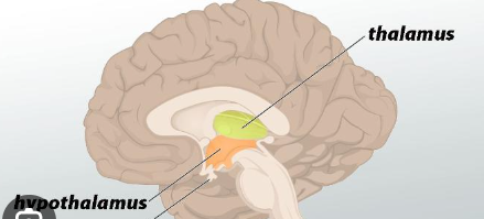
Thalamus
get sensory input, process & integrate sensory info BEFORE send to cortex
get info as it travels from the spinal cord

Hypothalamus
USES →
regulates endocrine functions w hormone release
regulate homeostasis systems
temp
body water
hunger/ food intake
cardiovascular
circadian clock
emotions
thirst
ANATOMY →
base of brain, anterior brain stem, under thalamus
use negative feedback system
stimuli trigger homeostasis change
sensor see info
control center sends info →
effector
mechanism (effector also uses this to send info back to stimuli integration center to regulate signaling)
Midbrain
mesencephalon
bridge lower brainstem & upper diencephalon
f(x)
eye movement
visual and auditory reflexes

Pons
USE → relay station w cerebellum & cerebral cortex
regulate breathing w medulla
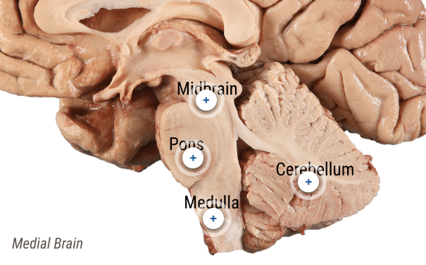
Medulla
main control 4 involuntary f(x)
breathing
blood pressure
swallowing
hear rate
corticospinal track fibers from motor cortex cross to opp sides of spinal cord →innervate muscles on opp sides

pituitary gland/hypophyse
controlled by hypothalamus
diff parts secrete diff hormones & diff anatomy
USE →
regulates endocrine organs
hormones secret differ based on each section
EX → stress, lactation, growth, dvpmt, rpxdtn
anterior pituitary → from epithelial tissue of pharynx
posterior pituitary → from neural tissue of hypothalamus
Hormones
chemicals cells use to comm over "long-distance" w blood stream
info → growth, stress, development, homeostasis regulation from higher integration centers to effector organs
EX→ skin, muscles and other tissues
Because myelin is required for fast saltatory conduction, damage significantly impairs communication throughout the nervous system. T/F?
T
The premotor cortex (motor association area) works with the prefrontal cortex to integrate movement information with other sensory inputs to generate perception (or interpretation) of stimuli. T/F?
T
synapse
where impulses passed by neurons to comm. w cells
(1) electrical
(2) chemical → presynaptic, synaptic clef, post synaptic neruon
electrical synapse
site of cell to cell comm
neurons directly exchange ions w channels → create AP in next cell
channels = 2 communicating cell long
chemical synapse
site cell to cell comm. w excitable cell release neurotransmitter to comm.
2 neurons X have channel → separated by synaptic cleft
components
presynaptic neuron
synaptic clef
post synaptic neuron
neurotransmitter
chem. released by neuron @ axon terminals
GOAL = comm. w other neurons
PROCESS
synthesized & stored w synaptic vesicles
when released w/ AP, diffuse synaptic cleft
bind to receptor/ ion channels on post synaptic cleft → ion influx in the cell
binding neurotransmitter to channel = electrical impulses that are EPSP, or IPSP
presynaptic neuron
transmit info → synaptic cleft (w axon & axon terminals) → dendrites next neuron
synaptic cleft
small space btwn axon terminals of 1 neuron & dendrites another
area where neurotransmitters released
post synaptic neuron
transmit info ← synaptic cleft from dendrites & toward soma
what is the exact process of synaptic transmission?
AP @ axon terminal → depolarizes pre-synaptic membrane
Voltage-gated Ca2+ channels open
voltage change by AP = channel open & Ca2+ enter
on synapse, membrane axon terminal
Ca2+ enters cell
trigger biochemical rxn w release neurotransmitter
synaptic vesicles fuse w pre-synaptic membrane w exocytosis
neurotransmitters released from synaptic vesicle → synaptic cleft
bind to receptors on the post-synaptic membrane → diffuse out of synapse down [gradients]
break down by enzymes on synaptic cleft→ re-uptake into pre-synaptic cell to be recycled
neurotransmitters bind = open ligand-gated receptors on post-synaptic membrane
ion channels OR trigger events that open ion channels
RESULT → graded potentials
neurotransmitter bind = receptors de/hyper polarization post-synaptic cell (based on which channel opens)
![<ol><li><p>AP @ axon terminal → depolarizes pre-synaptic membrane</p></li><li><p>Voltage-gated Ca<sup>2+</sup> channels open</p><ul><li><p>voltage change by AP = channel open & Ca2+ enter</p></li><li><p>on synapse, membrane axon terminal</p></li></ul></li><li><p>Ca<sup>2+</sup> enters cell</p><ul><li><p>trigger biochemical rxn w release neurotransmitter</p></li><li><p>synaptic vesicles fuse w pre-synaptic membrane w exocytosis</p></li></ul></li><li><p>neurotransmitters released from synaptic vesicle → synaptic cleft</p><ol><li><p>bind to receptors on the post-synaptic membrane → diffuse out of synapse down [gradients]</p></li><li><p>break down by enzymes on synaptic cleft→ re-uptake into pre-synaptic cell to be recycled</p></li></ol></li><li><p>neurotransmitters bind = open ligand-gated receptors on post-synaptic membrane</p><ul><li><p>ion channels OR trigger events that open ion channels</p></li><li><p>RESULT → graded potentials</p></li></ul></li><li><p>neurotransmitter bind = receptors de/hyper polarization post-synaptic cell (based on which channel opens)</p></li></ol><p></p>](https://assets.knowt.com/user-attachments/38e0cd2b-b070-4666-b3d3-0af6fe39d586.png)
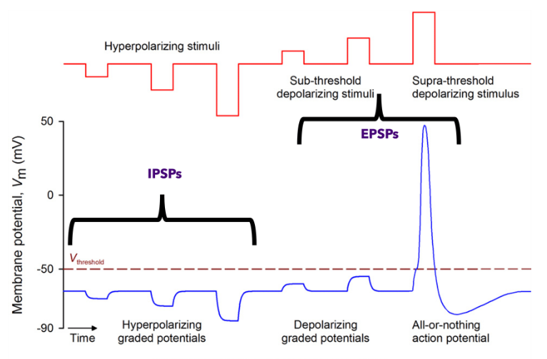
graded potential
small localized subthreshold depolarizations of membrane
diff size occur & amts stack up → based on stimuli magnitude
made by opening ligand gated ion channels
decay when farther from stimulation site
X make AP
(1) EPSP (depolarizing potentials)
(2) IPSP (hyperpolarizing potentials)
excitatory post synaptic potential (EPSPs)
X make AP
localized → depolarization 1 area on membrane
summed → stack to make INC depolarization & INC # bring closer to AP
graded → INC stimuli = INC depolarization
decay → when propagate across membrane (INC far depolarization from stimuli = smaller)
occur when neurotransmitter:
(1) open K+ channel → move out cell = inside INC (-) & depolarization occur
mainly Na move in & little K move out, both needed bc EPSP use non selective cation channel
(2) open Na+ channel → move in cell = inside INC (+) & depolarization occur
inhibitory post synaptic potential (IPSPs)
localized → hyperpolarized on 1 area membrane
graded → INC stimuli = INC hyperpolarization
summed → stack to make INC hyperpolarization & INC # farther from AP
decay → when propagate across membrane (INC far depolarization from stimuli = smaller)
decay → when propagate across membrane (INC far depolarization from stimuli = smaller)
occur when neurotransmitter:
(1) open K+ channel → move out cell = inside INC (-) & hyperpolarization occur
(2) open Cl- channel → move in cell = inside INC (-) & hyperpolarization occur
EPSP vs IPSP
EPSP → depolarize below threshold & INC inside (+)
turn on neuron
IPSP → hyperpolarize below RMP & INC inside (-) than RMP
shut off neuron
size of stimuli = INC change membrane potential
both graded potential b/c size based on stimuli
how do EPSP & IPSP affect AP production?
@ 1 time → 1 post synaptic cell get many inputs (EPSP or IPSP) from many presynaptic cells
vary based on →
(1) type of presynaptic neuron synapsed onto post synaptic neuron
(2) type of neurotransmitter released
AP formed based on sum of IPSP & EPSP when get to axon hillock
AP = all or nothing so need to reach threshold to occur
IMAGE
(1) IPSP & EPSP decay as get to axon hillock
(2) IPSP & EPSP summed there
(3) BUT threshold X met here so X AP made
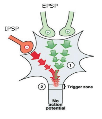
axon hillock
trigger zone that determines if AP will occur
AP generated based on sum IPSP & EPSP @ this location
why does depolarization have to occur at axon hillock?
dendrites & soma & have voltage gated channels → need for AP formation
voltage gated channels occur INC [ ] @ axon hillock & axon membrane
how does decay affect graded potentials ?
farther from site of stimuli = DEC intensity
for enough depolarization need strong & large enough current of EPSP to spread from synpase on axon hillock
use temporal & spatial summation
how do graded potentials activate APs?
use summation to ensure depolarization large & strong enough reach threshold
effect of EPSP & IPSP sun at axon hillock & if above threshold, AP fire
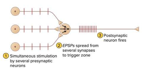
spatial summation
ADD effect
many EPSP made at diff synapses on SAME POSTsynaptic neuron at SAME time
many neurons fire same time
same for IPSP but opp effect → INC hyperpolarization
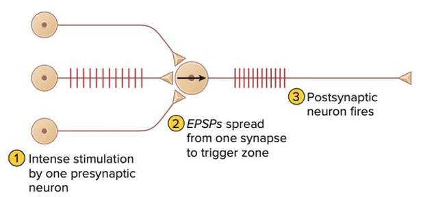
temporal summation
ADD effect
many EPSP made at SAME synapse by many high frequency APs on PREsynaptic neuron
1 neuron fire many times
same for IPSP but opp effect → INC hyperpolarization
what are the types of neurotramitters?
acetylcholine
acetyl choline
biogenic amines
catecholamine → epinephrine, norepinephrine
catecholamine → dopamine
serotonin
amino acids
glutamate & aspartate
GABA & glycine
neuropeptides
endogenous opioids → endorphins
vasoactive intestinal peptides
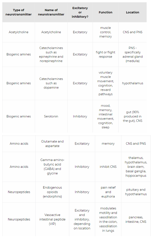
acetylcholine (ACh)
excitatory
CNS & PNS
muscles control & memory → w release @ NMJ
USES →
neurotransmitter @ NMJ
bind to nicotinic receptors in NMJ + autonomic ganglion
bind to muscarinic receptors @ target organ of PSYN
neurotransmitter of autonomic ganglion
epinephrine & norepinephrine
excitatory
biogenic amines → catecholamine
PNS → adrenal gland in medulla
fight/flight response