Pathology eye, +
 Call Kai
 Learn
Learn Practice Test
 Spaced Repetition
 Match
Match Flashcards
 Knowt Play
1/41

Earn XP
Description and Tags
this will be slowly updated with anything that will be part of exam 1. Its mainly going to be about the terms and discribing them and less about the picture because if you can make a good mental image of it them even if it is a different photo on the exam you got this!
Name | Mastery | Learn | Test | Matching | Spaced | Call with Kai | Chat |
|---|
No analytics yet
Send a link to your students to track their progress
42 Terms
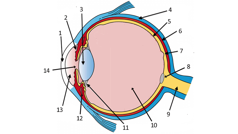
1.cornea 2.filtration angle 3.lens 4.slcera 5.retina 6.choroid 9.optic nerve 10.12.13
lable 1.2.3.4.5.6.9.10.12.13
makes the lipid layer of the tear film. is located within the upper and lower eyelid deepto the eyelashes.
meibomian gland function and location
lipid, aqueous, mucin (mucus)
layers of the tear film (in order from top to bottom)
makes the aqueous layer of the tear film. located in the lateral upper eyelid and lower medial eyelid
lacrimal gland function and location (main gland and 3rd eyelid one)
upper corniculous, nasolacrimal duct, gland of the 3rd eyelid, punctum lacrimal, 3rd eyelid, conjunctival fornix of the lacrimal gland
parts of the lacrimals apparatus
conformational abnormality. when the eyelids roll inwards. Shar-Pei and chow chow
describe entropion (type of abnormality, definition, common breeds affected[ at least 2])
conformational abnormality. drooping/sagging of the eyelids (very loose eyelids), breeds with loose skin, hounds
describe extropion (type of abnormality, definition, common breeds affected[breed description])
conformational abnormality, when an extra row of eyelashes grow out of the meibomian gland ducts, resulting in the eyelash touching the eye, spanials and retrievers
describe distichiasis (type of abnormality, definition, common breeds affected[ at least 2])