OPT 114 (Ocular Anatomy): ORBIT (BONES FORAMINA FISSURES ETC)
1/78
There's no tags or description
Looks like no tags are added yet.
Name | Mastery | Learn | Test | Matching | Spaced | Call with Kai |
|---|
No analytics yet
Send a link to your students to track their progress
79 Terms
Temporal Bones of the Skull
form the sides and base of the cranium
Two parts of the skull
Cranium and face
Sutures
These are the areas where bones meet (look like seams)
Sinuses
Air filled cavities within bones
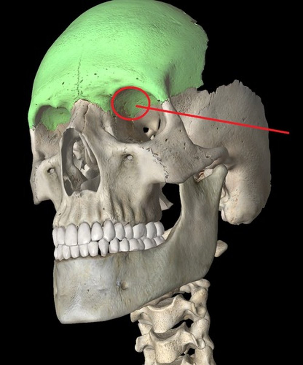
frontal bone of skull
Forms anterior portion of cranium, anterior floor, and superior part of the face
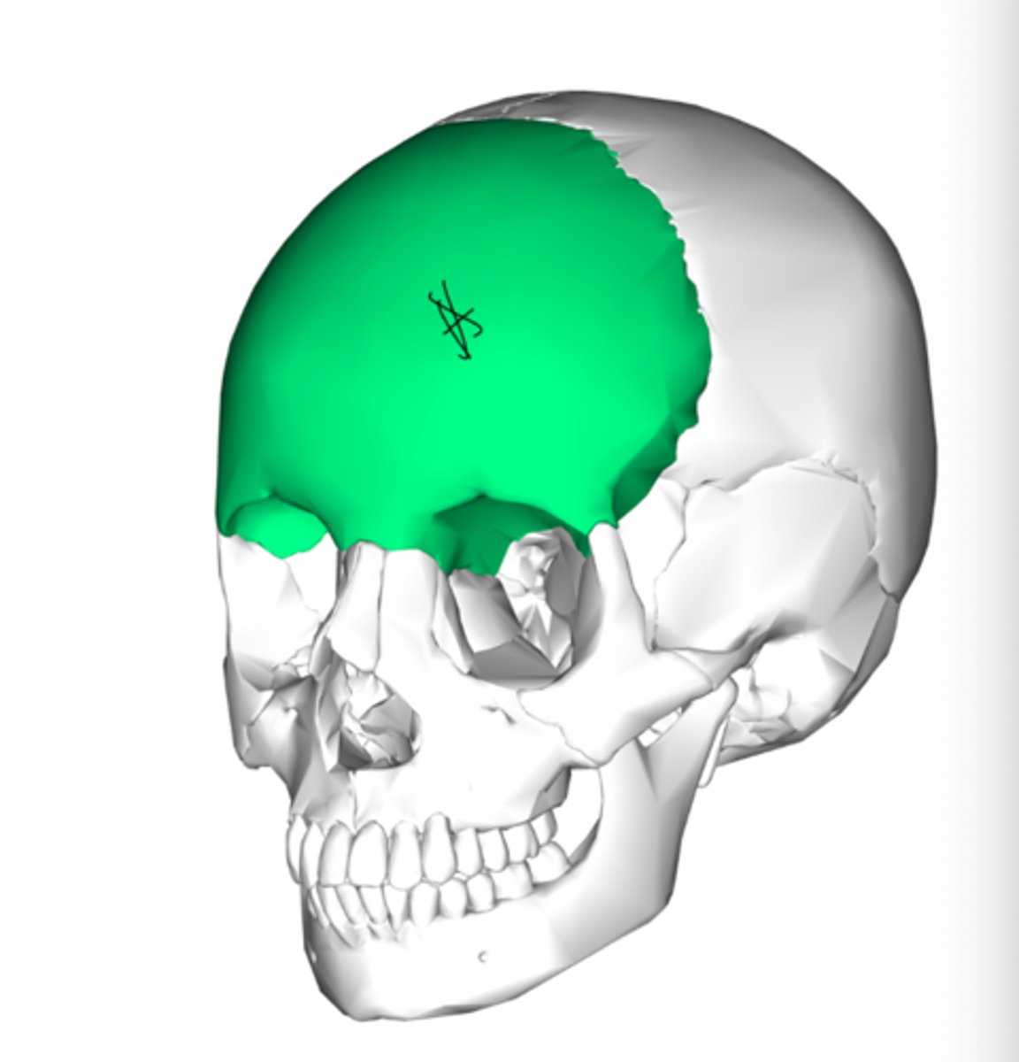
Parietal Bones
Form the roof and lateral sides of the skull
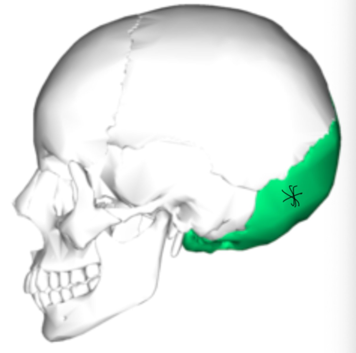
occipital bone (skull)
forms the back part of the skull and the base of the cranium
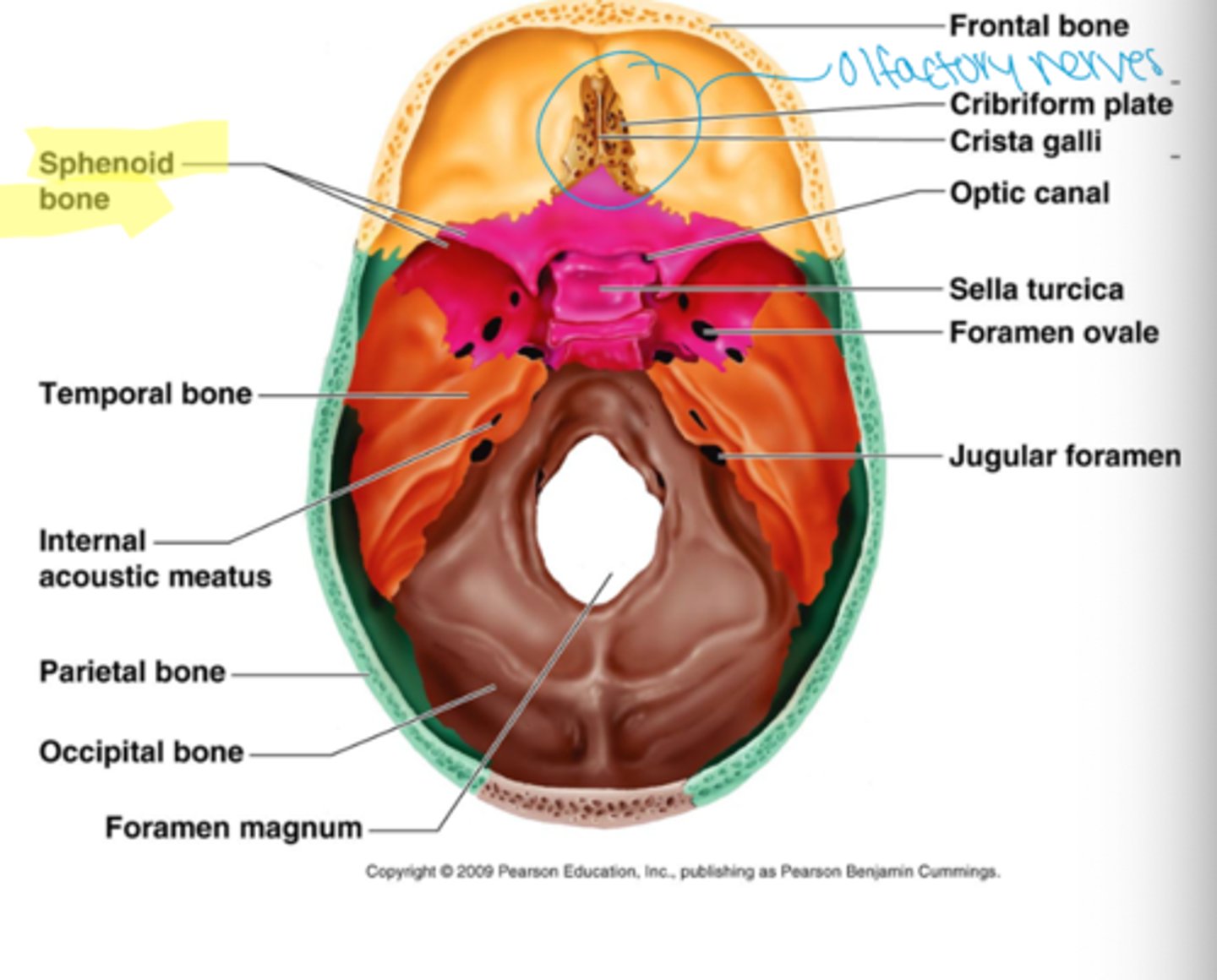
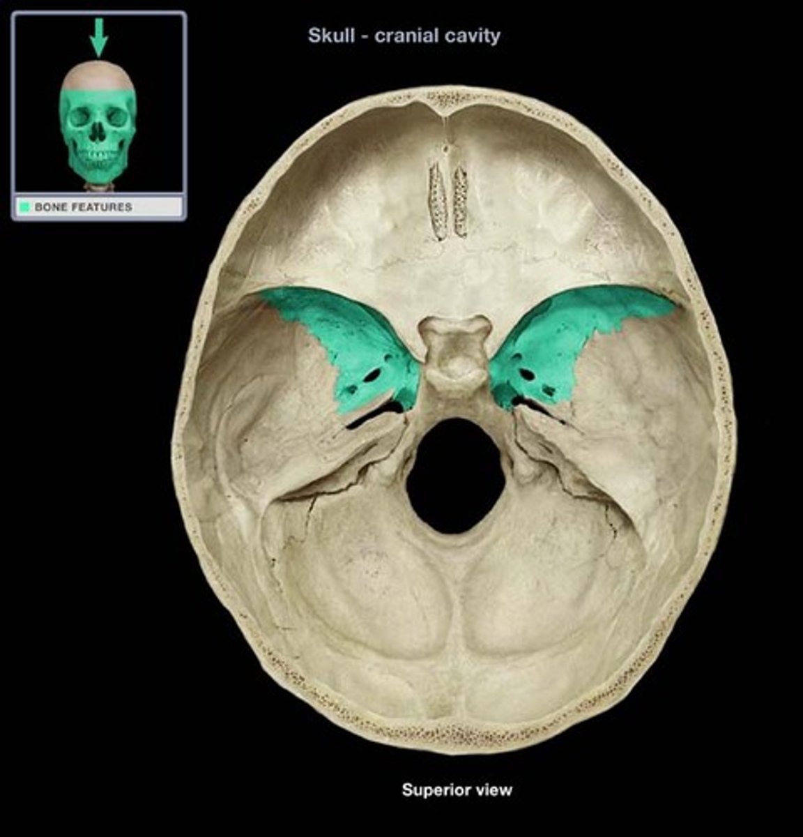
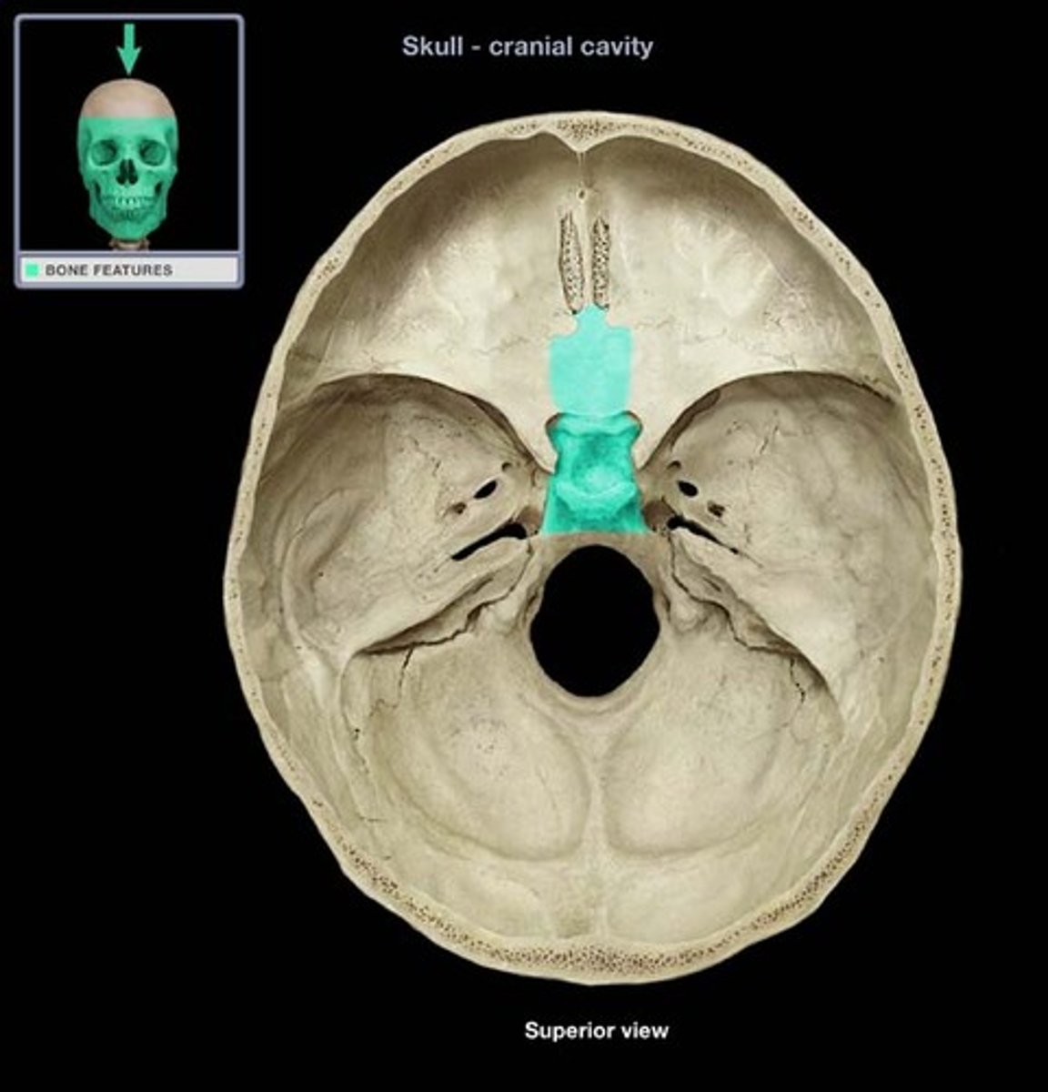
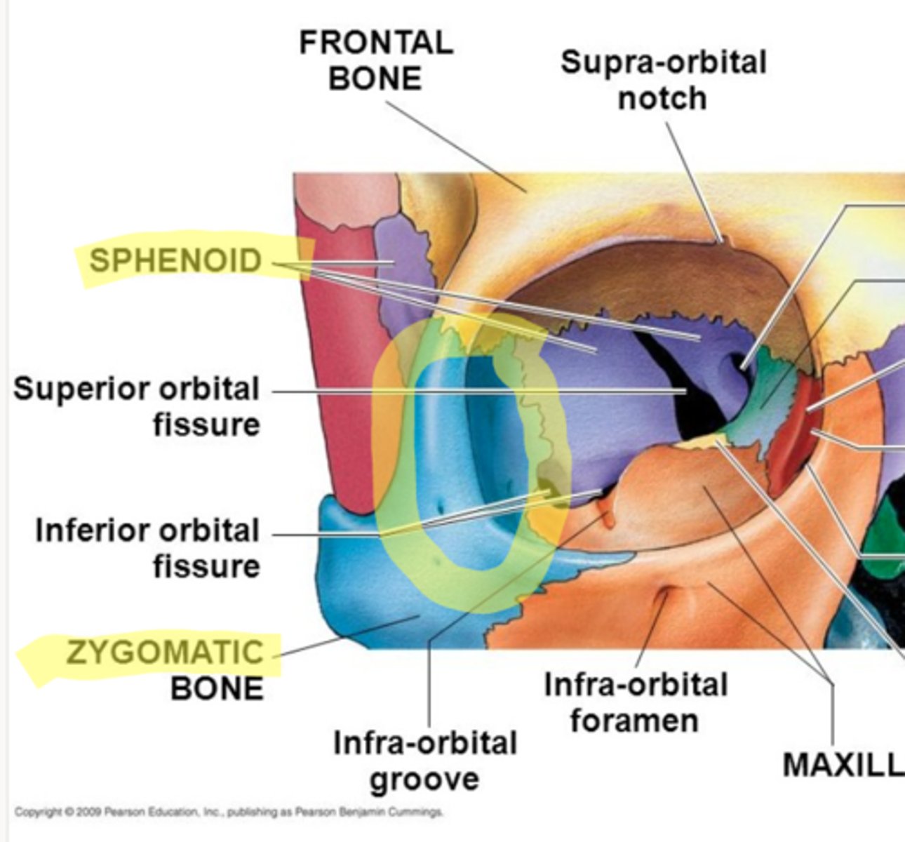
sphenoid bone
forms part of the base of the skull and parts of the floor and sides of the orbit. (Butterfly shape)
3 parts of sphenoid bone
Body of sphenoid, lesser wings, greater wings)
greater wing of sphenoid bone
Protects from lateral aspect of body
lesser wing of sphenoid bone
protects from anterior aspect of body

body of sphenoid bone
houses pituitary gland in a depression called the sella turcica
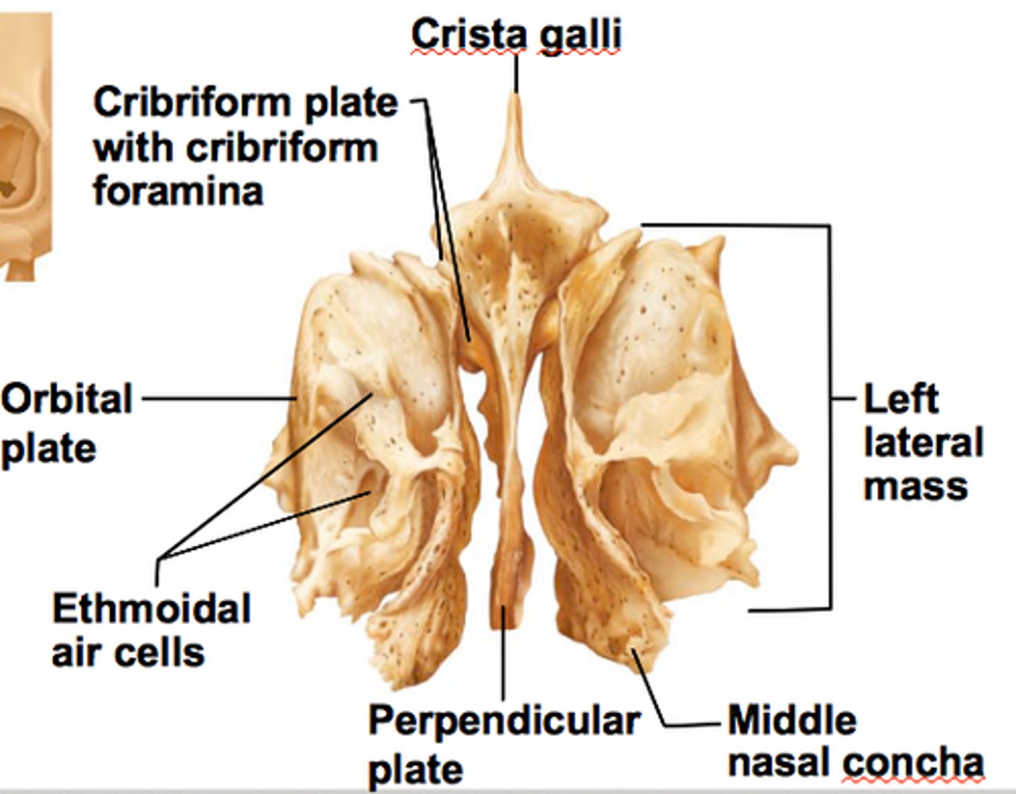
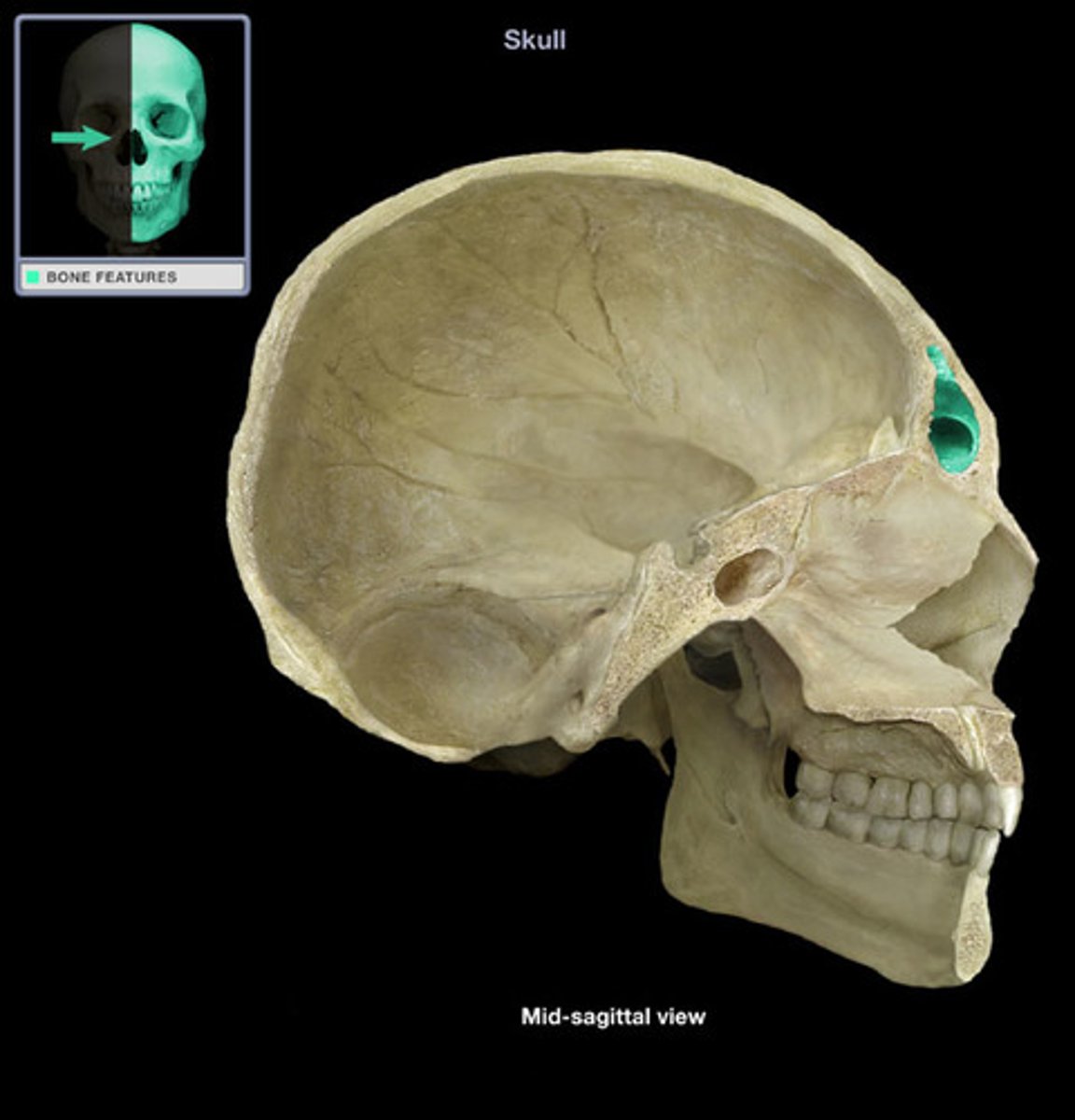
ethmoid bone
forms part of the posterior portion of the nose, the orbit, and the floor of the cranium
4 parts of the ethmoid bone
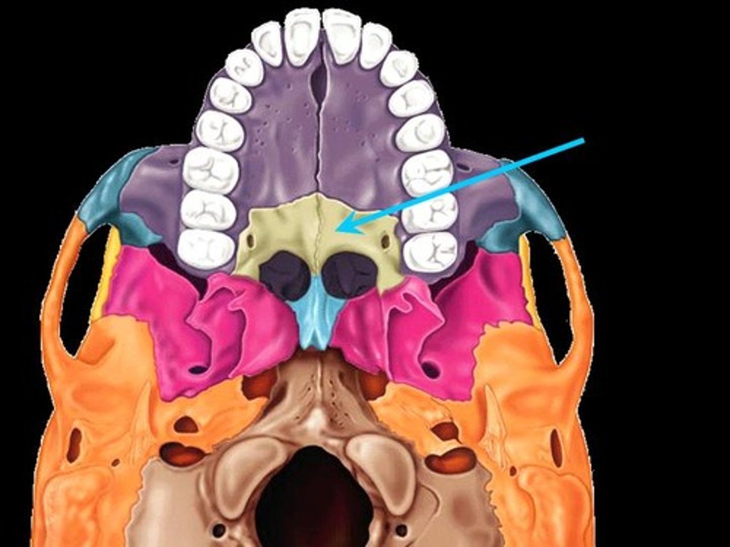
Vertical (perpendicular plate) - forms nasal septum
horizontal (cribriform plate)- olfactory nerves pass through
labyrinths (2)- house ethmoidal sinuses
palatine bone
Extends from hard palate to orbital floor
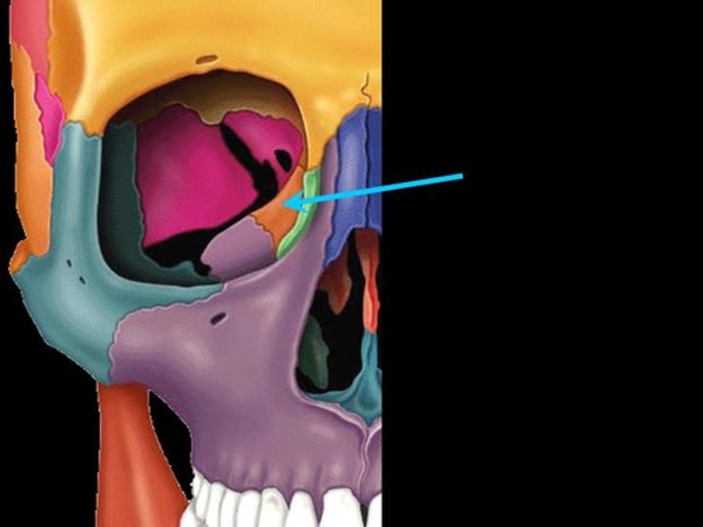
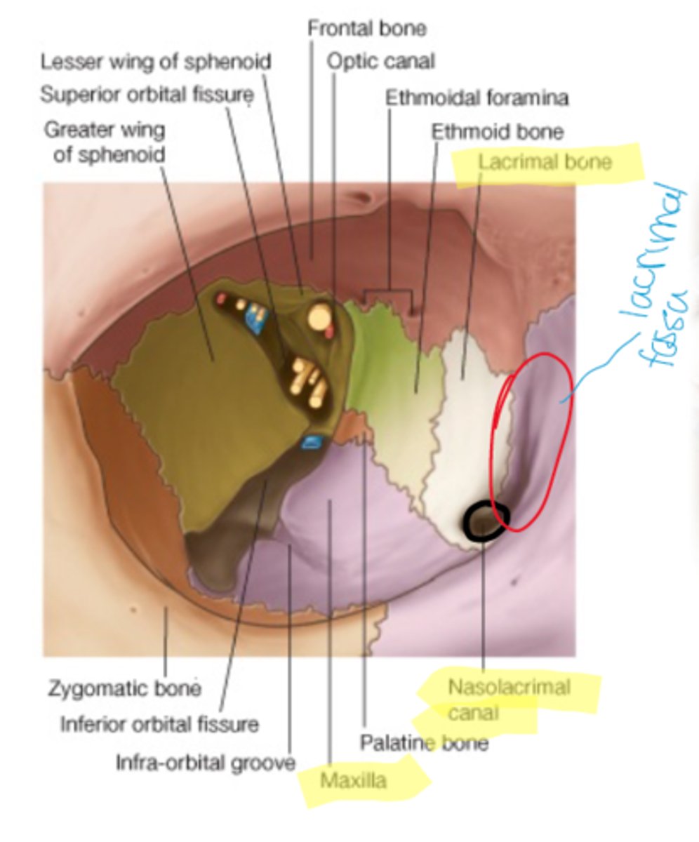
lacriminal bone
located along the medial wall of the orbit
nasal bone
forms the bridge of the nose
inferior conchae
Located along lateral wall of nasal cavity
Vomer
forms the posterior part of the nasal septum
Zygomatic Bone
forms lateral part of cheekbone and the lateral wall and floor of the orbit
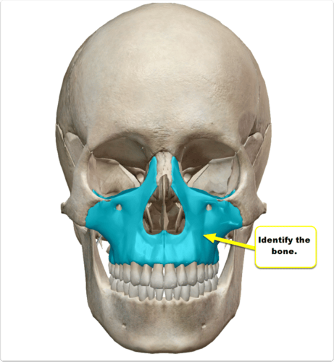
Maxillary Bone (maxilla)
Forms upper jaw, the cheek, hard palate, lateral wall of nasal cavity and the floor of the orbit
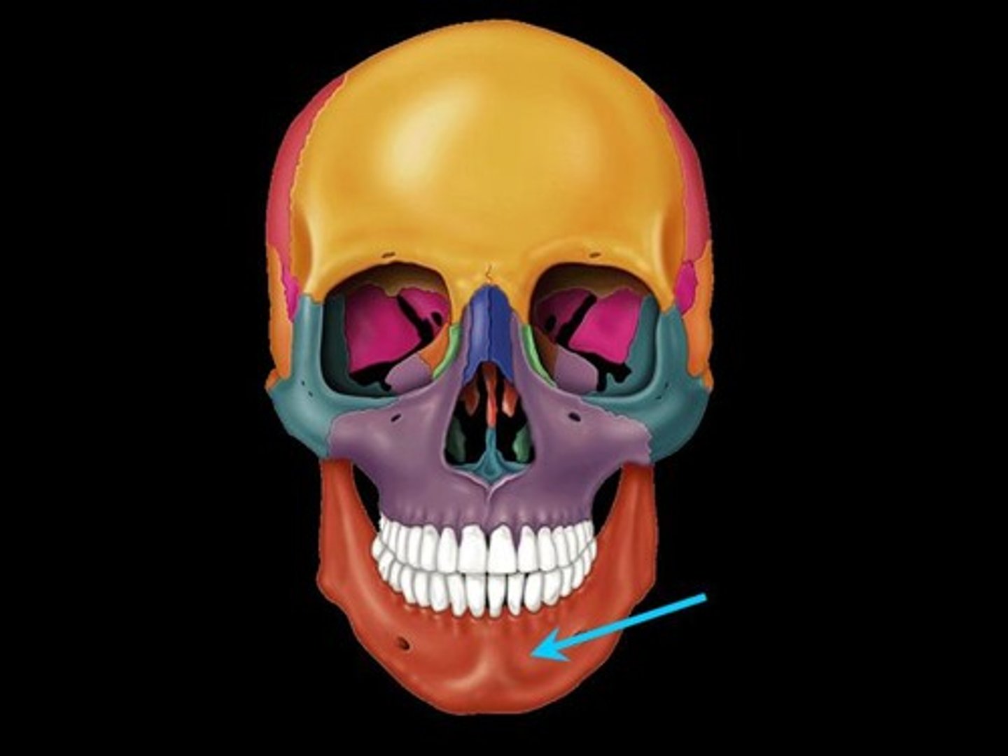
Mandible
Forms the movable lower jaw
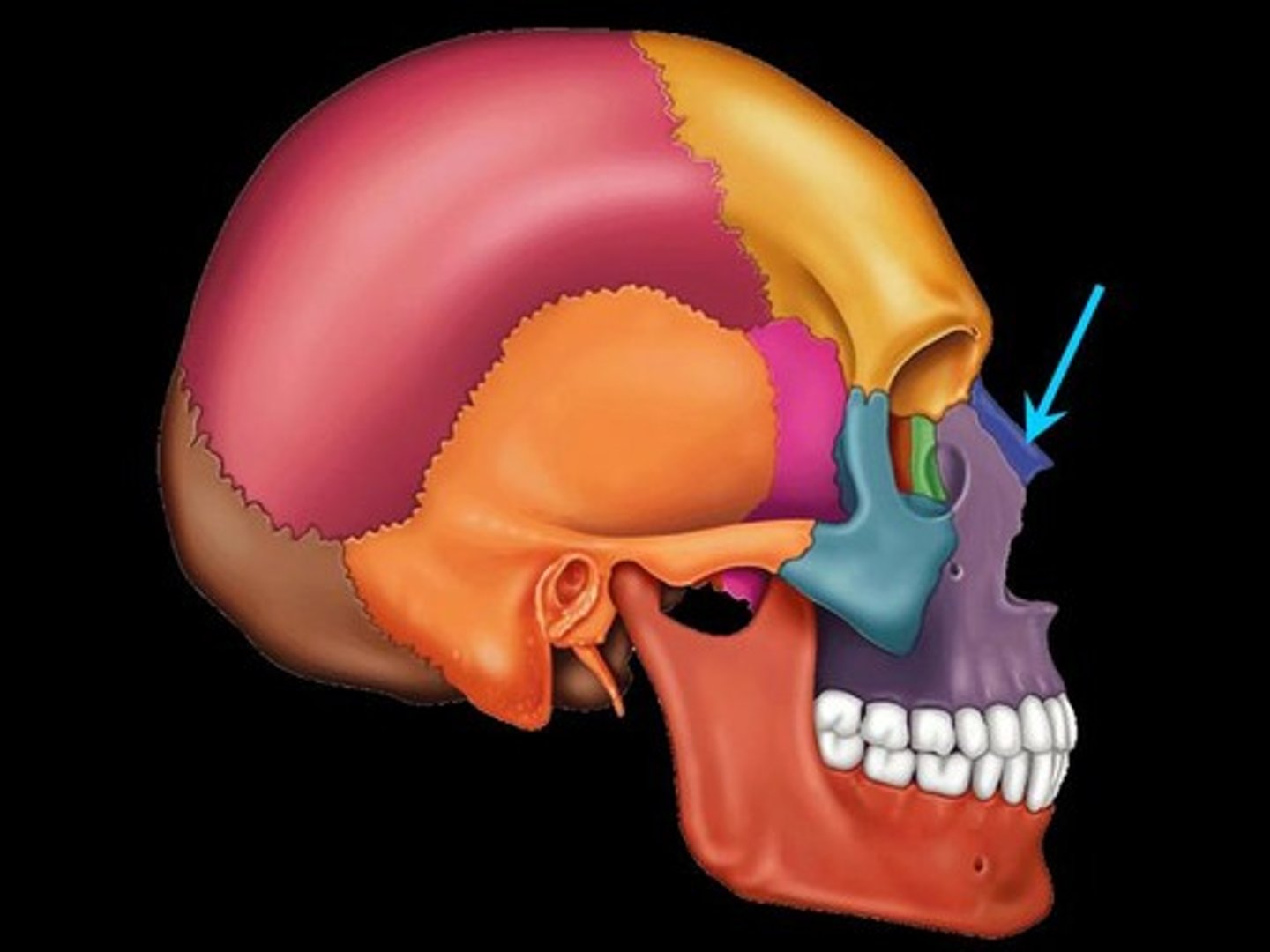
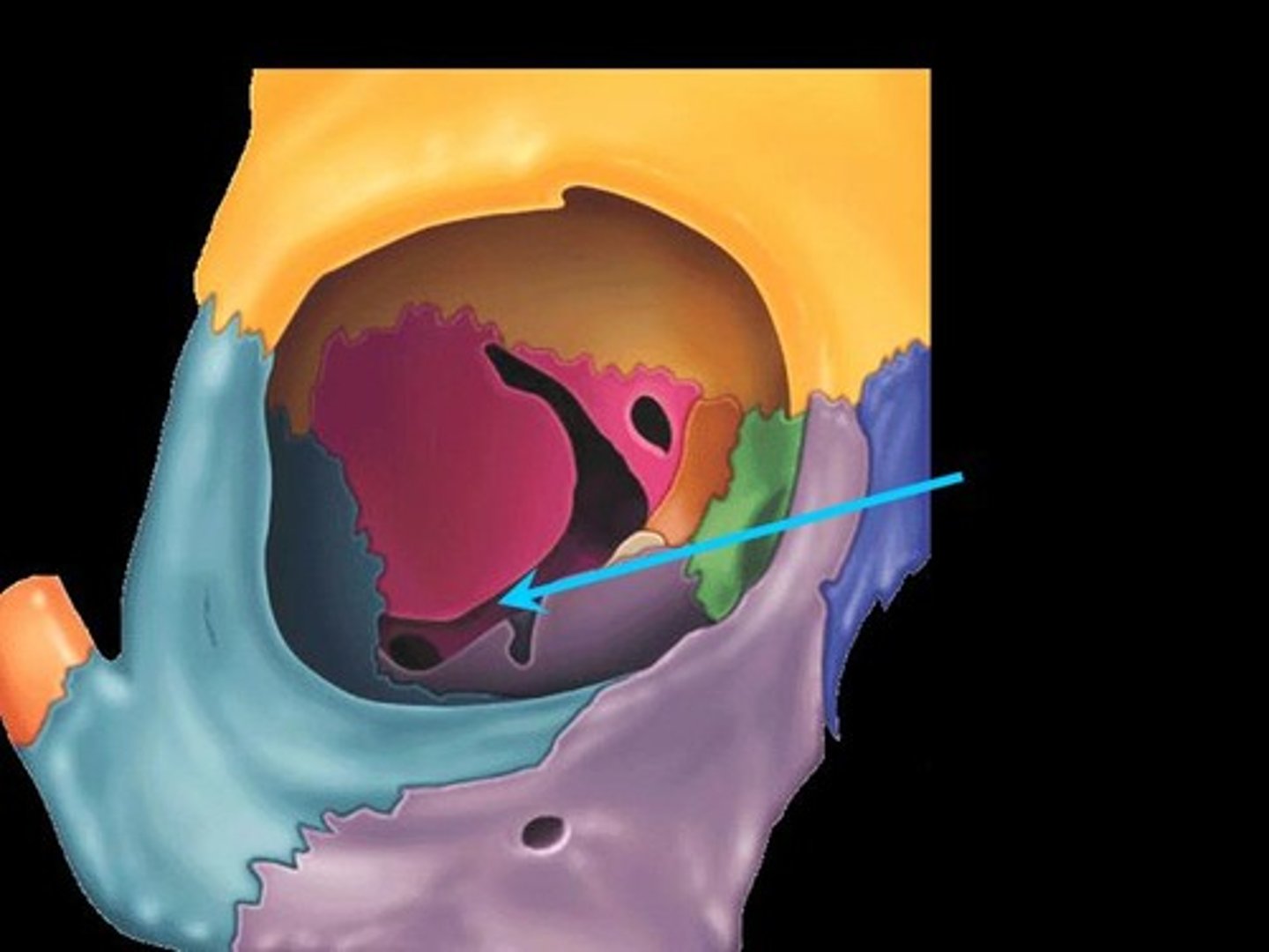
orbit
contains globe, connective tissue, extra ocular muscles, orbital nerves, blood vessels and fat
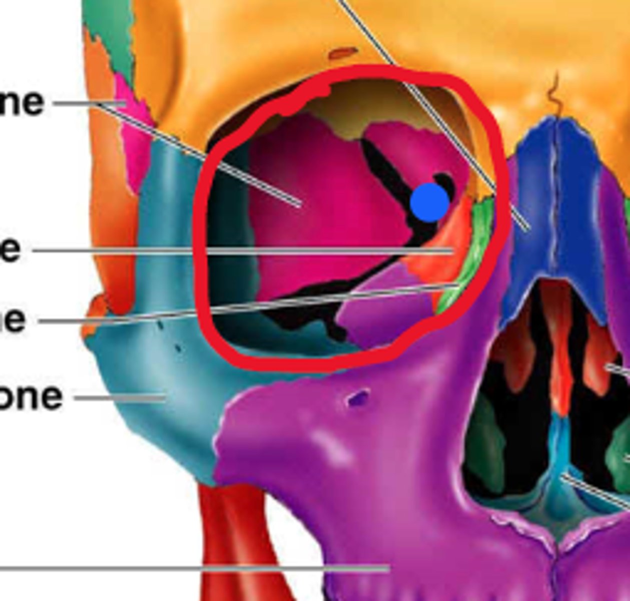
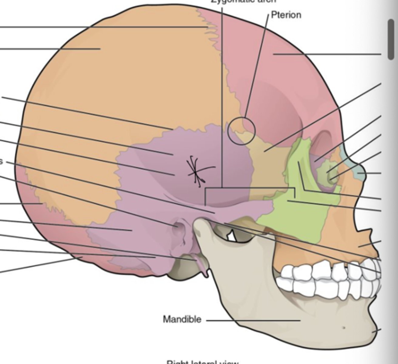
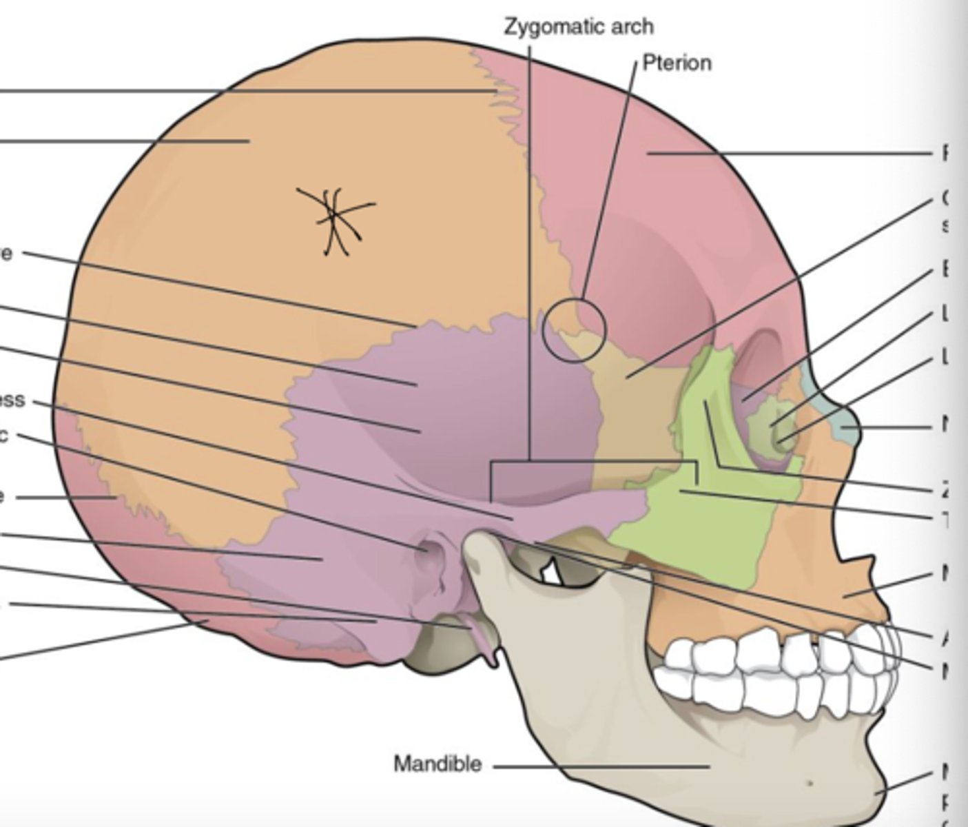
7 bones of the orbit
Frontal, palatine, lacrimal, zygomatic, maxillary, sphenoid, and ethmoid
roof of orbit
-lesser wing of sphenoid bone
-orbital plate of frontal bone
Floor of orbit (weakest wall in orbit)
-orbital process of palatine bone
-orbital surface of maxillary bone
-zygomatic bone
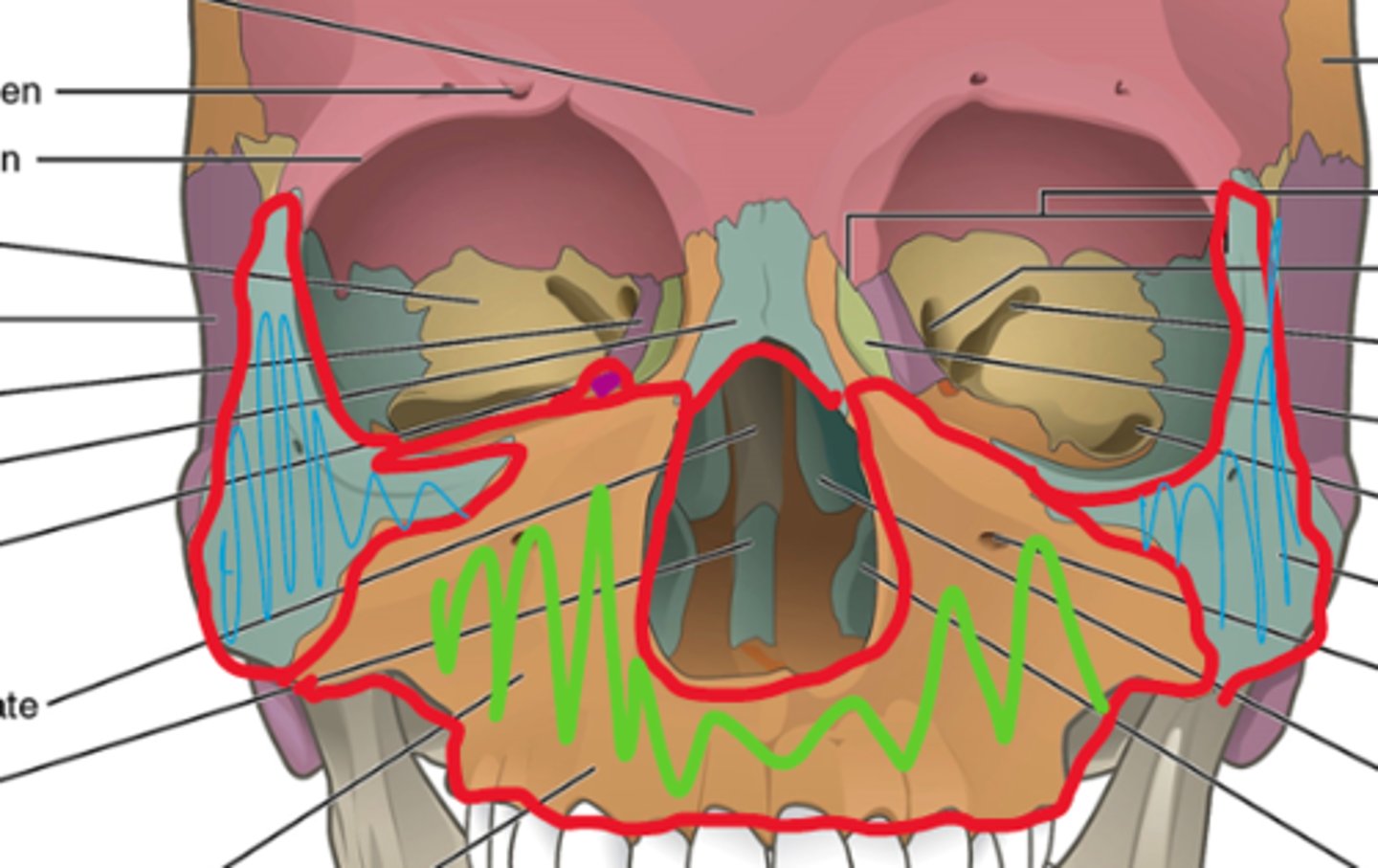
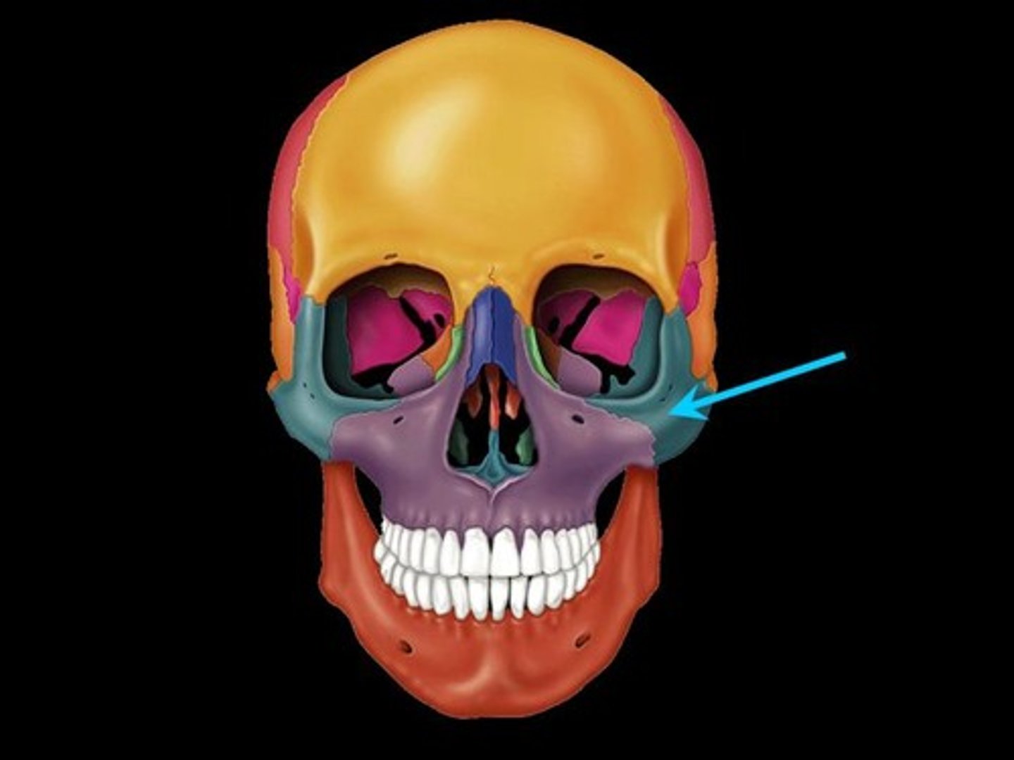
medial wall of orbit (thinnest wall in the orbit)
formed by ethmoid, maxillary, lacrimal, and sphenoid
lateral wall of orbit (strongest area in the orbit)
formed by the zygomatic bone and greater wing of sphenoid bone
paranasal sinuses
Air filled pockets that reduce the weight of the skull and filter the air we breathe
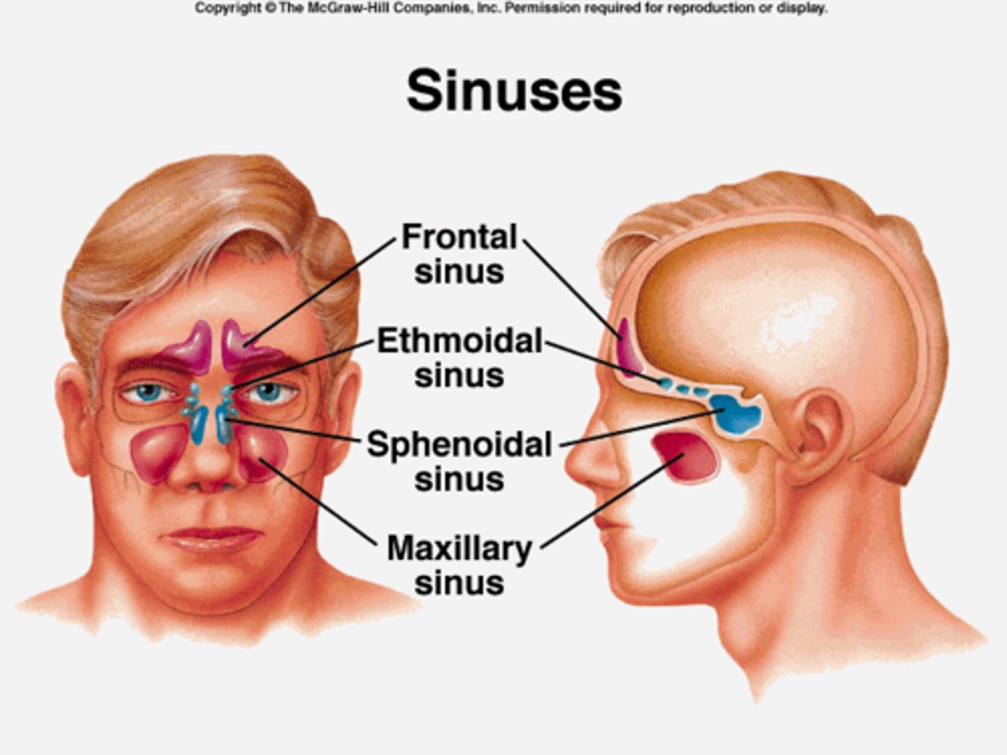
maxillary sinus
inferior to orbits, largest sinus
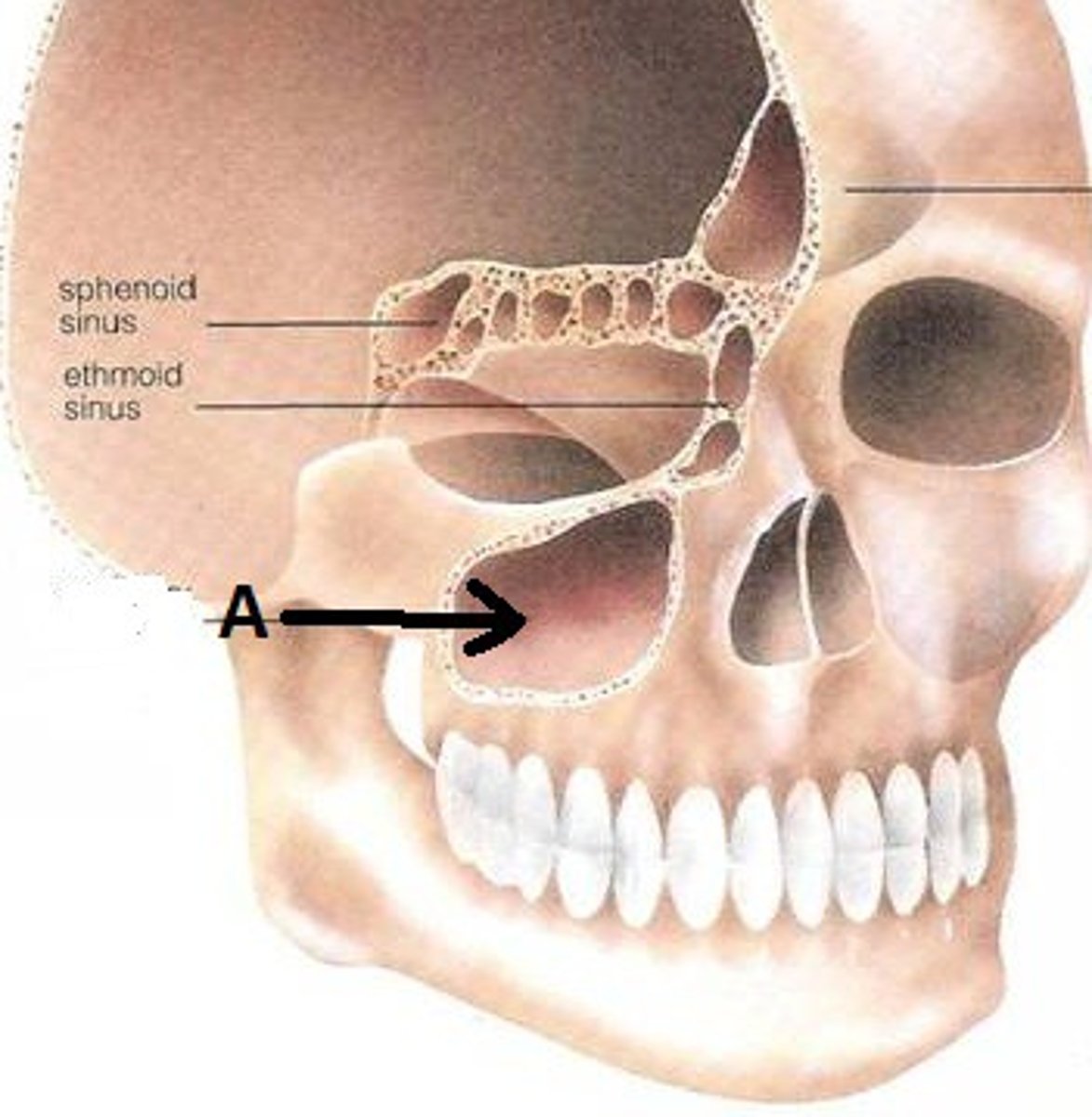
sphenoid sinus
Posterior and medial to orbits
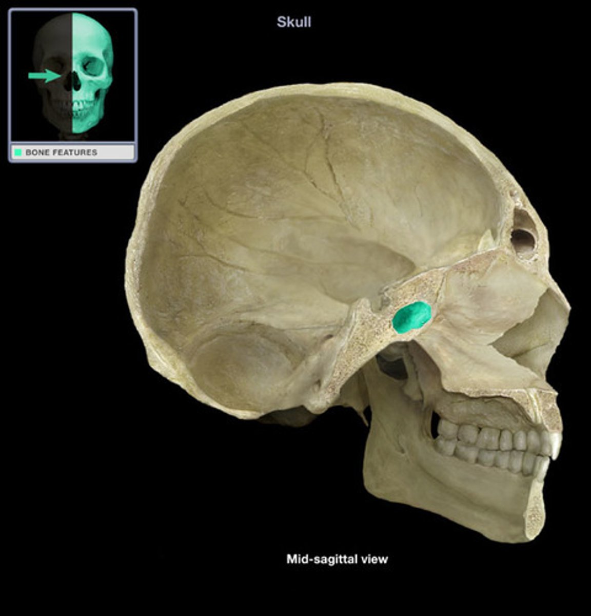
ethmoid sinus
medial to orbit
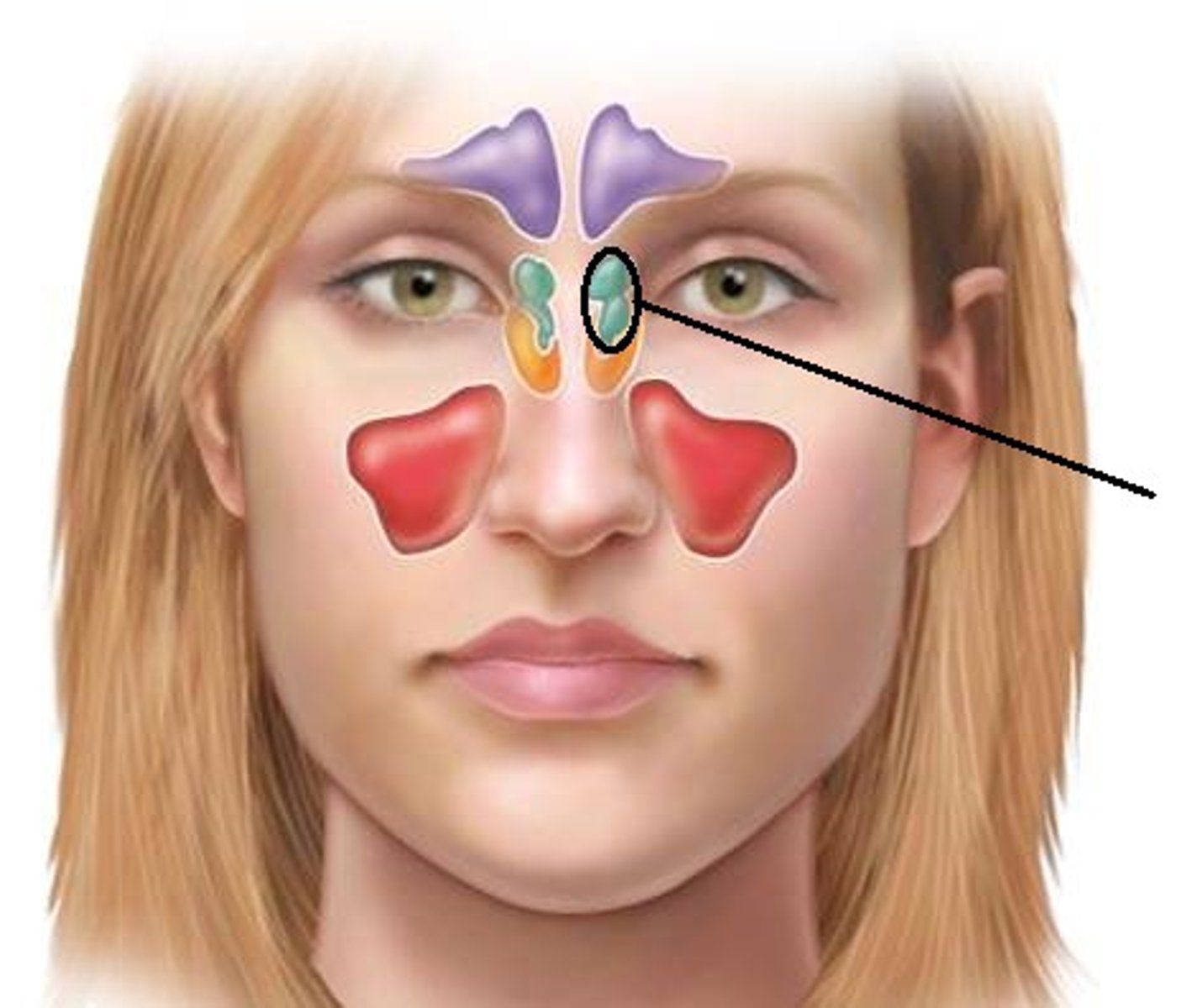
frontal sinus
superior to orbit
sinusitis
inflammation of the sinuses
foramen (foramina)
hole or opening in the bone
Function of the Foramen
allows entrance and exit of nerves and vessels in and out of orbit
optic foramen (canal)
between lesser wing and body of sphenoid bone. Opening between the orbit and cranial cavity
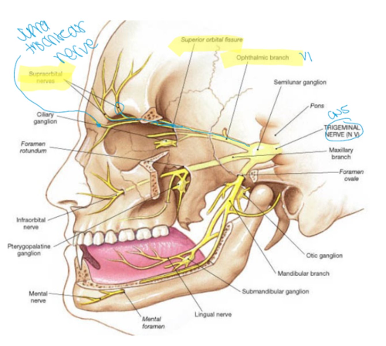
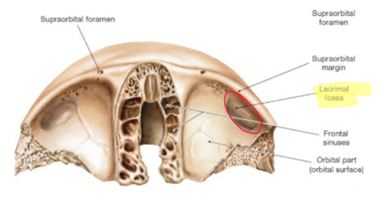
supraorbital foramen
opening above each orbit allowing blood vessels and nerves to pass
infraorbital foramen
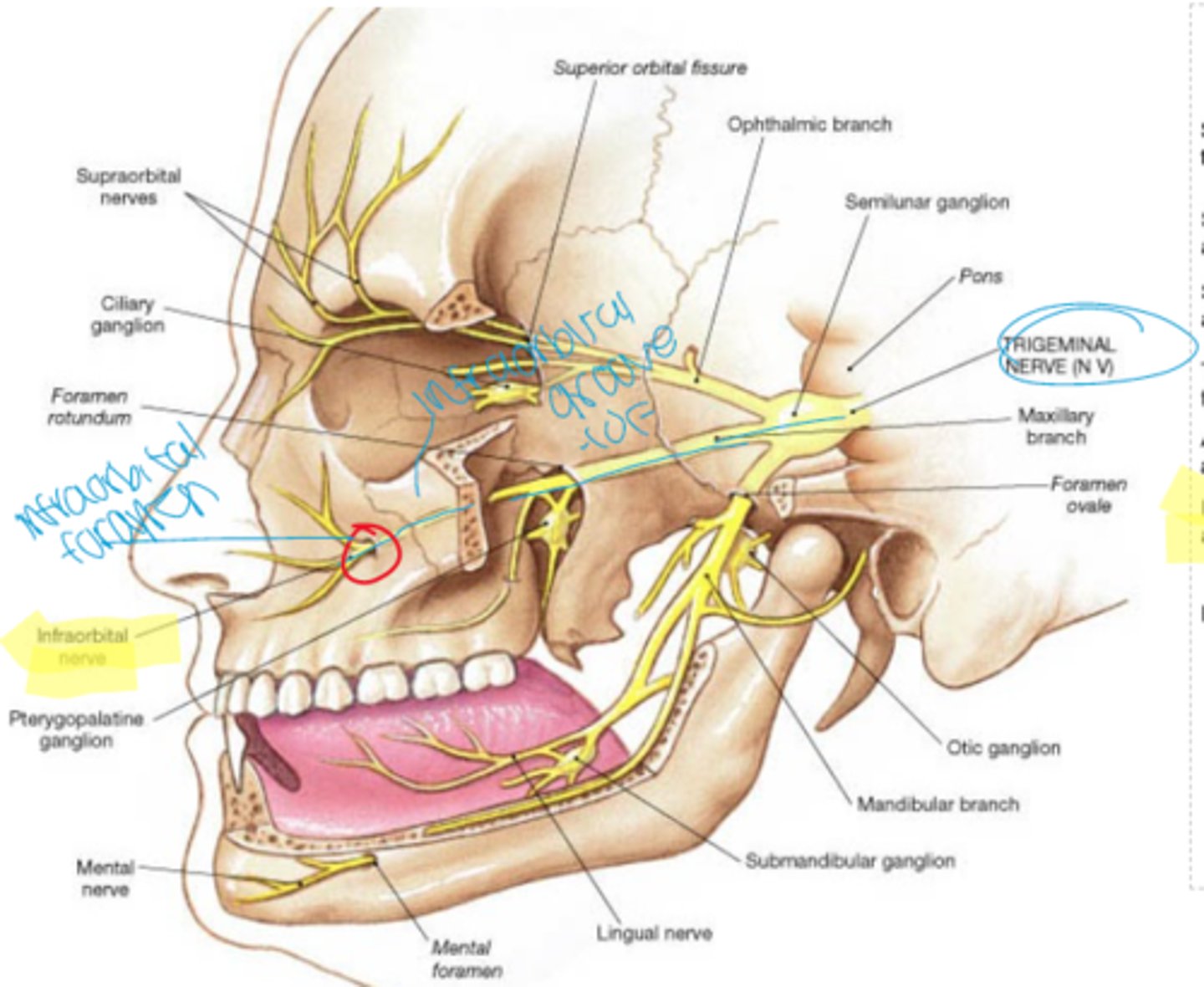
opening under the orbit carrying the infraorbital nerves and blood vessels the the nasal region
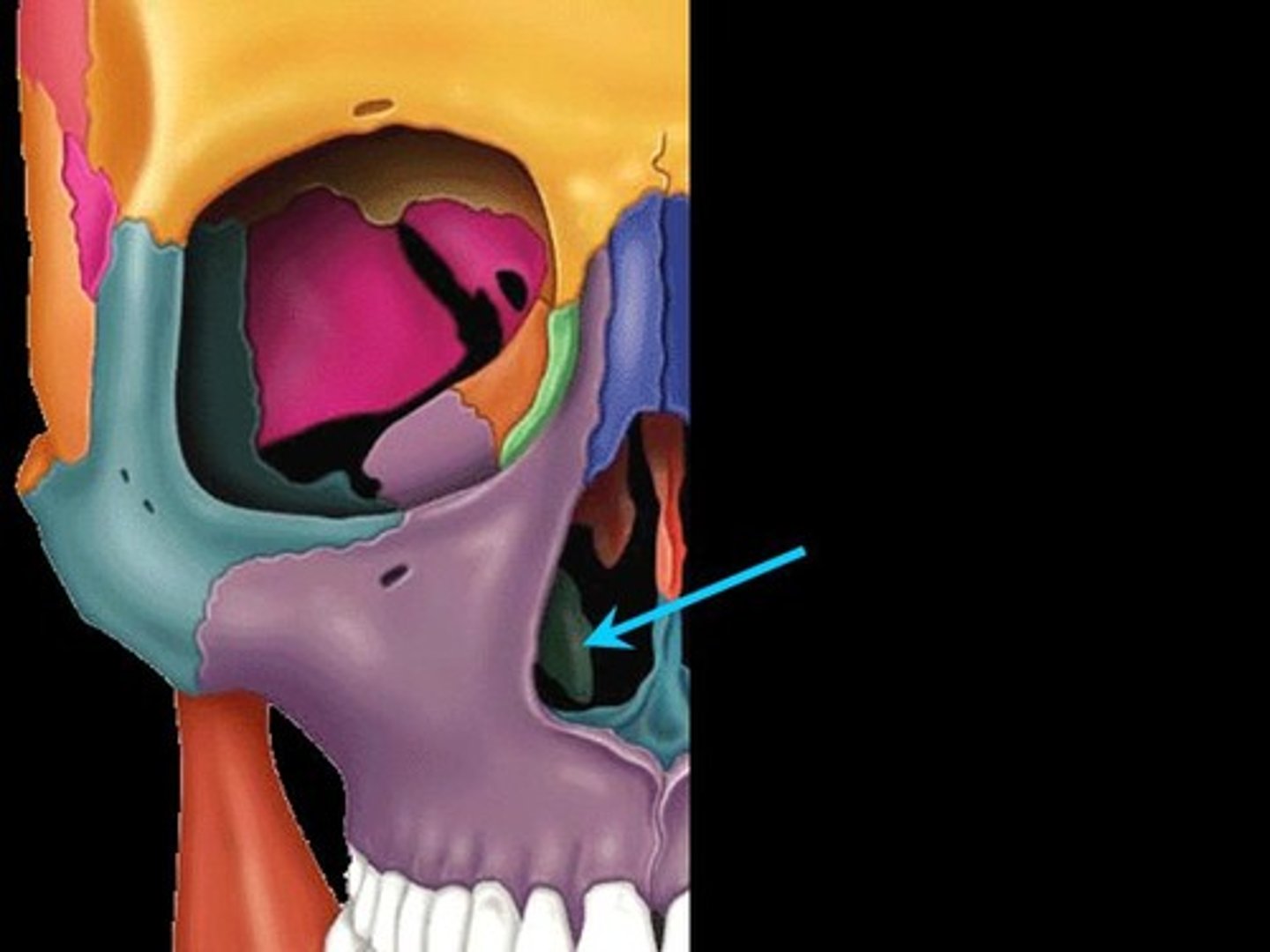
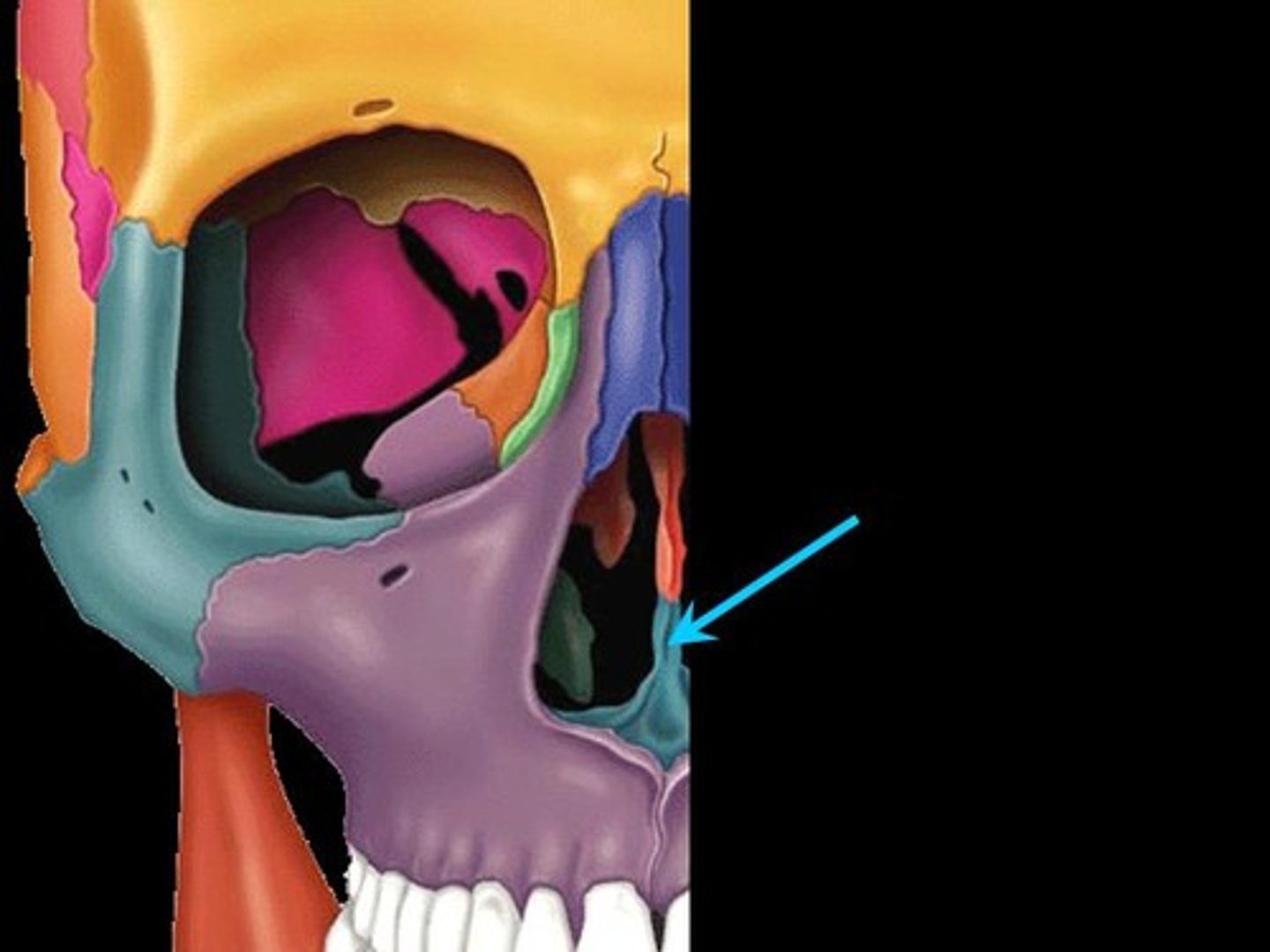
fissure
long, narrow opening in bone allowing entrance and exit of nerves and vessels in and out of the orbit
Fossa
shallow depression in bone
Traveling to the infraorbital foramen
Infraorbital nerve CN V2, artery and veins pass through the inferior orbital fissure and continue along the infraorbital groove to pass through the infraorbital canal and infraorbital foramen
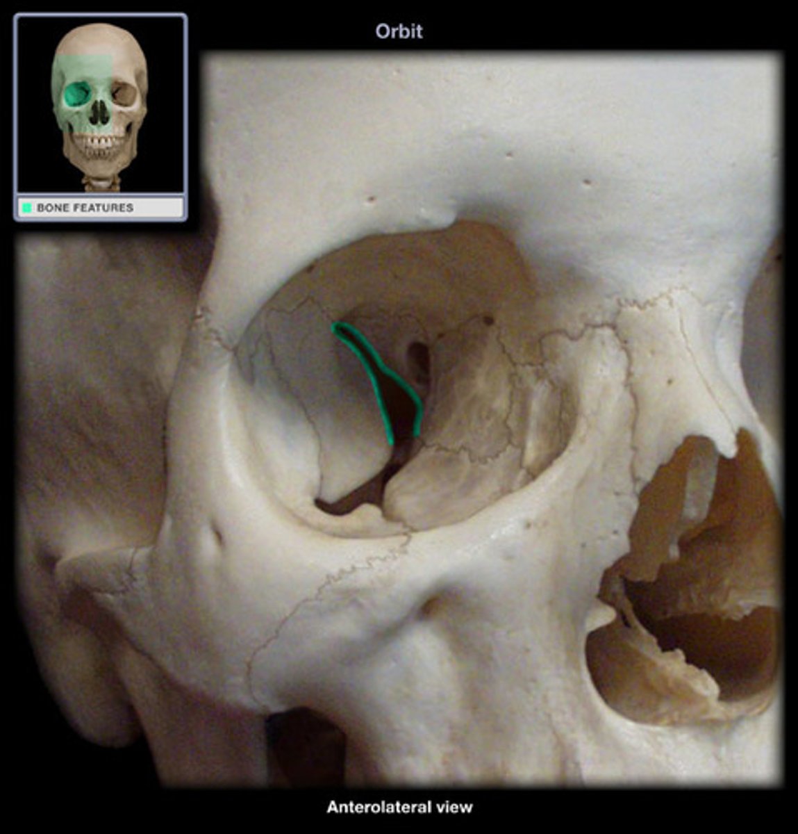
superior trochlear foramen/notch location
located in frontal bone, medial to the supraorbital foramen)
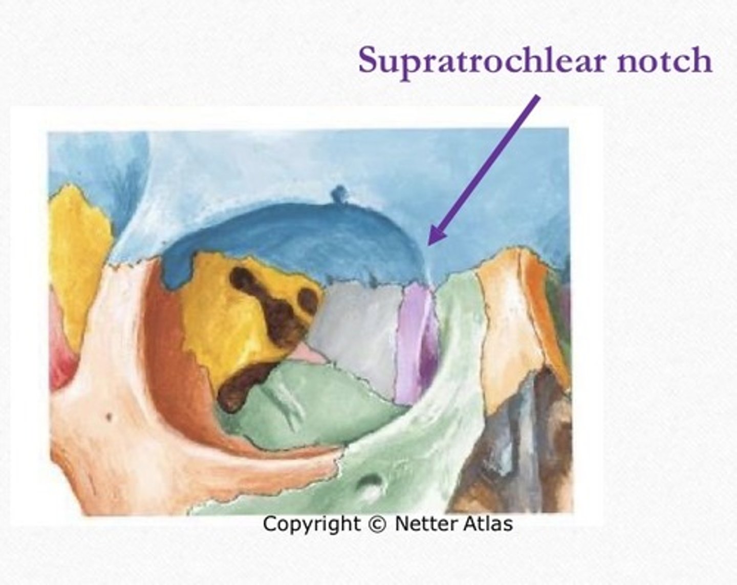
Superior trochlear foramen
CNV1 (supratrochlear nerve), artery, and vein pass through the foramen/notch
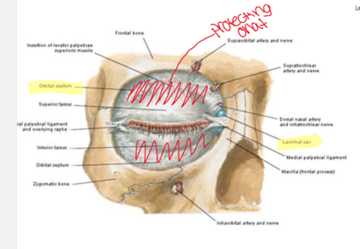
Lacrimal fossa (for the lacrimal sac)
located medially and formed by lacrimal bone and frontal process of maxilla. separated from the orbit by the orbital septum
Lacrimal fossa (for the lacrimal gland)
located in the frontal bone, house of the lacrimal gland
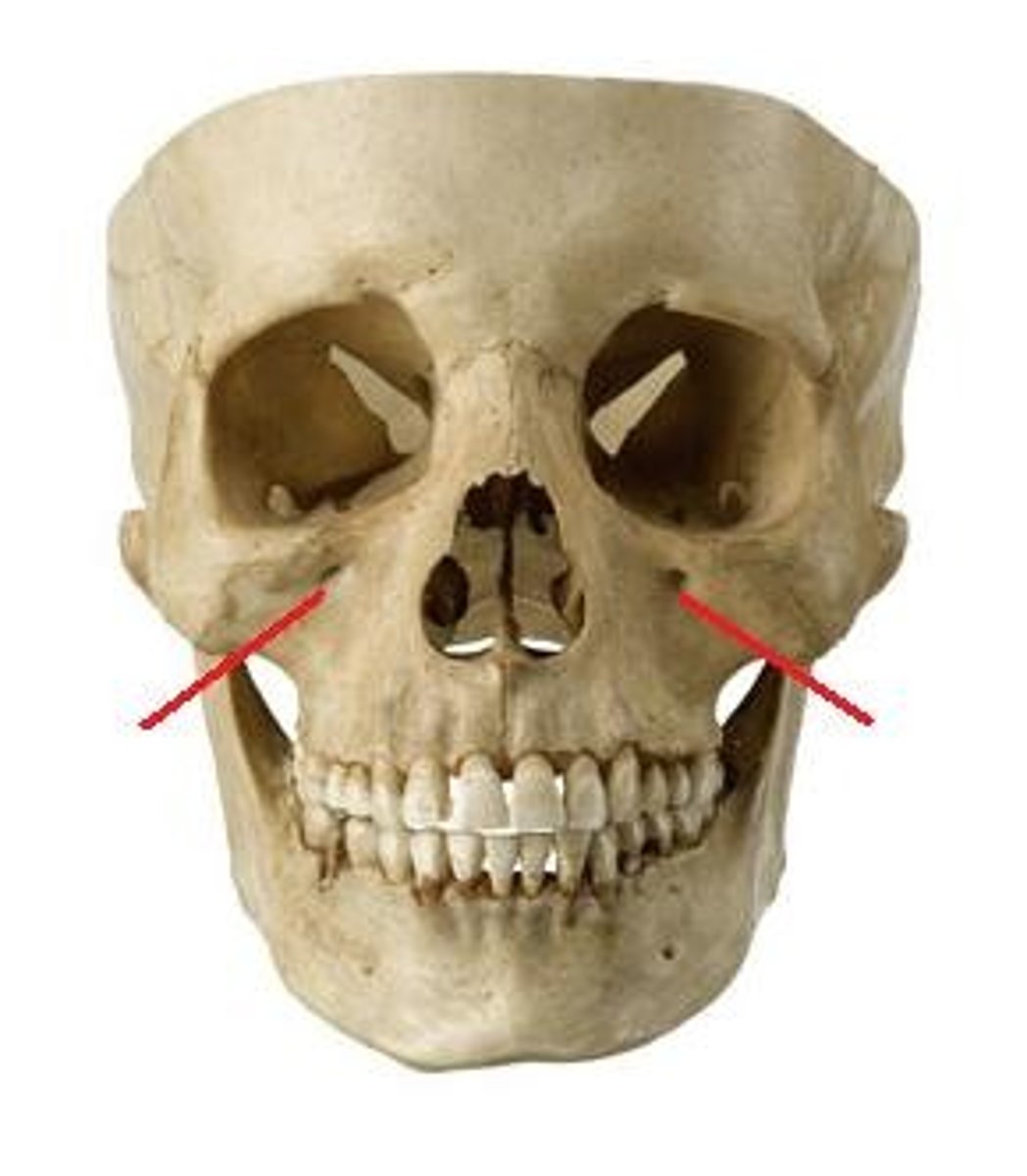
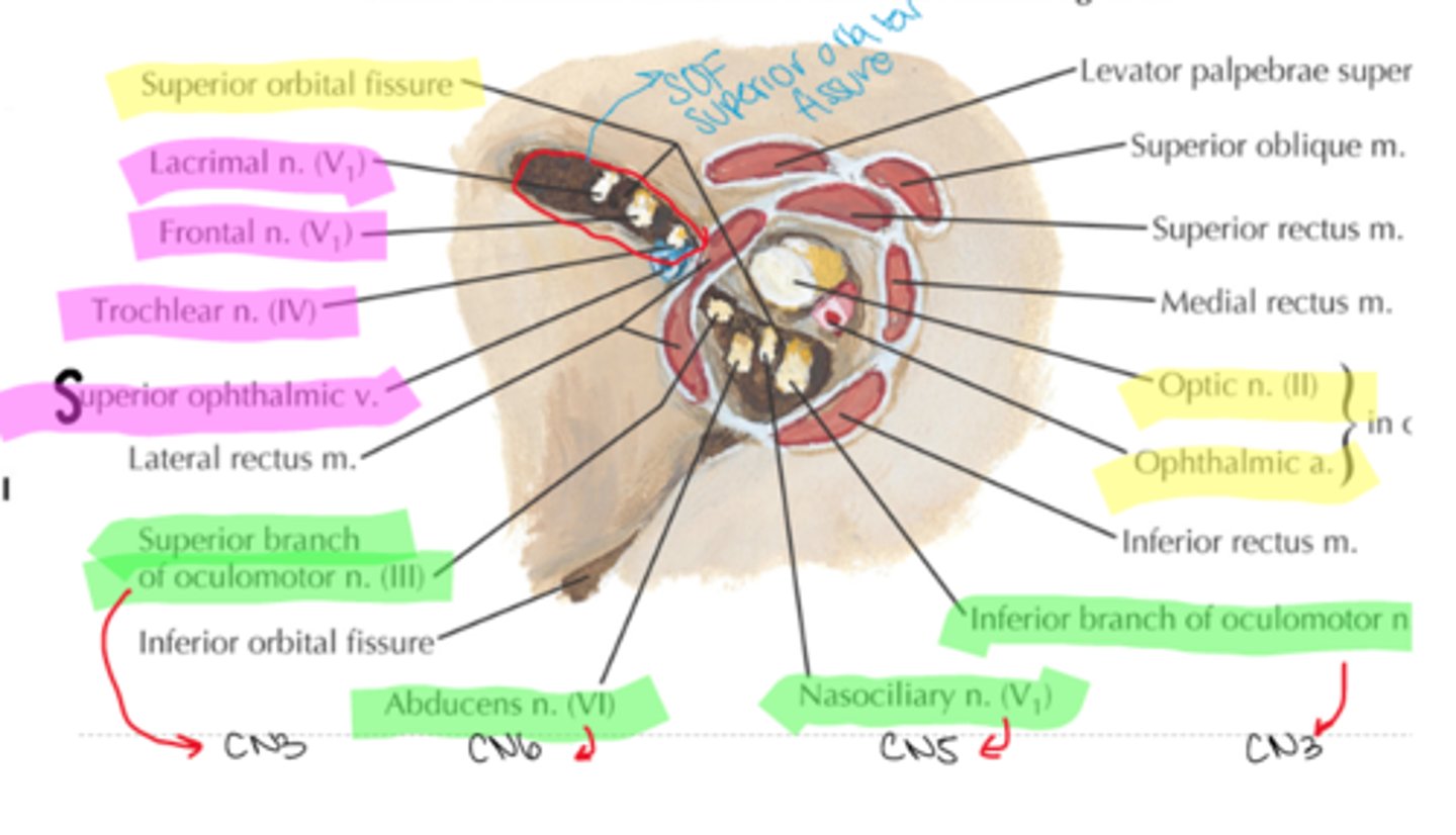
inferior orbital fissure
lies between floor of orbit and lateral wall
Nerves and vessels that pass through the infraorbital fissure
-Inferior ophthalmic vein
-zygomatic nerve (CN V2)
-infraorbital nerve, artery, and vein (CN V2)
Supraorbital Fissure
lies between the greater and lesser wings of sphenoid (slightly nasal and posterior of orbit)
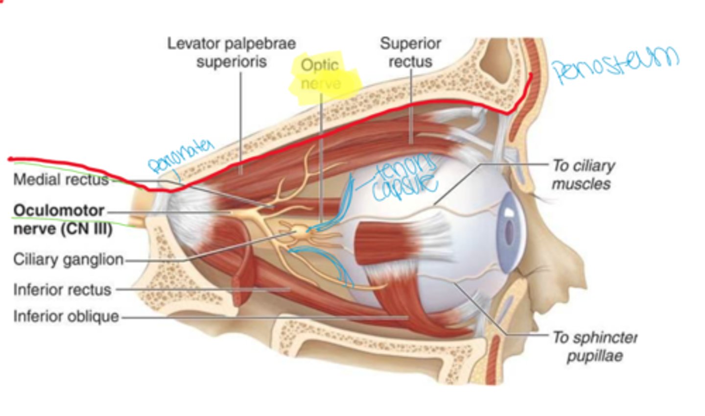
Common tendinous ring (Annulus of Zinn)
fibrous structure that the rectus muscles arise from
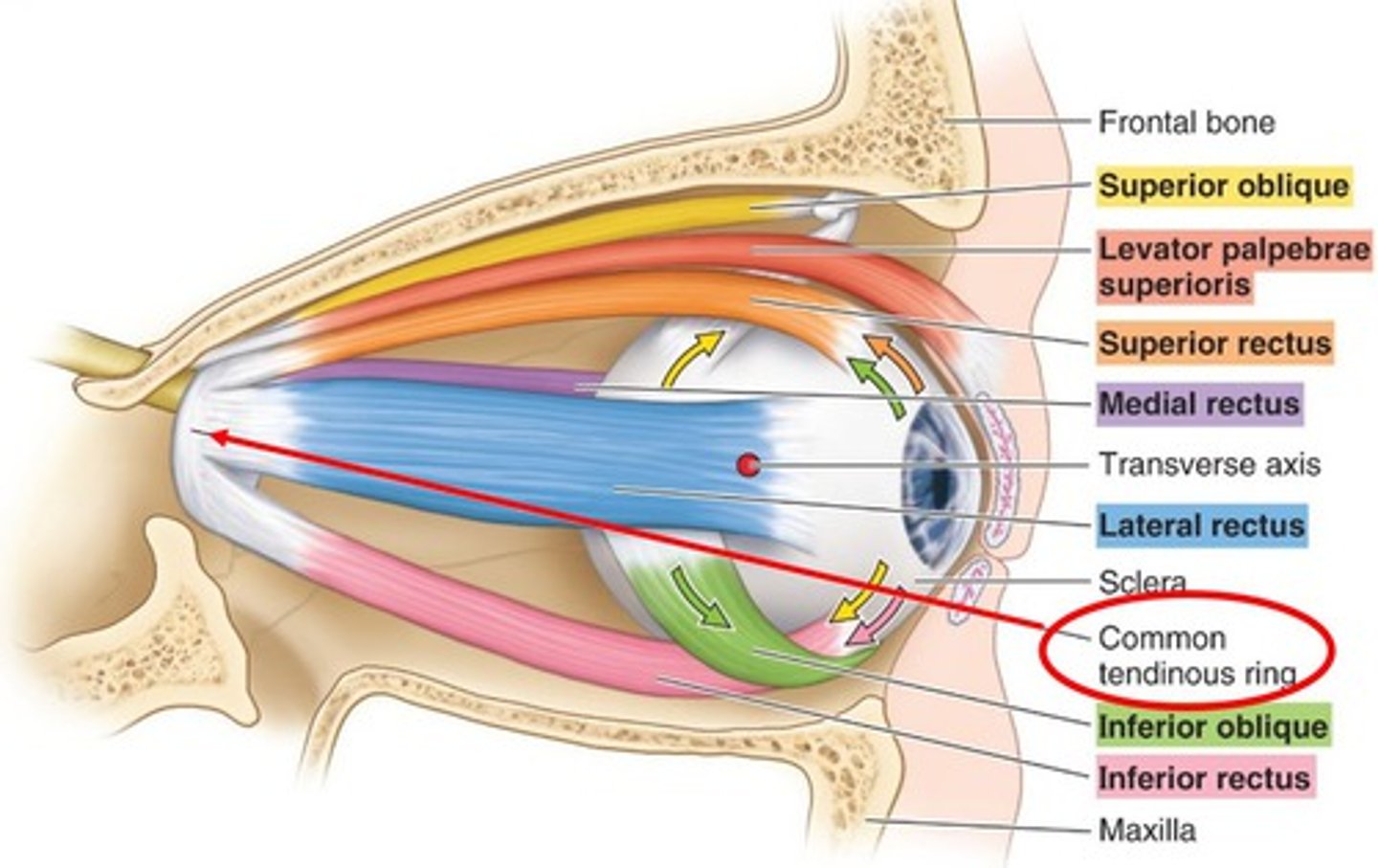
Nerves that pass through superior orbital fissure and common tendinous ring
-Superior and inferior division of oculomotor nerve CN III
- Nasociliary Nerve (CN V1)
-Abducens Nerve ( CNVI)
Nerves that pass through superior orbital fissue and above the annulus of zinn
-Superior Ophthalmic Vein
- Trochlear Nerve ( CN IV)
- Lacrimal Nerve (CN V1)
-Frontal Nerve (CN V1)
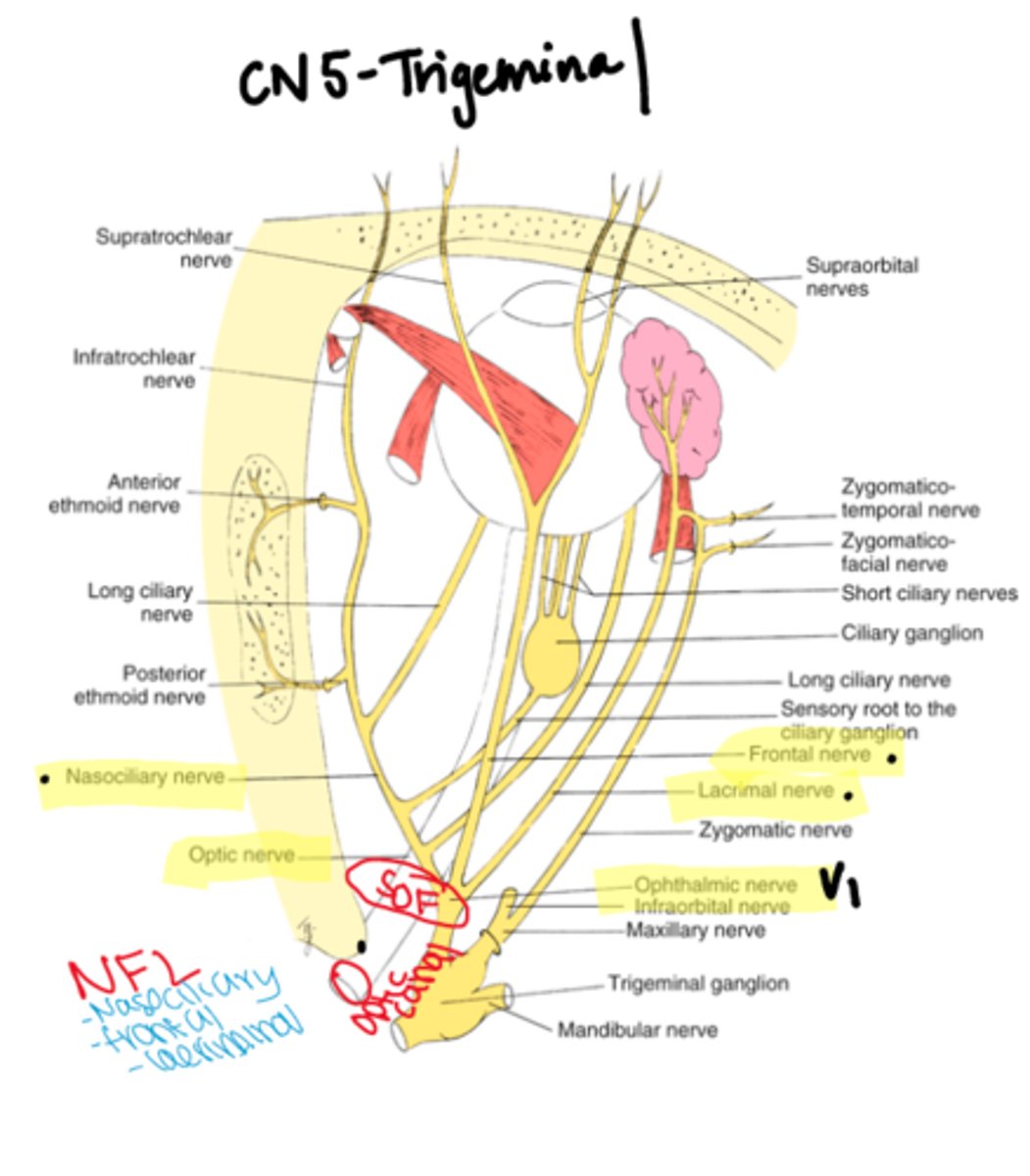
CN V- cranial nerve 5
V1- ophthalmic
V2- maxillary
V3- mandibular
Orbit contents
globe, connective tissue, extraocular muscles (EOMS), orbital nerves, blood vessels, and fat
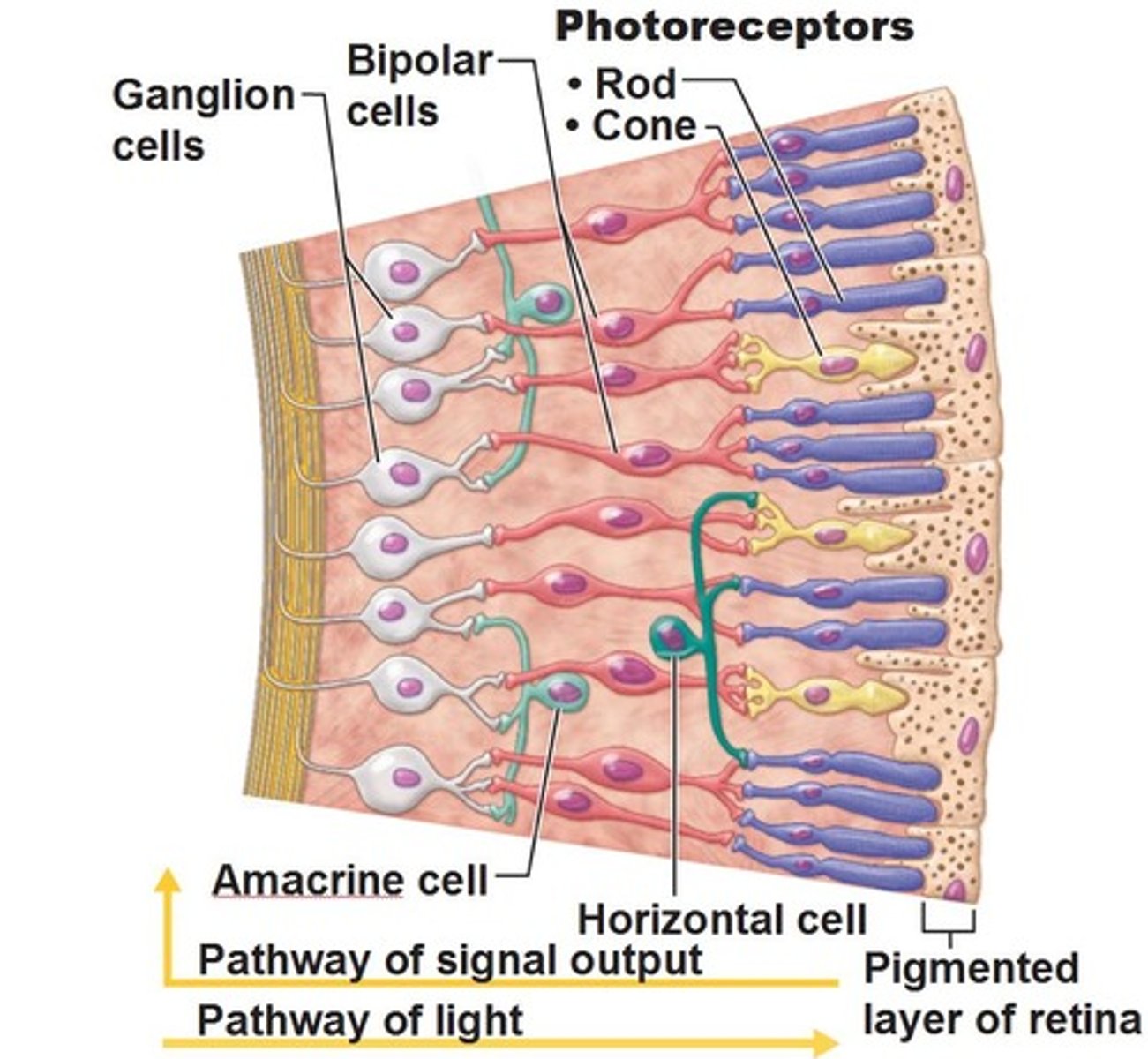
Globe (eyeball) 3 coats
outer fibrous layer
middle vascular layer
inner neural layer
outer fibrous layer of eye
sclera and cornea
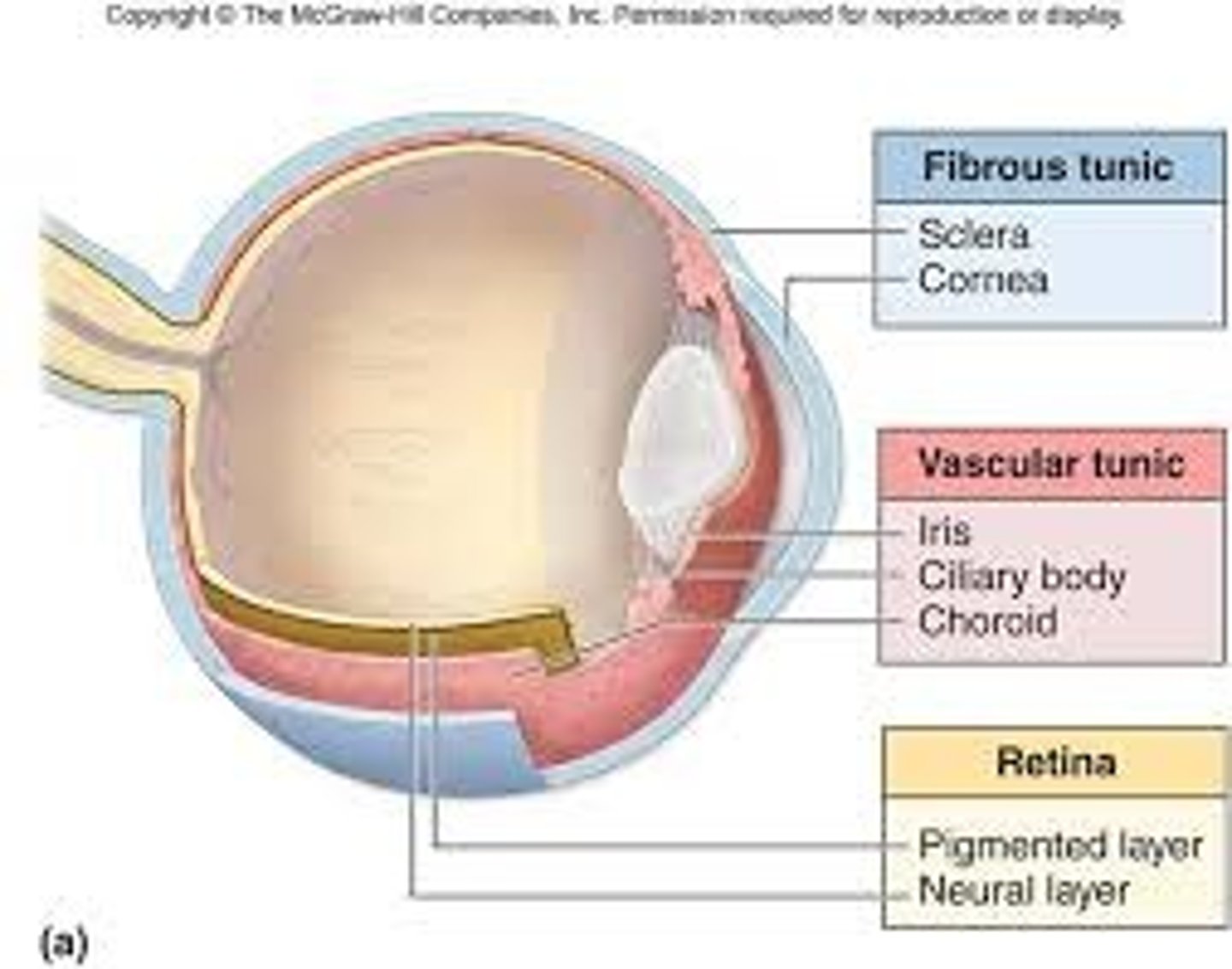
middle vascular area of eye
iris, ciliary body, choroid (uvea)
inner neural layer of eye
retina
anterior chamber of eye
between the iris and cornea

posterior chamber of eye
between ciliary body and lens
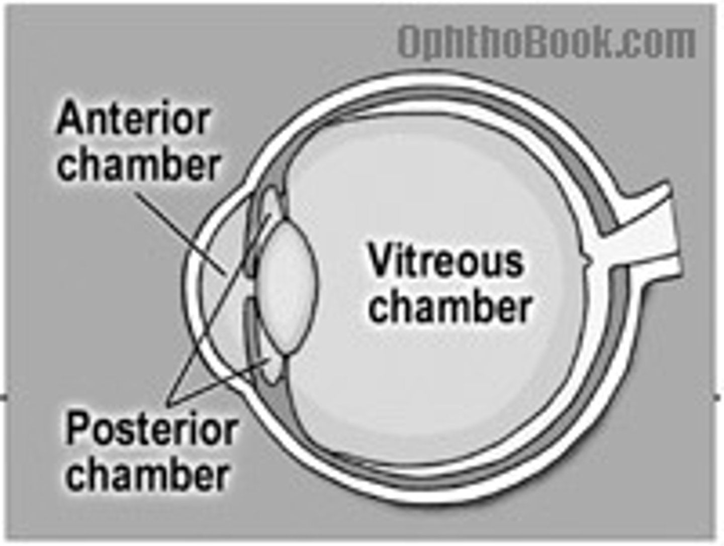
The vitreous chamber of the eye
back half of the eye

Tenon's Capsule/Bulbar Fascia
sheet of dense connective tissue that covers the sclera
anteriorly merges with the sclera and conjunctiva at limbus but posteriorly continuous with dural sheath of ocular nerve
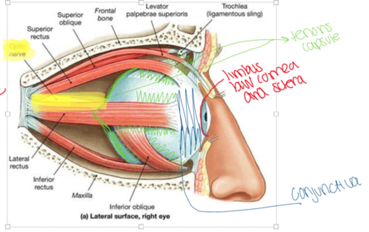
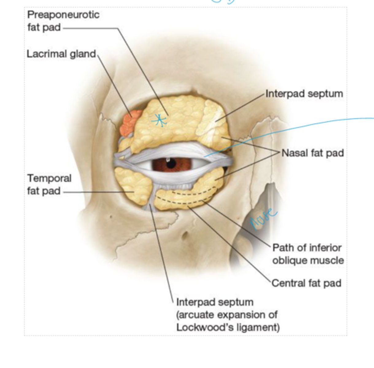
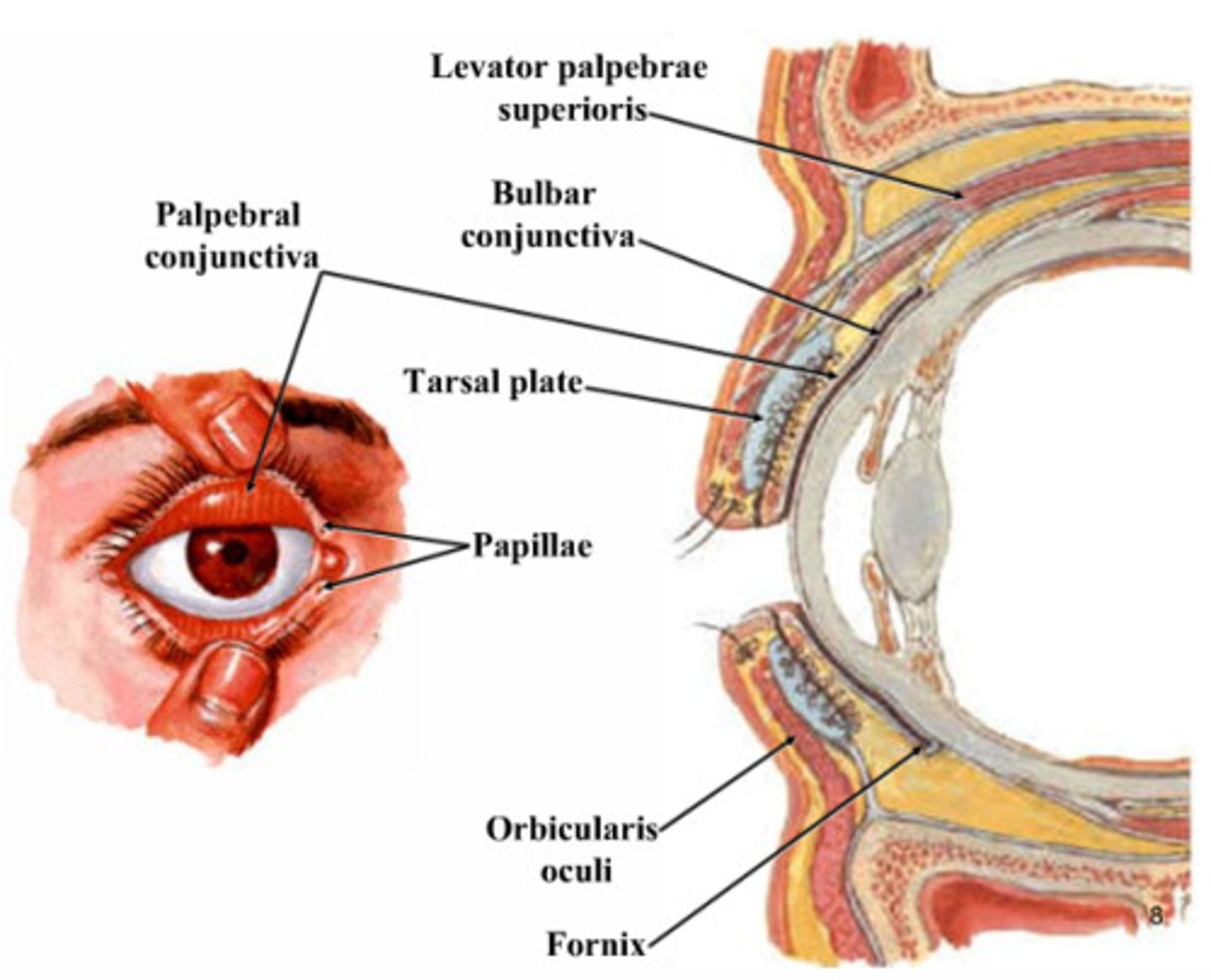
orbital septum (palpebral fascia)
sheet of dense connective tissue that extends the rim of the orbit to the tarsal plate . keeps orbital fat in place
-prevents facial, eyelid, nasolacrimal system infections from entering the orbit
The tarsal plate of the eyelid ________.
is connected to the levator palpebrae
Periorbita (Orbital fascia, Periosteum)
Sheet of dense connective tissue that covers the bones of the orbit
attachment site for muscles, tendons, and ligaments
provides support structure
Whitnall's ligament (superior transverse ligament)
transverse dense connective tissue provides support and maintains spatial relationships between anatomic structures in the superior orbit
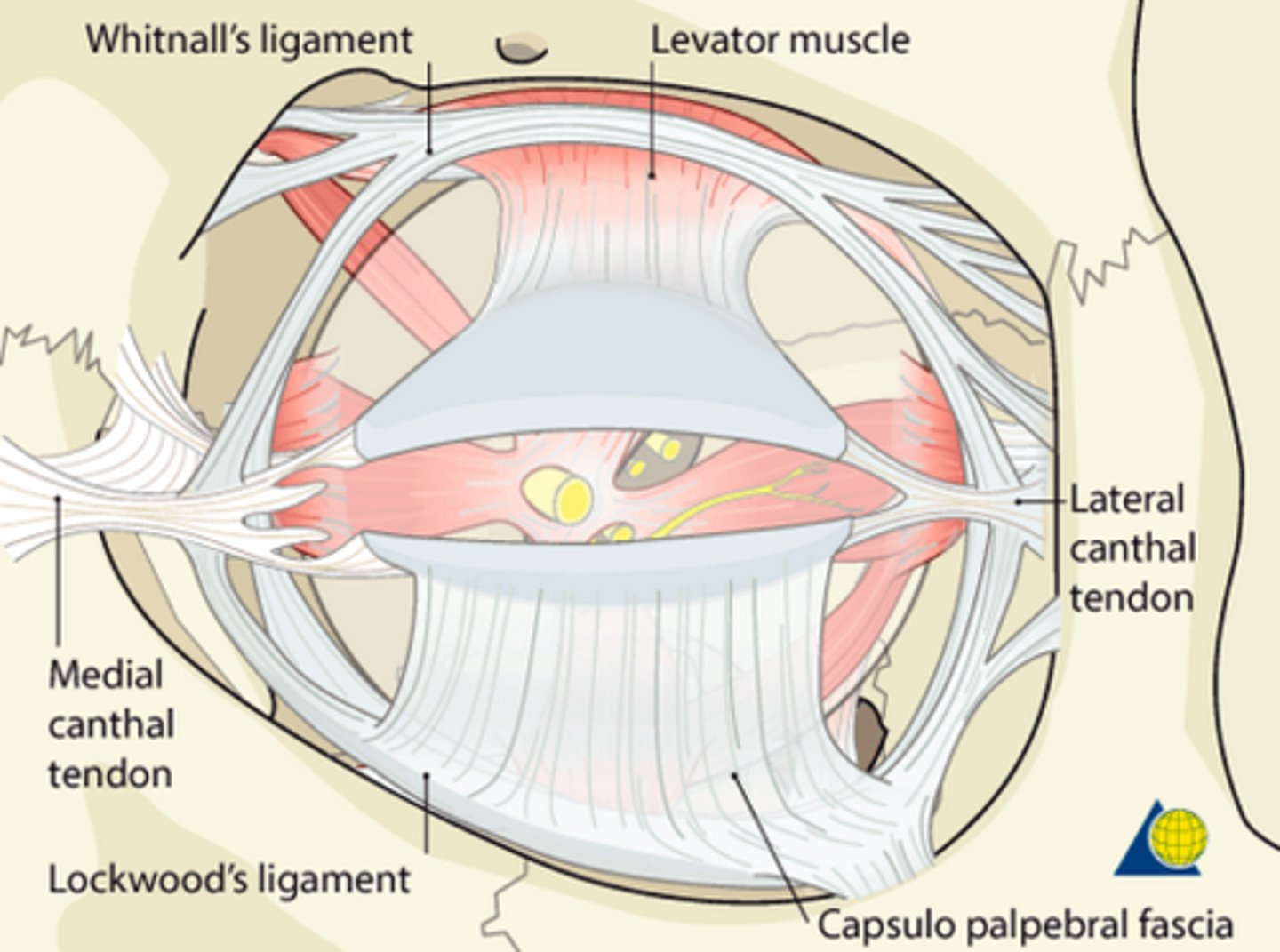
What forms the Whitnall's ligament?
condensation of the levator muscle
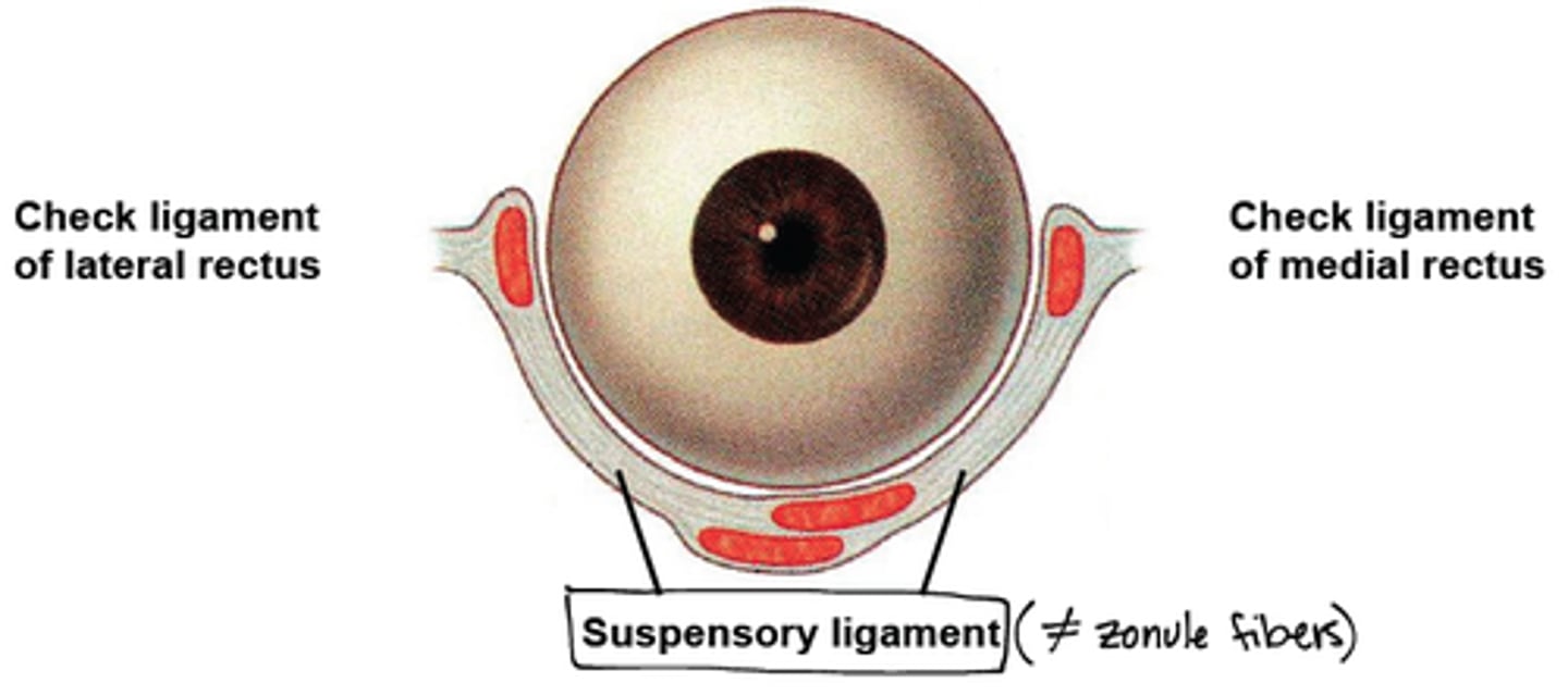
Lockwood's Ligament (Suspensory ligament)
transverse dense connective tissue located in the inferior orbit, providing support for anatomic structures in the inferior orbit
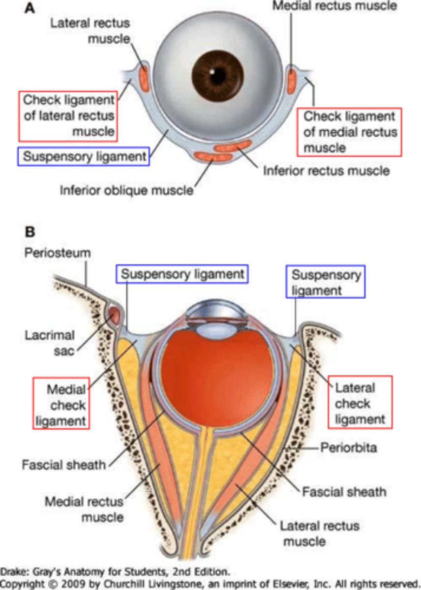
medial check ligament
- transverse dense connective tissue that is an expansion of the sheath of the medial rectus
- attaches to lacrimal bone
- prevents overaction of medial rectus
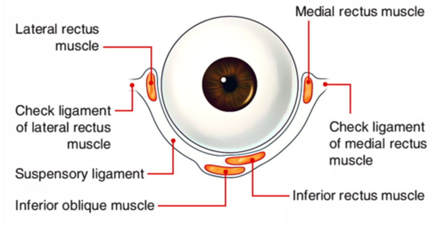
lateral check ligament
- transverse dense connective tissue that is an expansion of the sheath of the lateral rectus
- attaches to zygomatic bone
- prevents overaction of lateral rectus
orbital septum system
web of interconnecting connective tissue septa
-anchors and supports EOMs, nerves and blood vessels
orbital nerves
II optic nerve, III oculomotor, IV trochlear, V1 ophthalmic, V2 maxillary, VI abducens
extraocular muscles
control movement of globe
recti muscles of eye
Medial Rectus (MR)
Lateral Rectus (LR)
Superior Rectus (SR)
Inferior Rectus (IR)
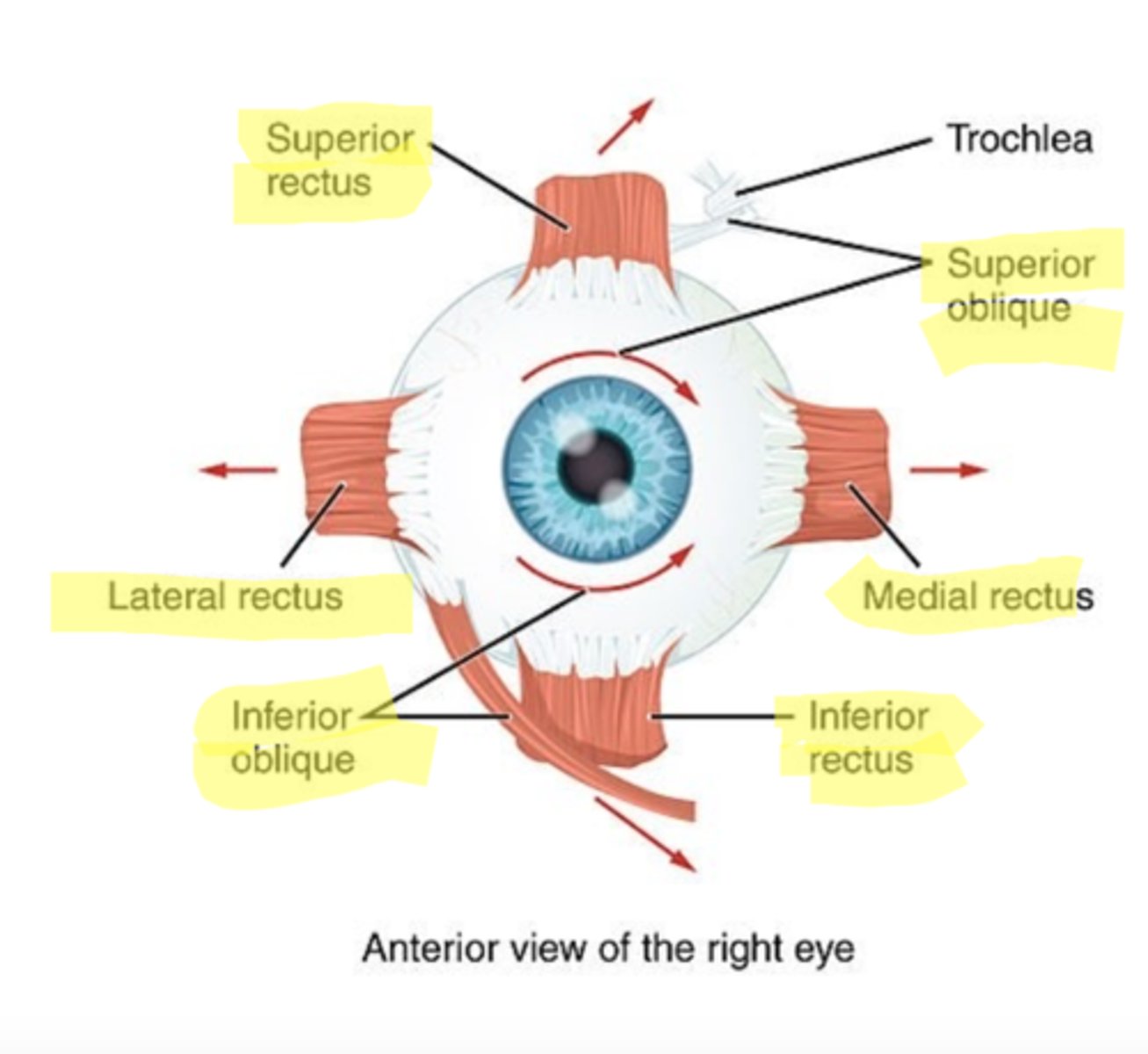
Oblique muscles of the eye
Superior Oblique (SO)
Inferior Oblique (IO)

levator palpebrae
elevates eyelid
Superior and inferior tarsal muscles
controls movement of eyelid
orbital fat
cushions eye, protects vessels and nerves of orbit
-surrounds ON
-separated muscles in the orbit from the orbital walls
Preaponeurotic Fat Pad
held in place by orbital septum