Ch 13 The Circulatory System

0.0(0)
Studied by 0 people
call kaiCall Kai
Locked
learnLearn
examPractice Test
spaced repetitionSpaced Repetition
heart puzzleMatch
flashcardsFlashcards
GameKnowt Play
Card Sorting

1/101

encourage image

There's no tags or description

Looks like no tags are added yet.

Last updated 9:02 PM on 8/13/26
Name
Mastery
Learn
Test
Matching
Spaced
Call with Kai
Chat

No analytics yet

Send a link to your students to track their progress

102 Terms

1
New cards
The main function of the circulatory system
Transports oxygen and nutrients to cells; removes carbon dioxide and waste products from cells.
2
New cards
The four main parts of the circulatory system
The heart, blood vessels, the blood, and the lymphatic system.
3
New cards
Pericardium
The fluid-filled membrane or sac that surrounds and protects the heart.
4
New cards
Systole
The contraction phase of the heart cycle.
5
New cards
Diastole
The relaxation phase of the heart cycle.
6
New cards
Coronary arteries and veins
Surface vessels responsible for supplying blood directly to and from the heart muscle.
7
New cards
The four internal chambers of the heart
Right atrium, left atrium, right ventricle, and left ventricle.
8
New cards
Atrioventricular (AV) valves
The bicuspid (mitral) and tricuspid valves, located between the atria and the ventricles.
9
New cards
Semilunar valves
The pulmonic and aortic valves, located between the ventricles and the major exiting blood vessels.
10
New cards

"Lub-dub" heart sounds

Sounds caused by the sequential closing of the heart valves.

  • Lub = closing of the bicuspid and tricuspid valves

  • Dub = closing of the semilunar valves

11
New cards
Pulmonary circulation pathway
Right atrium -> Tricuspid valve -> Right ventricle -> Pulmonic valve -> Pulmonary artery -> Lungs -> Pulmonary vein -> Left atrium.
12
New cards
Systemic circulation pathway
Left atrium -> Mitral/Bicuspid valve -> Left ventricle -> Aortic valve -> Aorta -> Body tissues.
13
New cards
Oxygen status of the pulmonary artery
Carries deoxygenated blood from the right ventricle to the lungs.
14
New cards
Oxygen status of the pulmonary vein
Carries oxygenated blood from the lungs back to the left atrium.
15
New cards
Portal circulation
A specialized circulatory pathway that serves only the internal organs, carrying substances on which those organs react.
16
New cards
Microcirculation flow pattern
Arteries -> Arterioles -> Capillaries -> Venules -> Veins.
17
New cards
Sinoatrial (SA) node
The natural pacemaker of the heart, located in the right atrium, which initiates the electrical impulse.
18
New cards
Atrioventricular (AV) node
The node that receives the electrical impulse from the SA node and transmits it to cause ventricular contraction.
19
New cards
Cardiac rhythm disorders
Conditions involving an interruption in the electrical impulse or a deviation in the regularity of the heartbeat.
20
New cards
Artificial pacemaker
A device used to cause the heart to contract effectively when drug therapy fails to correct rhythm disorders.
21
New cards
Vagus and accelerator nerves
The two primary nerves responsible for controlling the rate of the heart.
22
New cards
Three types of blood vessels
Arteries, veins, and capillaries.
23
New cards
Main systemic arteries
Aorta, gastric, splenic, renal, carotid, pulmonary, brachial, radial, ulnar, femoral, tibial, and dorsalis pedis.
24
New cards
Main systemic veins
Tibial, saphenous, femoral, common iliac, inferior and superior vena cava, jugular, brachial, cephalic, and pulmonary.
25
New cards
Superior vena cava
The major vein returning deoxygenated blood to the heart from the head and upper extremities.
26
New cards
Inferior vena cava
The major vein returning deoxygenated blood to the heart from the lower extremities and abdomen.
27
New cards
Components of blood
Plasma, red blood cells (erythrocytes), white blood cells (leukocytes), and platelets (thrombocytes).
28
New cards
Four main blood types
A, B, AB, and O.
29
New cards
Average adult blood volume
Between 8 to 10 pints.
30
New cards
Type and crossmatch
The mandatory testing process performed before a blood transfusion to ensure compatibility between donor and recipient.
31
New cards
Rh factor
An inherited antigen on red blood cells; its presence makes a person Rh-positive, and its absence makes them Rh-negative.
32
New cards
Cholesterol
A product of fat metabolism; total, LDL, and HDL levels are evaluated to assess the risk of a vascular event.
33
New cards
Triglycerides
Fats normally present in the body; high levels are clinically associated with heart disease and atherosclerosis.
34
New cards
Cardiovascular diagnostic tests
Arteriography, cardiac catheterization, stress testing, Doppler ultrasound, echocardiogram, ECG, heart scan, MUGA scan, stress Cardiolite, TEE, venogram, and carotid ultrasound.
35
New cards
Congestive Heart Failure (CHF)
A condition that impacts multiple body systems, specifically the circulatory, respiratory, integumentary, and digestive systems.
36
New cards
Leukemia
A cancer of the blood-forming tissues that involves the circulatory, integumentary, immune, and skeletal systems.
37
New cards
Arteriosclerosis vs. Atherosclerosis
Arteriosclerosis is the general hardening of the arteries; atherosclerosis is the specific buildup of fatty plaque deposits.
38
New cards
Carditis types
Pericarditis (outer sac inflammation), myocarditis (muscle inflammation), and endocarditis (inner lining inflammation).
39
New cards
Anemia
Lack of red blood cells and/or hemoglobin
40
New cards
Aneurysm
A bulging arterial wall that may produce palpitations or tear
41
New cards
Angina
Severe chest pain from a coronary artery spasm
42
New cards
Cardiac arrest
Complete and sudden cessation of heart action
43
New cards
Arrhythmia
Irregular heart rhythm
44
New cards
Arteriosclerosis
Rigid arterial walls; causes hypertension
45
New cards
Atherosclerosis
Fatty deposits on the lining of blood vessels
46
New cards
Athletic heart syndrome
Pounding heartbeats after exercise, enlarged heart, slow pulse
47
New cards
CVA
Confusion, weakness of one side, visual changes, paralysis, personality change
48
New cards
CHF
Congestion of blood in the circulatory system; edema of extremities and lungs
49
New cards
Coronary artery disease
Tightness of chest, substernal chest pain radiating down left arm, nausea and vomiting, perspiration, fainting
50
New cards
Embolus
A circulating foreign substance in a blood vessel
51
New cards
Endocarditis
Vegetative growths or inflammation of inner heart structures
52
New cards
Hypertension
Blood pressure consistently above normal
53
New cards
Hypotension
Consistently low blood pressure
54
New cards
Leukemia
Excessive WBCs, bruising, fatigue, painful lymph nodes
55
New cards
Murmur
A gurgling or swishing sound heard upon auscultation of the heart
56
New cards
MI
Severe, crushing pain radiating through chest to neck and jaw and down left arm; nausea, dyspnea
57
New cards
Myocarditis
Mild chest soreness, fever, dyspnea, palpitations, feeling of pressure
58
New cards
Pericarditis
Sharp, sudden pain at sternum radiating to back of shoulders and arms; decreases when erect or leaning forward
59
New cards
Phlebitis
Localized inflammation of a vein
60
New cards
Sickle cell anemia
Abnormally shaped red blood cells, enlarged liver, pallor, painful crisis periods
61
New cards
Stasis ulcer
Skin breakdown from inadequate circulation
62
New cards
Thrombophlebitis
Inflamed vein lining with thrombus formation, severe pain, fever, chills, and discoloration of involved extremity
63
New cards
Varicosities
Dilated, twisted veins, inefficient valves, leg cramps
64
New cards
Four major parts of the circulatory system
Heart; Blood vessels; Blood; Lymphatic system
65
New cards
Difference between pulmonary and systemic circulation
Pulmonary circulation flows blood from the right ventricle through the right pulmonary artery to the lungs and back to the left atrium. Systemic circulation flows blood from the left ventricle through the rest of the body to the right atrium.
66
New cards
Where the "Lub" heart sound is auscultated
Apex of the heart
67
New cards
Where the "Dub" heart sound is auscultated
Second intercostal space on each side of the sternum
68
New cards
Location and action of the natural pacemaker (SA node)
Located in the right atrium. It initiates the heartbeat; causing both atria to contract and sending impulses to the AV node; bundle of His; and Purkinje fibers to contract the ventricles.
69
New cards
Heart block
Occurs when the impulse from the SA node does not carry over to the AV node. Atria beat at a normal rate while ventricles contract slowly at about 40 beats per minute.
70
New cards
Fibrillation
A life-threatening condition caused by a breakdown of rhythm and contraction strength due to pacemaker malfunction; resulting in an uncoordinated and ineffective heartbeat.
71
New cards
Basic cause of arrhythmia
An area of the heart (known as an ectopic pacemaker) "sparks" and stimulates an abnormal contraction of the myocardium.
72
New cards
Purpose and function of an artificial pacemaker
It generates an electrical impulse through an electrode catheter connected to a battery-powered pulse generator to cause a contraction when the SA node fails.
73
New cards
Pathway of blood through the body (tracing)
Superior/inferior vena cava -> right atrium -> tricuspid valve -> right ventricle -> semilunar valve -> pulmonary arteries -> lung arterioles/capillaries -> venules -> pulmonary veins -> left atrium -> bicuspid valve -> left ventricle -> semilunar valve -> aorta -> body arterioles/capillaries -> venules/veins -> back to vena cava.
74
New cards
Source and destination of portal circulation
Comes from single organs (stomach; pancreas; large/small intestines; spleen) and goes to the liver to be processed by liver cells.
75
New cards
Five things transported by blood through the body
Oxygen; Nutrients; Cell waste; Hormones; Minerals
76
New cards
What is plasma?
A straw-colored liquid that makes up over half the volume of blood.
77
New cards
What are some things that can be found circulating in plasma? (List 1-6)
Water, Calcium, Sodium, Potassium, Phosphorus, Bicarbonates.
78
New cards
What are some things that can be found circulating in plasma? (List 7-12)
Amino acids, Vitamins, Hormones, Enzymes, Glucose, Fatty acids.
79
New cards
What are some things that can be found circulating in plasma? (List 13-17)
Oxygen, Carbon dioxide, Fibrinogen, Serum albumin, Serum globulin.
80
New cards
What is the function of red blood cells?
They have hemoglobin, which carries oxygen to cells.
81
New cards
What is the function of white blood cells?
They destroy bacteria within tissue fluid, consume toxic substances, and provide immunity.
82
New cards
What is the function of platelets?
To clot blood.
83
New cards
Why is blood typed and crossmatched before being given to a patient?
To ensure compatibility. Agglutinin and agglutinogens must be different to prevent agglutination (clotting) and possible death.
84
New cards
Why is the Rh factor important in a transfusion?
If an Rh-negative person receives Rh-positive blood, their body produces antibodies. A second exposure will cause serious, reactive complications.
85
New cards
Why is the Rh factor important in pregnancy?
An Rh-negative mother with an Rh-positive baby may develop antibodies. In subsequent pregnancies, these antibodies can destroy the baby's RBCs, causing profound anemia.
86
New cards
What is the purpose of an Arteriograph?
Injecting dye to indicate the status of blood flow, malformed vessels, aneurysm, or hemorrhage.
87
New cards
What is the purpose of Cardiac catheterization?
To visualize by fluoroscope the internal heart structure and activity and to visualize the coronary arteries.
88
New cards
What is the purpose of Doppler ultrasonography?
To evaluate major blood vessels to determine deep vein thrombosis, peripheral aneurysms, and occluded carotid arteries.
89
New cards
What is the purpose of an Echocardiograph?
To evaluate cardiac function and structure, and to detect defects by means of sound waves.
90
New cards
What is the purpose of an Electrocardiogram (ECG/EKG)?
To identify heart rhythm, electrolyte imbalance, and conduction.
91
New cards
What is the purpose of a Holter monitor?
To detect irregularly occurring symptoms or evaluate the status of a recovering cardiac patient.
92
New cards
What is the purpose of a Venogram?
Detects the condition of deep veins of the legs, especially deep vein thrombosis.
93
New cards
What is the purpose of a MUGA scan?
To determine the condition of the myocardium.
94
New cards
Arteries
Structure: Have elastic fibers in their walls to expand and recoil to produce a pulse. Function: Carry oxygenated blood away from the heart.
95
New cards
Arterioles
Structure: Same as arteries, only smaller. Function: Carry blood from artery to capillary.
96
New cards
Capillaries
Structure: One-cell-thick walls, microscopic blood vessels. Function: Exchange of oxygen and carbon dioxide with the cells; permit WBCs to leave to attack bacteria and allow plasma to leave to add to the tissue fluid.
97
New cards
Venules
Structure: Small veins. Function: Carry blood from capillary to vein.
98
New cards
Veins
Structure: Have thinner walls than arteries and lack an elastic layer; have valves to assist the return flow of blood. Function: Carry deoxygenated blood back to the heart.
99
New cards

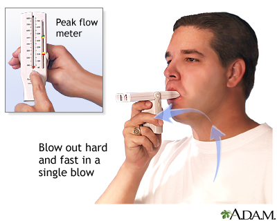
What does a peak flow meter measure? How do you record it?

peak expiratory flow (PEF), or how quickly a patient can force air out of their lungs in L/min

  • common to monitor asthma

  • helps to identify worsening airway obstruction before symptoms cecome severe

How to Perform a Peak Flow Measurement

Correct technique is essential for obtaining a useful peak flow value. Because the test is highly effort dependent, the respiratory therapist or clinician must explain and demonstrate the maneuver clearly.

The basic steps include:

  1. Set the peak flow meter to zero.

  2. Have the patient sit or stand upright.

  3. Instruct the patient to take the deepest breath possible.

  4. Have the patient place the mouthpiece in the mouth with the tongue out of the way.

  5. Make sure the lips form a tight seal around the mouthpiece.

  6. Instruct the patient to blow out as hard and fast as possible.

  7. Record the value.

  8. Repeat the maneuver until three acceptable efforts are obtained.

  9. Record the highest value.

The patient does not need to exhale completely for several seconds. Peak flow focuses on the fastest burst of air at the beginning of expiration, not the total amount of air exhaled.
<p>peak expiratory flow (PEF), or how quickly a patient can force air out of their lungs in L/min</p><ul><li><p>common to monitor asthma</p></li><li><p>helps to identify worsening airway obstruction before symptoms cecome severe</p></li></ul><p></p><p><strong>How to Perform a Peak Flow Measurement</strong></p><p>Correct technique is essential for obtaining a useful peak flow value. Because the test is highly effort dependent, the respiratory therapist or clinician must explain and demonstrate the maneuver clearly.</p><p><strong>The basic steps include:</strong></p><ol><li><p>Set the peak flow meter to zero.</p></li><li><p>Have the patient sit or stand upright.</p></li><li><p>Instruct the patient to take the deepest breath possible.</p></li><li><p>Have the patient place the mouthpiece in the mouth with the tongue out of the way.</p></li><li><p>Make sure the lips form a tight seal around the mouthpiece.</p></li><li><p>Instruct the patient to blow out as hard and fast as possible.</p></li><li><p>Record the value.</p></li><li><p>Repeat the maneuver until three acceptable efforts are obtained.</p></li><li><p>Record the highest value.</p></li></ol><p>The patient does not need to exhale completely for several seconds. Peak flow focuses on the fastest burst of air at the beginning of expiration, not the total amount of air exhaled.</p><p></p>
100
New cards

What does a pulse oximeter measure? How do you record it?

arterial oxygen saturation (SpO2) and pulse rate (PR)

  • normal is 95% or higher



