Lab Exam 3
 Call Kai
 Learn
Learn Practice Test
 Spaced Repetition
 Match
Match Flashcards
 Knowt Play
1/188

There's no tags or description
Looks like no tags are added yet.
Name | Mastery | Learn | Test | Matching | Spaced | Call with Kai | Chat |
|---|
No analytics yet
Send a link to your students to track their progress
189 Terms
DEJ
Name the structure at E
Carries spread along this area
Inner enamel epithelium
Name the structure (innermost line)
Separated from connective tissue by basal lamina
Enamel rods
Name the structure
Rows arranged circumferentially around long axis of tooth
Enamel rods
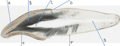
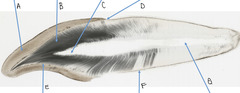
Name the structure at A
Can be described as: hexagonal, paddle, fish

Stria of Retzius
Name the structure at B
Developmental growth lines that create perikymata

Enamel
Name the structure at A
96% hydroxyapatite
Dentin
Name the structure at B
Transduction theory explain tooth sensitivity here

Pulp Chamber
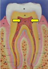
Name the structure at C
Greater amounts of secondary dentin at roof and floor of the coronal portion
CEJ
Name the structure at D
Rods run in horizontal direction
Cementum
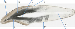
Name the structure at F
Cells forming this arise from the dental sac
Pulpal canal
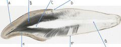
Name the structure at G

Dentinal tubules
Name the structure
Starts at right angles from pulpal surface

Cementocyte lacunae
Name the structure

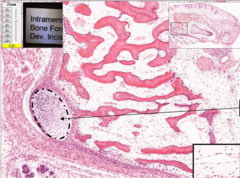
Intramembranous bone formation, developing incisor
Name the process
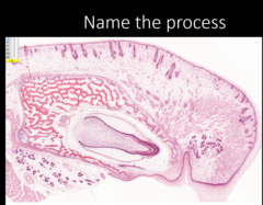
Alveolar bone
Name the tissue
Not present in histodifferentiation of epithelial cells

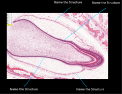
Developing tooth
Name the structure
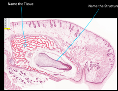
Enamel Organ
Name the structure in red
Center cells are called stellate reticulum
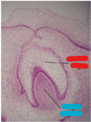
Dental papilla
Name the structure in blue
Undifferentiated mesenchymal cells
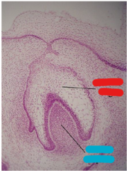
Pulp cavity
Name the structure (left line)
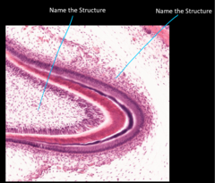
Enamel organ
Name the structure (right arrow)
Dental papilla induces oral epithelium to invaginate into this
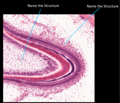
Outer enamel epithelium
Name the structure (outermost arrow)

Cervical loop
Name the structure (circle)
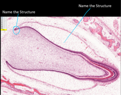
Stellate reticulum
Name the structure (line)
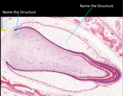
Odontoblasts
Name the Structure (1)
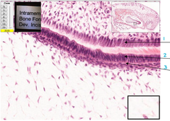
Ameloblasts
Name the structure (2)
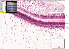
Stratum intermedium
Name the structure (3)
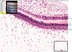
Stratum intermedium
Name the structure

Pulp cavity
Name the structure (1)

Odontoblast
Name the structure (2)
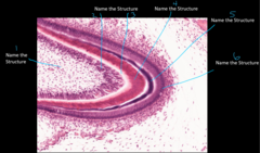
Predentin
Name the structure (3)
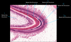
Dentin
Name the structure 4
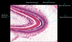
Enamel
Name the structure at 5
Major inorganic constituent by weight: phosphate
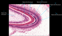
Ameloblast
Name the structure (6)
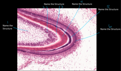
Stratified squamous epithelium
Name the structure the arrow is pointing to

Dental Follice
What is the arrow in circle 1 pointing to
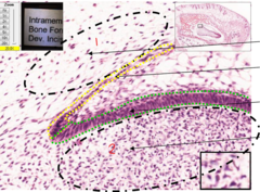
Outer enamel epithelium
What is the name of the structure in the yellow circle

inner enamel epithelium
What is the name of the structure in the green circle
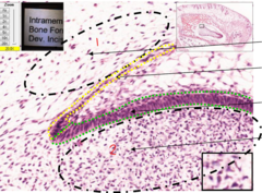
Dental papilla
What is the arrow in circle 2 pointing to
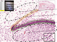
Merkel's cartilage
Name the structure the arrow is pointing to
Cervical loop
What is arrow 1 pointing at
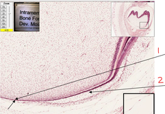
Stellate reticulum
What is arrow 2 pointing at
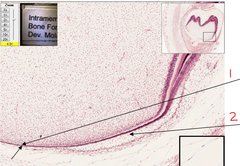
Dentinal tubules
What is the structure at 1
Ends perpendicular to DEJ & DCJ
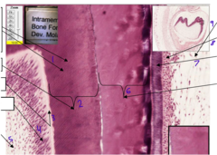
Dentin
What is the structure at 2
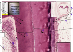
Predentin
What is the structure at 3

Odontoblasts
What is the structure at 4

Pulp
What is the structure at 5
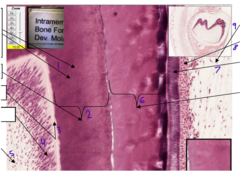
Enamel
Name the structure at 6
Defective formation: Amelogenesis imperfecta
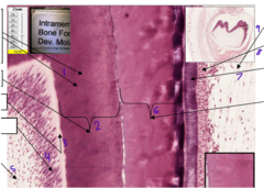
Ameloblasts
What are the cells at 7
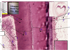
Stratum intermedium
What is the structure at 8
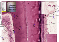
Stellate reticulum
What is the structure at 9
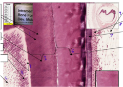
Tome's processes
What are the three arrows pointing to at 1
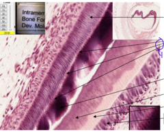
Enamel rods
What is the structure at 1
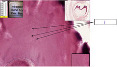
Developing alveolar bone
What is the structure at 1
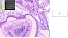
Ameloblasts
What is structure 1

Enamel
Name the structure at 2
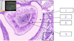
Dentin
What is structure 3
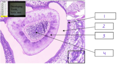
Pulp
What is structure 4
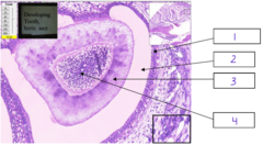
Tomes' processes
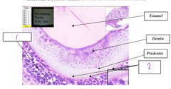
What is structure 1
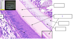
Odontoblasts
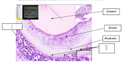
What is the structure 1
Dentinal tubules
What is the structure 2
Further apart and smaller in diameter at periphery
Striated duct
Name the structure

Excretory duct and stratified columnar epithleium
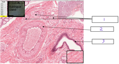
What is the structure at 1 and what epithelium lines it?
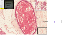
Auriculotemporal nerve branch
What is the structure at 1
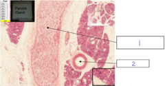
Muscular artery
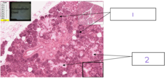
What is the structure at 2

Serous glands
What is the structure at 1
Mucous glands
Name the structure

Excretory duct
Name the structure at A
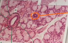
Muscular artery
What is the structure at 2

Striated duct
Name the structures
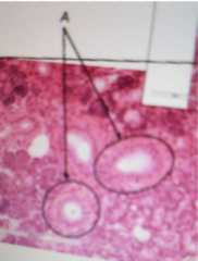
Serous demilunes
Name the structures
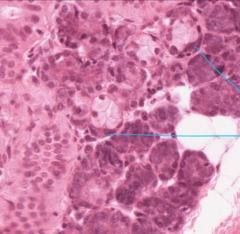
Mucous gland
What is the structure at 1
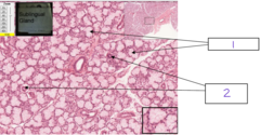
Intercalated ducts
What is the structure at 2

Nerve ganglia
What is the structure at 1
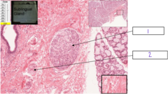
Peripheral nerve
What is the structure at 2
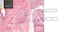
Peripheral nerve
What is the structure at 1

Muscular artery
What is the structure at 2
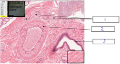
Excretory duct
What is the structure at 3
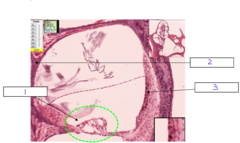
Cochlea
What is the structure at 1
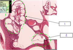
Scala vestibuli
What is the structure at 1
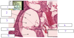
Reissner's (vestibular) membrane
What is the structure at 2
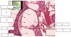
Scala media (cochlear duct)
What is the structure at 3

Spiral ligament
What is structure at 4
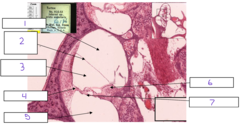
Organ of corti
Name the organ at 1
Has stereocilia hair cells that are connected to each other by ankle and tip links which bind microvilli to kinocilium
Scala tympani
What is the structure at 5
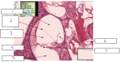
Spiral limbus
What is the structure at 6

Basilar membrane
What is the structure at 7
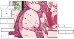
Spiral lamina
What is the structure at 2

Stria vascularis
What is the structure at 3
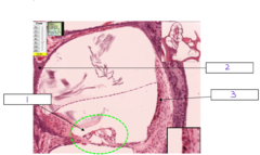
Pillar cells (surround inner tunnel)
What is the structure at 1
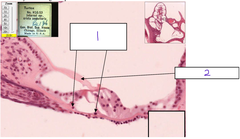
Mucous acini
Name
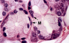
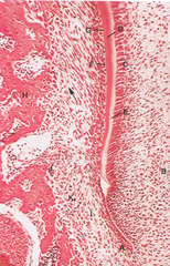
Dental papillae
B
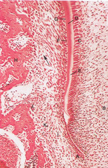
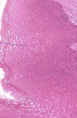
stratified squamous epithelium, non-keratinized
Identify the tissue
Odontoblasts
C
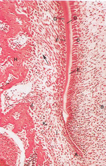
HERS
A
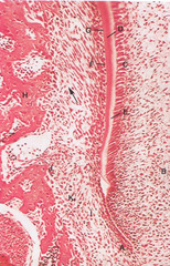
Dentin of root
D
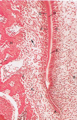
Predentin
E
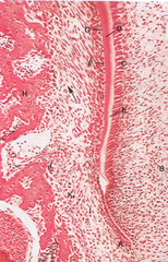
Cementoblasts
F

Developing cementum
G
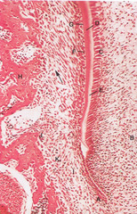
Alveolar bone
Name the structure at H
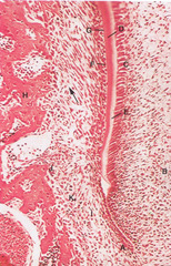
Follicle
I