19. Skin Cancer
 Call Kai
 Learn
Learn Practice Test
 Spaced Repetition
 Match
Match Flashcards
 Knowt Play
1/36

Earn XP
Description and Tags
https://www.youtube.com/watch?v=vqq7X3XB7Do
Name | Mastery | Learn | Test | Matching | Spaced | Call with Kai | Chat |
|---|
No analytics yet
Send a link to your students to track their progress
37 Terms
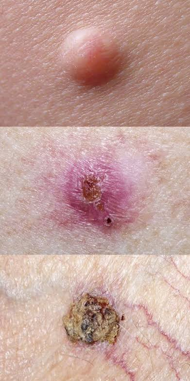
basal cell carcinoma
pearly papule with telangiectasias
rolled borders central ulceration ("rodent ulcer")

basal cell carcinoma
Most common skin cancer!
usually slow-growing and locally invasive, with metastasis being extremely rare.
orgin: Basal cells of the epidermis
#1 RF: sun exposure
Older fair-skinned patient + sun-exposed face/nose + slowly enlarging pearly papule with rolled borders and telangiectasias
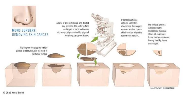
Surgical excision;
Mohs surgery — (face, recurrent, high-risk lesion)
first line treatment for BCC
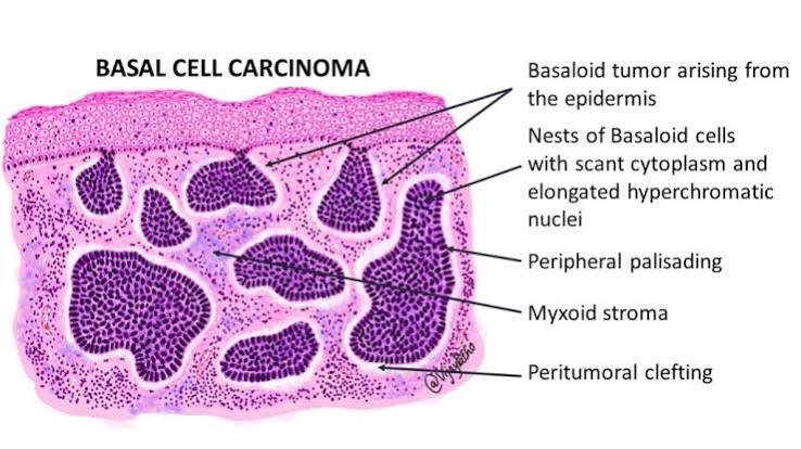
BCC = 3 P's:
Pearly papule
Palisading cells
Poor metastatic potential
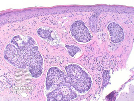
histo: Nests of basaloid cells with peripheral palisading
Topical | MOA | High-yield use |
|---|---|---|
Imiquimod ⭐ | TLR-7 agonist → stimulates local immune response | Superficial BCC |
5-Fluorouracil (5-FU) | Inhibits thymidylate synthase → ↓ DNA synthesis | Superficial BCC |
topicals for basal cell carcinomas superficial lesions (2)
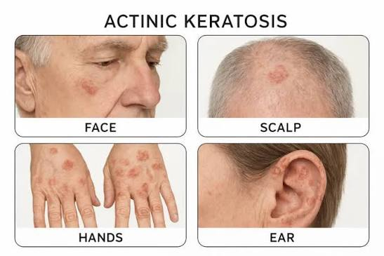
Actinic = related to sunlight / UV radiation ☀
Actin- = ray/radiation
-ic = pertaining to
Keratosis = abnormal keratin/thickening of the outer skin
Kerat- = keratin / horny tissue
-osis = abnormal condition
➡ Actinic keratosis = UV-induced abnormal keratinization/thickening of the skin.
Older patient + sun-exposed skin + rough, gritty/sandpaper lesion
& is a precursor of skin SCC
One AK → cryotherapy (liq. nitrogen)
Many AKs → topical 5-FU
treatment for actinic keratosis
#1 cumulative Sun exposure (UVB)
Actinic keratosis (precursor)
Immunosuppression (transplant patients)
Chronic wounds/scars (Marjolin ulcer)
HPV (especially genital SCC)
Smoking (lip/oral SCC)
risk factors of skin SCC
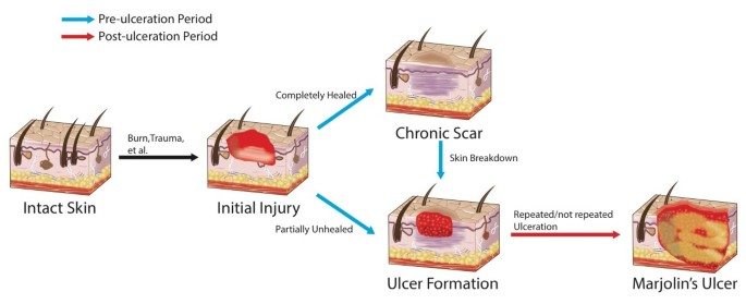
Marjolin ulcer
SCC arising from chronic wound such as previous burn scar, tends to be very aggressive
HPV 5 & 8 → skin SCC (epidermodysplasia verruciformis)
HPV 16 & 18 → cervical/anogenital SCC
HPV is a risk factor for this type of skin cancer

clinical appearance of SCC
Scaly, erythematous plaque or nodule
May ulcerate or bleed
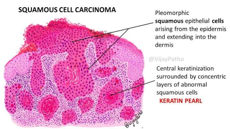
2 classic histologic findings are:
Keratin pearls
Concentric layers of keratinizing malignant squamous cells
Look like round, pink/eosinophilic “onion rings”
Intercellular bridges
Connections between adjacent squamous cells caused by desmosomes
Other findings include atypical keratinocytes, pleomorphism, hyperchromatic nuclei, mitotic figures, and invasion through the basement membrane into the dermis.
#1 UV exposure, especially intermittent intense sunburns
Fair skin, light eyes
Multiple or atypical nevi
major risk factors for melanoma
CDKN2A (p16)
Loss → impaired cell-cycle regulation → ↑ melanoma risk
Also associated with increased pancreatic cancer risk
genetic marker associated with familial melanoma
BRAF V600E
mutation associated with sporadic melanoma
pancreatic cancer
cancer associated with familial melanoma syndrome (CDKN2A (p16))
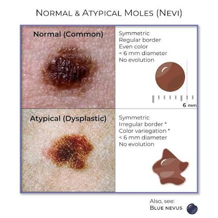
dysplastic nevi
atypical moles that can develop into skin cancer
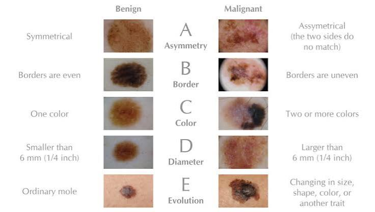
define the ABCDE appearance of melanoma
>6 mm
(1/4 inch)
diameter associated with melanoma
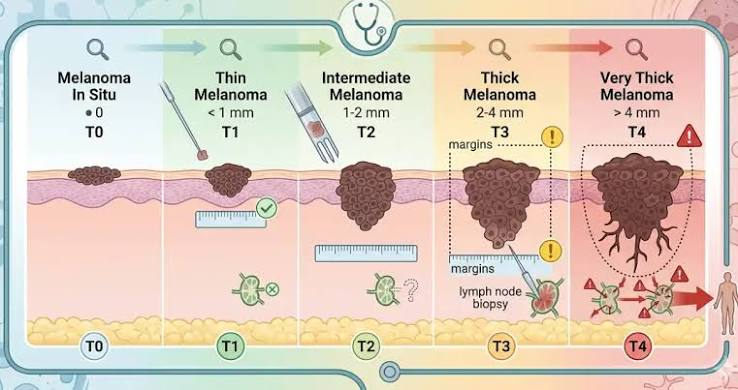
Breslow thickness
measures depth of tumor invasion in mm
breslow thickness (depth)
most important prognostic factor for melanoma
<1mm
low risk breslow depth
1-2 mm
intermediate risk breslow depth
>4mm
high risk breslow depth

Surgical margins &
Need for sentinel lymph node biopsy
clinical implications of breslow depth
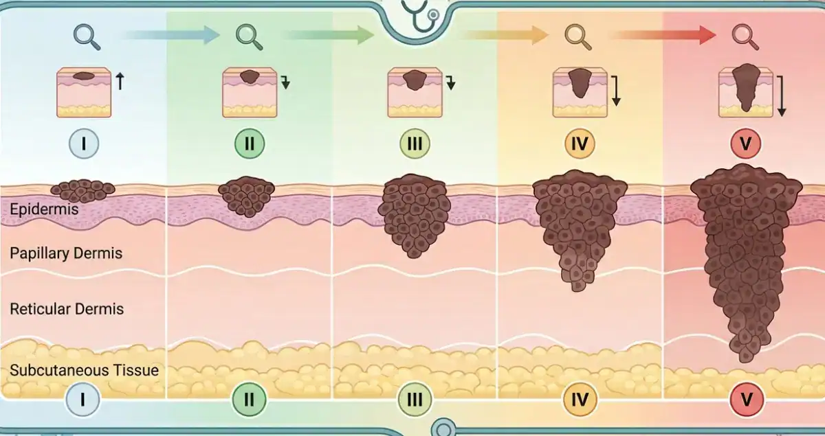
Clark Levels of Melanoma Staging
I: intraepidermal
II: in papillary dermis
III: fills papillary dermis
IV: reticular dermis
V: enters subcutaneous tissue
0.5-1 cm
recommended surgical margins for in situ melanoma
2cm
recommended surgical margins for >2mm depth
Mohs is NOT standard (except select in situ cases on face)
is Mohs standard therapy for all skin cancers?
Breslow thickness | Sentinel lymph node biopsy? |
|---|---|
<0.8 mm AND no ulceration | ❌ Usually NO |
<0.8 mm + ulceration | ⚠ Consider SLNB |
0.8–1.0 mm | ⚠ Discuss/consider SLNB |
>1.0 mm | ✅ Recommend/offer SLNB ⭐ |
& >0.8 —> consideration
indications for sentinel node biopsy in skin cancer
Immune checkpoint inhibitor–based therapy is a major first-line approach. ⭐
Drug/class | MOA |
|---|---|
Pembrolizumab / Nivolumab ⭐ | Anti–PD-1 |
Nivolumab + ipilimumab | PD-1 + CTLA-4 blockade |
Nivolumab + relatlimab | PD-1 + LAG-3 blockade |
If the melanoma has a BRAF V600 mutation, targeted BRAF + MEK inhibition (e.g., dabrafenib + trametinib) is another important first-line option.
Board answer:
Metastatic melanoma → immune checkpoint inhibitor (anti–PD-1) ⭐
BRAF V600+ metastatic melanoma → BRAF inhibitor + MEK inhibitor is a key targeted option.
first line medical treatment of metastatic melanoma
Immunosuppressed patients = dramatically increased risk
-esp with SCC
why do transplant patients have a higher risk of skin cancer
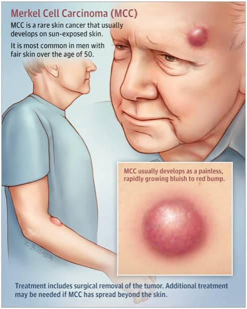
merkel cell carcinoma
rare, aggressive neuroendocrine skin cancer
Often on sun-exposed areas (head/neck)
Rapidly growing, painless, firm red/pink/purple nodule
“Painless, rapidly growing violaceous nodule”
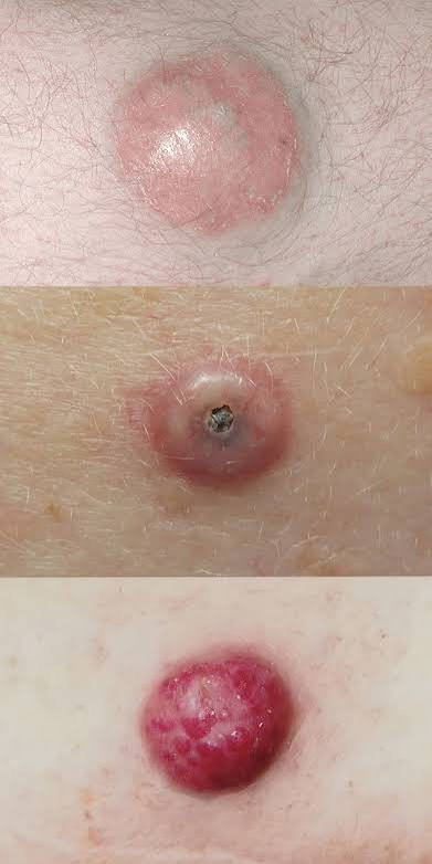
Merkel cell polyomavirus (MCPyV)
what virus maybe associated with Merkel Cell Carcinoma?

Merkel Cell Carcinoma
Small round blue cells |
markers: CK20+ classically in a perinuclear dot-like pattern; neuroendocrine markers such as synaptophysin+
Wide local excision + SLNB ⭐
Radiation therapy (often used)
Immunotherapy for advanced disease
treatment for merkel cell carcinoma
systemic therapy, NOT automatic dissection
mgmt of a positive sentinel lymph node biopsy