Hip: goniometry and muscle testing
 Call Kai
 Learn
Learn Practice Test
 Spaced Repetition
 Match
Match Flashcards
 Knowt Play
1/91

Earn XP
Description and Tags
Fundamentals of musculoskeltal assessment techniques chapter 12
Name | Mastery | Learn | Test | Matching | Spaced | Call with Kai | Chat |
|---|
No analytics yet
Send a link to your students to track their progress
92 Terms
Range of hip flexion
From 0 to 115 to 125 degrees into a position of hip flexion with knee flexion.
Position for measuring hip flexion
Preferred: Subject lies supine with the opposite lower limb flat on the table top (Fig. 12-1). e Alternate: Subject is in sidelying position on the opposite side (Fig. 12-2).
Goniometric alignment for hip flexion
Axis. Placed on the lateral aspect of the hip approximately a fingerbreadth anterior and superior to the greater trochanter of the femur.
Stationary Arm. Placed parallel to the long axis of the trunk, in line with the greater trochanter of the femur.
Moving Arm. Placed along the lateral midline of the femur toward the lateral epicondyle.
What happens to the femoral head as the hip moves into flexion?
As the hip moves into flexion, the head of the femur glides in a posterior and inferior direction.
Start position for measuring hip flexion
The start position for measuring hip flexion is when the subject is supine, with the knees extended and the feet positioned flat on the table.
End position for measuring hip flexion
The knee is brought toward the chest until the hip is flexed maximally, which indicates the endpoint of the movement.
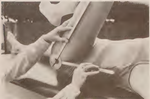
End position for alternate side lying position of hip flexion
The knee is drawn toward the chest while lying on the alternate side, achieving maximal hip flexion without rotation of the pelvis.
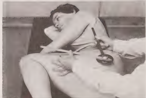
Precautions when testing hip flexion
Precautions: Allow the knee to flex to prevent a stretch on the hamstring muscles. Keep the opposite lower limb flat on the table to control posterior pelvic tilt. e Avoid lumbosacral motion.
Expected range of motion with hip extension
The expected range of motion for hip extension typically spans from 0 degrees to about 30 degrees, occurring when the hip moves behind the body in a straight line.
When measuring hip extension the ASIS…
must remain flat on the table to ensure that the motion is occuring at the hip joint and not at hte lumbar vertebrae to maintain proper alignment and isolate hip flexion.
What happens to the acetabulum during extension and hyperextension of the hip
The head of the femur glides in an anterior and inferior directionwithin the acetabulum, allowing for increased stability and range of motion in the hip joint.
Hyperextension of the hip shows that
it is an extension of the lumbar vertebrae with forward tilt of the pelvis.
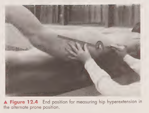
What is the end position for measuring hip hyperextension in prone
is when the knee is extended and the hip is positioned in the maximal extension with the pelvis remaining neutral.
What is the end position for measuring hip hyperextension in side lying
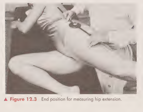
Precautions when measuirng hip extension
Avoid lumbar extension. Keep the knee joint extended to prevent stretch on the rectus femoris muscle. Avoid anterior pelvic tilt.
What is the goniometric alignment for hip extension?
Same as for hip flexion
Axis. Placed on the lateral aspect of the hip approximately a fingerbreadth anterior and superior to the greater trochanter of the femur.
Stationary Arm. Placed parallel to the long axis of the trunk, in line with the greater trochanter of the femur.
Moving Arm. Placed along the lateral midline of the femur toward the lateral epicondyle.
What happens to the head of the femur in hip abduction
the head of the femur gliding in an inferior direction in the acetabulum of the pelvis. Accompanied by a contralateral pelvic tilt, which is not included in the measurement.
What is the normal range of hip abduction?
Typically ranges from 0 to 45 degrees in adults.
Correct position to test hip abduction
Position. Subject lies supine with the lower limb to be tested in the anatomical position
Goniometric alignment of measuring hip abduction
is achieved by placing the axis of the goniometer over the anterior superior iliac spine, aligning the stationary arm with the opposite ASIS, and the moving arm along the midline of the femur.
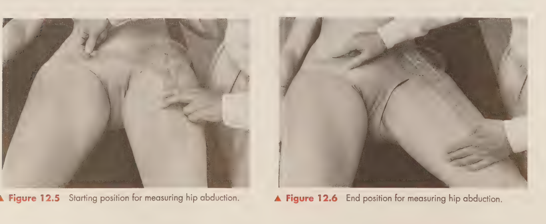
Precautions when testing abduction of the hip
Precautions Prevent lateral rotation at the hip joint. Prevent lateral tilt of the pelvis (hip hiking).
What happens to the femoral head during adduction?
The head of the femur glides in a superior direction in the acetabulum. ipsilateral tilt of the pelvis accompanies this, which is not included in the measurement.
What is the normal range of hip adduction?
The normal range of hip adduction is typically considered to be between 20 to 30 degrees, measured from the midline of the body.
Gonionmetric alignment during hip adduction
is achieved by placing the axis of the goniometer over the anterior superior iliac spine, aligning the stationary arm with the opposite ASIS, and the moving arm along the midline of the femur.
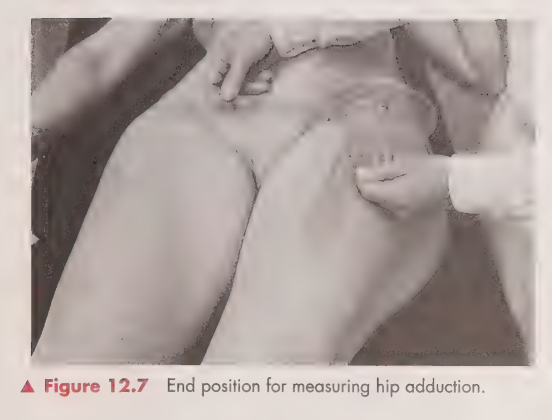
What happens to the head of the femur during medial rotation?
Glides in the posterior direction of the acetebulum
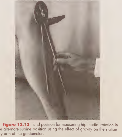
What are the positions for medial rotation? (Supine)
Preferred: Subject lies supine with the hip joint in the anatomical position and the knee flexed to 90 degrees over the edge of the table. The opposite hip and knee are flexed, and the foot lies flat on the table
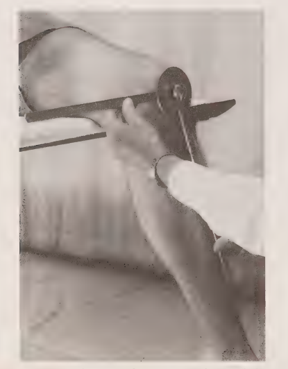
What are the positions for medial rotation? (sitting)
Subject sits with the knee flexed over the edge of the table. The femur is parallel with the table top, and the hip joint is flexed 90 degrees.
What are the positions for medial rotation? (prone)
Subject lies prone with the hip joint in the anatomical position and the knee joint flexed 90 degrees
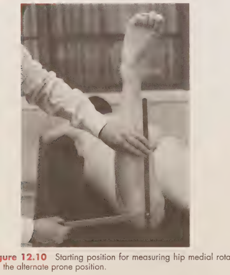
Goniometric alignment for medial rotation of the hip?
Stationary Arm Preferred:
Placed parallel to the table top.
Alternate: Placed perpendicular to the floor or parallel to the midline of the tibia.
Moving Arm. Placed along the crest of the tibia to a point midway between the malleoli.
Precautions when testing medial rotation?
Precautions » Avoid rotation and lateral tilting of the pelvis toward the same side. Prevent the pelvis from lifting off the table.
In sitting:
In the sitting position, prevent contralateral trunk flexion. e Prevent adduction at the hip joint.
In lateral rotation, where does the head of the femur glide?
As the motion of hip lateral rotation occurs, the head of the femur glides in an anterior direction in the acetabulum.
Goniometric alignment for lateral rotation of the hip
Stationary Arm Preferred:
Placed parallel to the table top. Alternate: Placed perpendicular to the floor or parallel to the midline of the tibia. Moving Arm: Placed along the crest of the tibia to a point midway between the malleoli.
Position for measuring lateral rotation in sitting
Subject lies supine with the stationary arm of the goniometer perpendicular to the floor
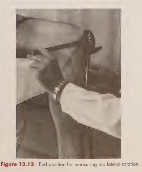
Position for measuring lateral rotation in prone
Subject lies prone with the test knee joint flexed to 90 degrees
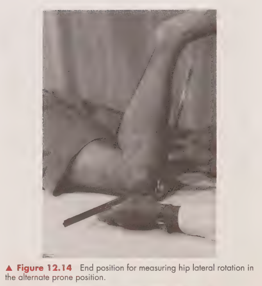
Position for measuring hip lateal rotation in supine
Preferred: Subject is supine, with the knee flexed to 90 degrees over the edge of the table. The nontest hip and knee joint are flexed, and the foot is flat on the table
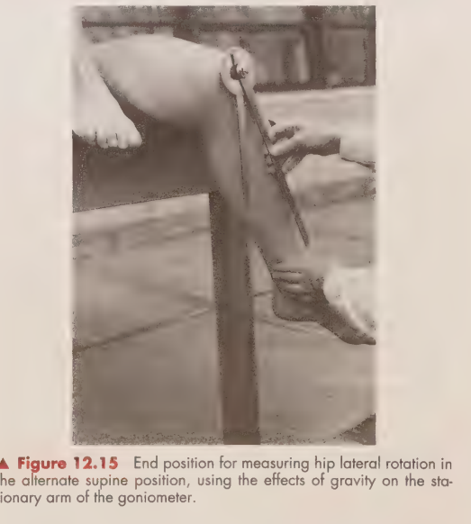
Precautions during medial rotation
Avoid rotation of the pelvis toward the opposite side. e Prevent hip joint adduction or flexion. e Avoid contralateral tilt of the pelvis. ¢ Prevent ipsilateral trunk flexion or rotation.
What level of flexion does the psoas majpor and the illiacus muscles produce?
30 degrees from a starting position of 90 degrees
How would you palpate the psoas major?
With the subject seated and bent forward to relax the abdominal muscles, place fingers deep into the abdomen below the ribs and above the iliac crest, gently pushing toward the posterior abdominal wall
How would you palpate the ilacus?
It is difficult to palpate the iliac muscle, which lies on the iliac fossa.
Position for MMT of the psoas major
Against gravity (AG): Subject sits with the knees flexed and the pelvis in posterior tilt. Hands should be holding on to the edge of the table to maintain balance and stabilization. The position should also allow for deep abdominal access, ensuring the fingers can reach the muscular area.
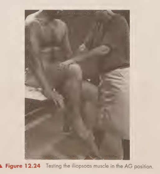
Position for testing psoas major
Against gravity (AG): Subject sits with the knees flexed and the pelvis in posterior tilt. Hands should be holding on to the edge of the table
Applying resistance to the psoas major msucle
Hand on oppsite hip. Other hand pressed on the knee and then that knee bends up and lifts. start in a lunge kind of position
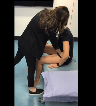
Manual muscle testing of the iliopsoas position
Lying supine position, riase their hip to flex it so their knee and hip is in a 90 degrees position. Abduction with lateral rotation and get them to bring their knee to their chest- while you resist pulling down
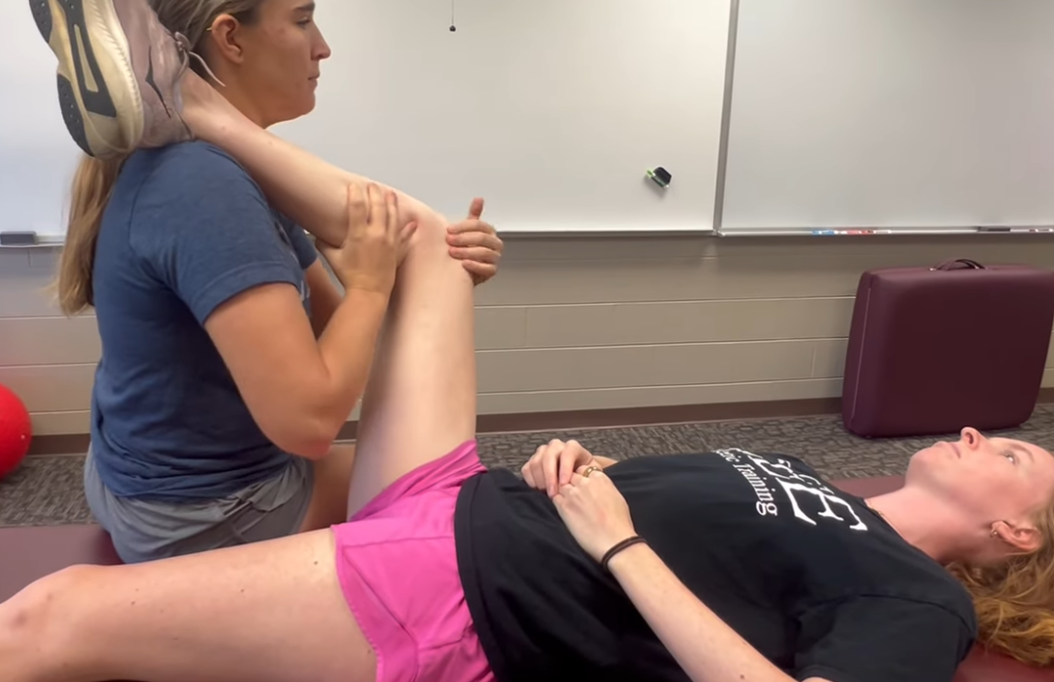
Muscle substitutions for iliopsoas
Substitutions: The hip abducts and laterally rotates as the sartorius flexes.The hip abducts and medially rotates as the tensor fasciae latae flexes. Rectus femoris is an accessory muscle to hip flexion.
How would you palpate the sartorius muscle?
Go just below the ASIS
Position for MMT of sartorius
AG: Subject sits with the pelvis in posterior pelvic tilt and both knees flexed to 90 degrees off the edge of the table (Fig. 12-27). GM: Subject lies supine with the heel of the test limb on the opposite ankle; the nontest limb is in the anatomical position (Fig. 12-28).
Movement for testing sartorius
Subject brings the plantar surface of the heel to the opposite knee.
How would you apply resistance to the sartorius?
Applied to the medial malleolus to resist hip lateral rotation and on the lateral surface of the thigh proximal to the knee to resist flexion from other muscles
Observed substitutions for sartorius
Substitutions: Iliopsoas and rectus femoris produce straight hip flexion without abduction or lateral rotation. ¢ Tensor fascia lata produces hip flexion and abduction with medial rather than lateral rotation.
How would you palpate the gluteus maximus?
Palpate between the sacrum and the greater trochanter, with the hip in lateral rotation
What is the range of motion for the gluteus maximus?
125 to 140 degrees.
Position for MMT of gluteus maximus
Subject stands with trunk flexed over a table and knee flexed
Movement for testing the gluteus maximus?
Extension of the hip from a flexed position (knee is in the donkey position)
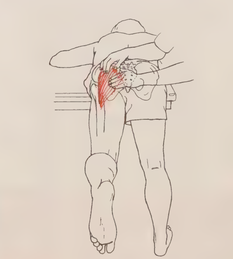
How would you apply resistance to the gluteus maximus?
Applied proximal to the knee joint on the posterior thigh. Starting in a lunge positiona and going up and the other arm is sitting over the hip
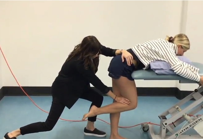
Stabilisation of the gluteus maximus
The pelvis is stabilized and hyperextension of the lumbar spine is prevented.
Observed substitutions for gluteus maximus
Hamstrings extend the hip without any action from the gluteus maximus. > Stabilize the lumbar vertebrae to prevent an increase in lordosis.
test range for gluteus medius and minimus
typically through to 45 degrees
How would you palpate the gluteus medius and maximus?
Gluteus Medius. Only the anterior and middle portions are palpable, either laterally below the crest of the ilium or immediately proximal to the greater trochanter (Fig. 12-32). Gluteus Minimus. It lies deep to the gluteus maximus and the gluteus medius and therefore is not palpable.
Position for MMT of gluteus medius and minimus?
Patient positioned on the side with the tested leg on top, hip extended and abducted.
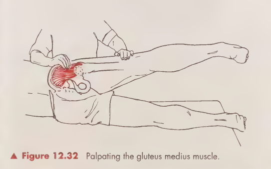
resistance for the medius and minimus
Applied proximal to the knee joint on the lateral side of the thigh.
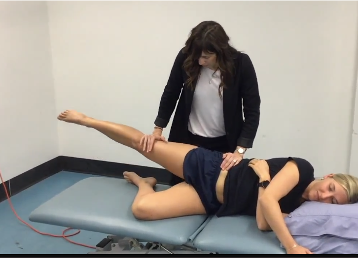
Subsitutions for testing medius and minimus
The quadratus lumborum and the lateral abdominal muscles tilt the pelvis laterally, giving the appearance of abduction. ° If the subject rolls slightly to the supine position, the tensor fasciae latae is in a more favorable position to abduct the hip because it will be in a flexed position. ¢ Gluteus maximus (superior portion) is an accessory to hip abduction.
How would you palpate the TFL?
Slightly below and alteral to the ASIS
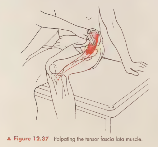
position for MMT of TFL?
Subject is in the sidelying position with the resting on the table in front of the nonnontest limb in the anatomical position.The test limb is in 45 degrees of hip flexion the knee is extended with the limb resting on the table in front of the non-lest limb. Make sure that the top is flexed
Movement, resistance and stabilisation
Movement. Abduction of the hip joint while maintaining 45 degrees of hip flexion. Resistance. Applied to the distal thigh on the lateral side. Stabilization. The pelvis is stabilized.
Substitutions observed with TFL
Prevent substitution by stabilizing the pelvis. The hip flexors produce only flexion at the hip joint with no abduction.
ADDUCTOR LONGUS, MAGNUS, AND BREVIS AND GRACILIS AND PECTINEUS MUSCLES
The adductor muscles as a group produce adduction of the hip with the hip extended through a test range of 20 to 25 degrees.
Adductor longus palpation
Adductor muscles group
Includes adductor longus, magnus, brevis, gracilis, and pectineus, which promote hip adduction with the hip extended through a test range of 20 to 25 degrees.
Function of adductor longus and brevis
These muscles may act as synergists in the flexion of the hip joint.
Role of adductor magnus (vertical portion)
Acts as a synergist to hip joint extension.
Palpation of adductor longus
Located on the medial side of the thigh immediately below the pubic arch.
Palpation of adductor magnus
Found along the medial aspect of the thigh in the middle to lower half.
Palpation of adductor brevis
Too deep for accurate assessment.
Palpation of gracilis
Located on the medial aspect of the knee.
Palpation of pectineus
Difficult to palpate and can be uncomfortable for the subject.
Position for MMT of adductor muscles (AG)
Subject is in the sidelying position with the nontest limb supported in 25 degrees of abduction.
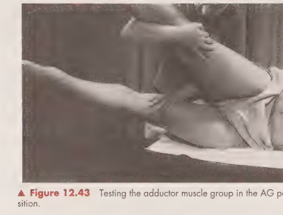
Position for MMT of adductor muscles (GM)
Subject lies supine with the opposite limb in 25 degrees of abduction and the test limb in slight abduction on a friction-free surface.
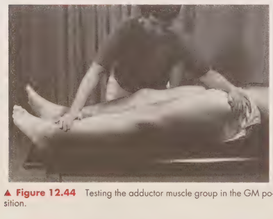
Movement for testing adductor muscles
Adduct the test hip toward the supported nontest limb approximately 25 degrees.
Resistance application for adductor muscles
Applied proximal to the knee joint to resist abduction.
Stabilization during adductor muscle testing
The pelvis is stabilized to prevent substitutions.
Substitutions in hip adduction testing
Hamstrings can substitute; hip adduction may be accompanied by lateral rotation.
Lateral rotator muscles
Includes obturator internus and externus, superior and inferior gemelli, quadratus femoris, and piriformis.
Function of lateral rotator muscles
They produce lateral rotation of the hip through a range of 45 degrees.
Palpation of piriformis
The tendon may be palpated approaching the greater trochanter posteriorly.
Position for testing lateral rotation (AG)
Subject lies supine with the knee flexed over the table, opposite hip and knee flexed.
Position for testing lateral rotation (GM)
Subject lies supine with the test limb’s knee extended and hip medially rotated.
Movement during lateral rotation testing
Lateral rotation of the hip, with knee moving beyond the midline in GM position.
Resistance application for lateral rotation
Applied to the distal leg proximal to the medial malleolus or proximal to the knee joint. You need to lunge up with them
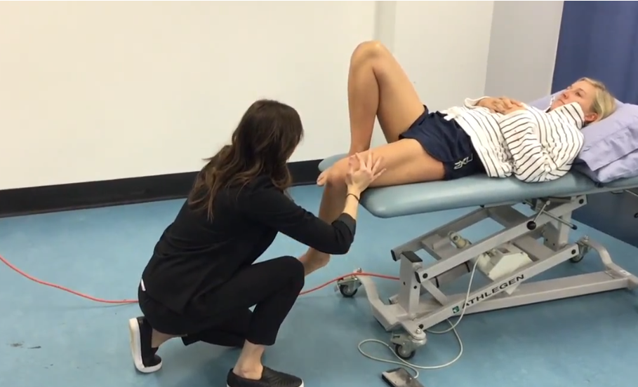
Stabilization during lateral rotation testing
The knee joint is stabilized on the lateral side.
Substitutions observed in lateral rotation testing
Elevation of the opposite buttock, foot inversion, knee flexion, or hip adduction may appear as lateral hip rotation.