Neuroscience: Brains, Neurons, Neural Conduction, Transmission, and the Blood-Brain Barrier
 Call Kai
 Learn
Learn Practice Test
 Spaced Repetition
 Match
Match Flashcards
 Knowt Play
1/39

Earn XP
Description and Tags
Flashcards testing core principles of neuronal function, membrane biophysics, neural conduction, synaptic transmission, and blood-brain barrier anatomy.
Name | Mastery | Learn | Test | Matching | Spaced | Call with Kai | Chat |
|---|
No analytics yet
Send a link to your students to track their progress
40 Terms
What are the main functional reasons why brains evolved? (3)
Brains evolved to generate adaptive behavior appropriate to current and predicted circumstances,
which includes managing the body energy budget to optimize functioning and stay alive.
Brain learns about circumstances by processing sensory
information from outside itself
What are the three basic jobs of the nervous system and the corresponding neuron classes that perform them?
Gather information about internal and external environments (performed by sensory neurons in the peripheral nervous system).
Process and integrate new information with stored information to produce actions (performed by interneurons in the central nervous system).
Produce actions in the internal or external environment (performed by motor neurons in the peripheral nervous system).
What is the difference between afferent and efferent neural pathways?
Afferent (toward): carries sensory information from the peripheral nervous system toward the central nervous system (CNS)
Efferent (away): carries motor information away from the CNS to muscles or glands (PNS)
How do neural conduction and neural transmission differ?
Neural conduction is the movement of an electrical signal within a single neuron, whereas neural transmission is the movement of a chemical signal from one neuron to another across a synapse.
What are the functions of microtubules and cytoplasm within a neuron?
Microtubules act as a structural skeleton to support neuron shape and provide tracks to transport materials; cytoplasm is the crowded, jelly-like watery fluid inside the cell.
How does the plasma membrane's selective permeability treat lipid-soluble versus water-soluble substances?
Most lipid-soluble (fat-soluble) substances dissolve directly through the membrane's phospholipid bilayer, whereas water-soluble substances cannot dissolve through the bilayer and require specialized protein doors (channels or transporters) to cross.
What are the quantitative values defined for a Dalton (Da) and a mole?
A Dalton (Da) equals 1.66×10⁻²⁴g, and a mole equals 6.022×10²³ particles.
What is the difference between a cation and an anion?
A cation is an ion with fewer orbiting electrons than protons in its nucleus, giving it a net positive electrical charge.
An anion is an ion with more orbiting electrons than protons, giving it a net negative electrical charge.
Why are ions water-soluble?
Water (H2O) is polar because shared electrons spend more time around the oxygen nucleus, making the oxygen end negative and the hydrogen end positive. This polarity exerts electrostatic pressure on ions, surrounding and pulling them apart to dissolve them.
Why are lipids not water-soluble?
Lipids are non-polar, so there is no electrical attraction between lipid molecules and polar water molecules. Because water molecules are strongly attracted to each other, lipids cluster together away from water.
How do phospholipids arrange themselves in an aqueous solution to form a plasma membrane?
They spontaneously form a bilayer ("fat sandwich") where the hydrophilic, weakly negatively charged phosphate heads face outward toward water, and the hydrophobic fatty acid tails face inward toward each other.
What is Electrostatic pressure?
The invisible force ions exert on each other
What is the typical resting membrane potential of a neuron?
The resting membrane potential is approximately −60mV to −70mV inside relative to the outside (or +60mV to +70mV outside relative to the inside).
What is the concentration distribution of sodium (Na+), chloride (Cl−), and potassium (K+) ions across a neuronal membrane at rest?
There is 10× more Na+ and 40× more Cl− outside the cell, and 50× more K+ inside the cell, along with trapped organic protein anions inside.
How does diffusion pressure drive particle movement across a membrane?
Particles constantly jiggle and bounce randomly off one another, resulting in a net drift from areas of higher concentration toward areas of lower concentration until concentration is equalized.
What are the specific movements and energy consumption of the sodium-potassium (Na+/K+) pump?
The pump uses ATP to continuously move three sodium ions (3Na+) out of the cell for every two potassium ions (2K+) it moves into the cell.
How do potassium (K+) and sodium (Na+) leak channels affect the resting membrane potential?
Many K+ leak channels allow potassium to leak out by diffusion, driving potential toward −90mV, while a few Na+ leak channels allow sodium to leak in, bringing the potential back to about −70mV.
What are the three functional shapes/states of voltage-gated sodium (Na+) channels?
Closed (reset state at resting potential)
Open (permeable to Na+ upon depolarization to the threshold of excitation)
Inactivated (refractory state triggered by depolarization to ∼+30mV to +40mV, preventing reopening until closed/reset).

Why do action potentials originate at the axon hillock rather than on the soma or dendrites?
Soma and dendrites lack voltage-dependent Na+ channels (containing only neurotransmitter-dependent channels), whereas the axon hillock contains the high concentration of voltage-dependent Na+ channels needed to initiate an action potential.
What is the difference between the absolute refractory period and the relative refractory period?
During the absolute refractory period, voltage-gated Na+ channels are inactivated and cannot reopen, preventing another action potential; during the relative refractory period, another action potential can occur but requires greater depolarization to overcome hyperpolarization.
Why is saltatory conduction faster and thriftier than conduction in unmyelinated axons?
In saltatory conduction, depolarization moves rapidly beneath insulating myelin sheaths by passive conduction, regenerating action potentials only at the nodes of Ranvier. This increases speed and reduces total ion movement, requiring less work from the Na+/K+ pump.
What is the typical ratio of myelin segment length to axon diameter?
Myelin segment length averages 100× the diameter of the axon.
What is the All-or-None Law of neural communication?
The All-or-None Law states that all action potentials in a given neuron have the same amplitude/size and, once triggered, propagate completely down the entire axon and all its branches.
What is the Rate Law, and why is it necessary?
Because action potential magnitude is constant (All-or-None Law), stimulus or response intensity cannot be represented by action potential size; instead, the Rate Law states that intensity is represented by the frequency or rate of action potentials fired per unit time.
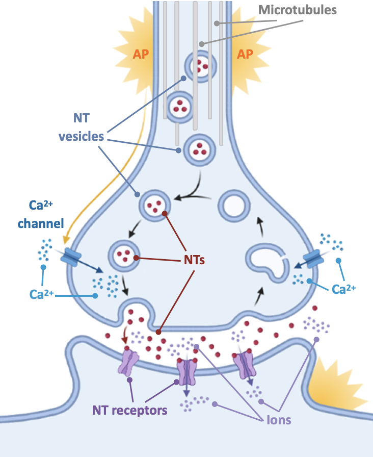
What cellular event directly triggers the release of neurotransmitters into the synaptic cleft?
Depolarization by an arriving action potential opens voltage-gated calcium (Ca2+) channels, allowing Ca2+ to enter the terminal bouton and trigger neurotransmitter vesicles to fuse with the presynaptic membrane.

What structure is formed when a synaptic vesicle fuses with the presynaptic membrane during neurotransmitter release, as seen in electron micrographs?
An omega figure, which represents the omega-shaped remnant of a synaptic vesicle fusing with the presynaptic membrane to release neurotransmitters into the synaptic cleft.
What are the three structural classifications of synapses?
Axodendritic (terminal bouton attached to a smooth dendrite or dendritic spine)
Axosomatic (terminal bouton attached to a soma)
Axoaxonic (terminal bouton attached to another terminal bouton)

How do ionotropic and metabotropic neurotransmitter receptors differ in structure and speed?
Ionotropic receptors contain a neurotransmitter binding site directly on an ion channel, opening the channel quickly for short-term effects (tens of milliseconds).
Metabotropic receptors are separate from ion channels and act via G-proteins and second messengers, producing slower, longer-lasting effects (hundreds of milliseconds to tens of seconds).
How do specific ion movements produce EPSPs versus IPSPs across the postsynaptic membrane?
Influx of Na+ or Ca2+ causes depolarization, generating an Excitatory Postsynaptic Potential (EPSP). Efflux of K+ or influx of Cl− causes hyperpolarization, generating an Inhibitory Postsynaptic Potential (IPSP).
What is the difference between spatial summation and temporal summation?
Spatial summation combines two or more postsynaptic potentials generated simultaneously at different synapses. Temporal summation combines two or more postsynaptic potentials generated in rapid succession at the same synapse.
Why do synapses located closer to the axon hillock have a stronger impact on action potential firing?
Postsynaptic potentials spread by passive conduction and undergo decremental decay (decrease in voltage over space and time); potentials generated closer to the axon hillock undergo less decay before reaching the trigger zone.
What are the three mechanisms for deactivating neurotransmitters in the synaptic cleft?
- Reuptake (transporters rapidly move NT back into the presynaptic terminal)
- Enzymatic breakdown (specialized enzymes degrade NT in the cleft)
- Diffusion (slow and inefficient passive drift away from the synapse)
What are autoreceptors, where are they located, and how do they function?
Autoreceptors are metabotropic receptors located on presynaptic terminal boutons that bind the neuron's own released neurotransmitter, providing a negative feedback signal to reduce future neurotransmitter synthesis and release.
What are five major structural patterns of neural circuits and networks?
- Divergence
- Convergence
- Serial processing
- Parallel processing
- Reverberation
Why is neural excitation or inhibition not equivalent to behavioral excitation or inhibition?
Behavior is controlled by complex neural circuits; exciting an inhibitory neuron can suppress a behavior, whereas inhibiting an inhibitory neuron (disinhibition) can cause expression of a behavior.
What are the three levels of protein structure emphasized in relation to functional shape (excluding secondary structure)?
- Primary structure: Specific linear sequence of amino acids.
- Tertiary structure: Twists and folds of the amino acid chain held by weak side-chain attractions.
- Quaternary structure: Combinations of tertiary protein subunits forming a functional molecule.
What are the primary support functions of astrocytes, oligodendrocytes, and microglia?
Astrocytes provide physical, nutritional, and biochemical support, contribute to the glymphatic system, and form part of the blood-brain barrier.
Oligodendrocytes form myelin sheaths around central nervous system axons.
Microglia manage immune defense.
What are the three components that make up the structure of the blood-brain barrier (BBB)?
- Continuous tight junctions between endothelial cells of capillary walls
- Astrocyte end-feet surrounding the capillary walls
- Plasma membranes of endothelial cells and astrocytes populated with specialized active transporters
How do brain capillaries differ structurally from typical capillaries elsewhere in the body?
Typical body capillaries have gaps between cells, fenestrations ("windows"), and pinocytotic vesicles allowing free substance exchange. Brain capillaries have continuous tight junctions with no intercellular gaps, no fenestrations, and no pinocytotic vesicles.
Why is the blood-brain barrier permeable at the area postrema?
The blood-brain barrier is permeable at the area postrema so the brain can detect toxins in the bloodstream and trigger vomiting to protect the organism.
Because most natural toxins are water-soluble, BBB prevents them from getting into brain
