D2.1 Cell and Nuclear Division
 Call Kai
 Learn
Learn Practice Test
 Spaced Repetition
 Match
Match Flashcards
 Knowt Play
1/38

There's no tags or description
Looks like no tags are added yet.
Name | Mastery | Learn | Test | Matching | Spaced | Call with Kai | Chat |
|---|
No analytics yet
Send a link to your students to track their progress
39 Terms
Cell division in prokaryotes
binary fission (asexual reproduction) & cytokinesis

Cell division in eukaryotes
Eukaryotes: mitosis (nuclear division) & cytokinesis
Overlaps the most at telophase (nuclear and cytoplasm dividing)
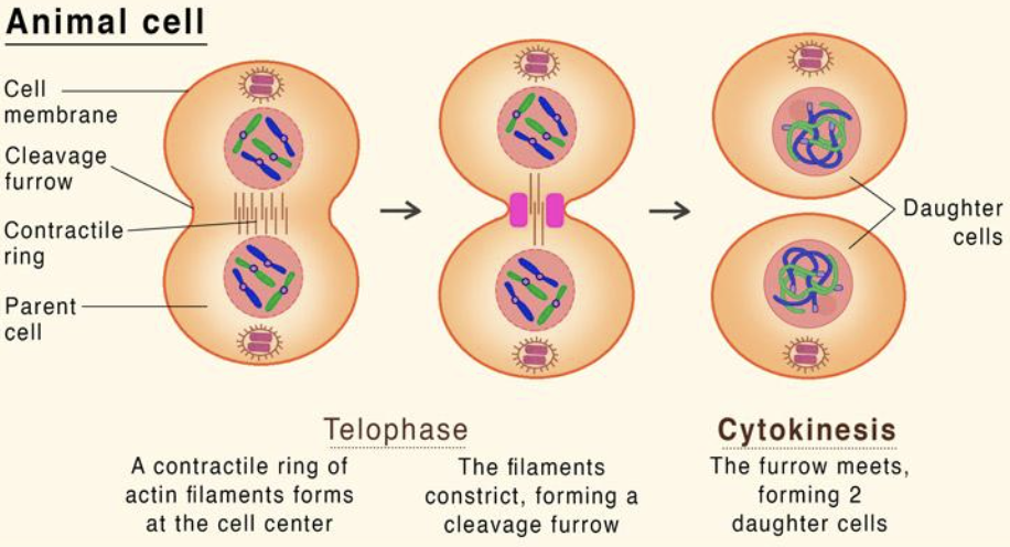
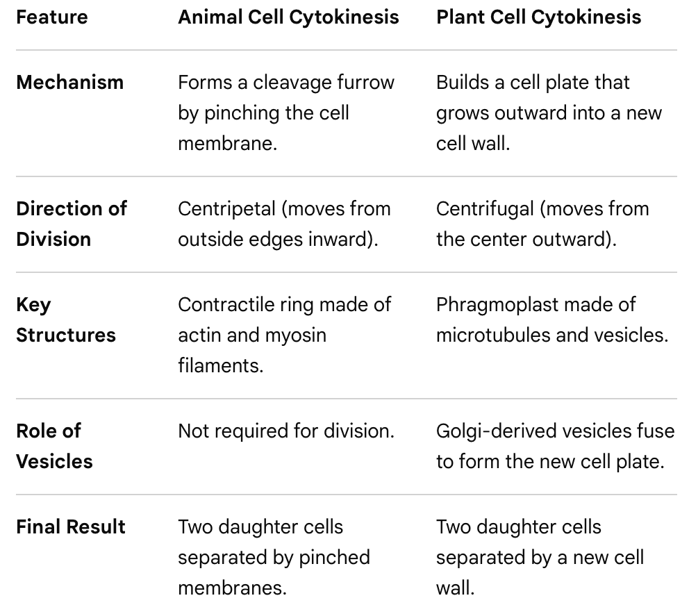
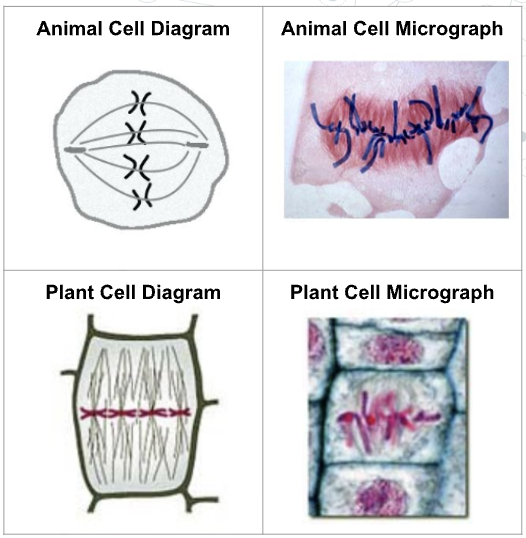
Compare and contrast cytokinesis in plant and animal cells (eukaryotes)
IN ANIMAL CELLS:
IN PLANTS CELLS:

State the difference between the definition of mitosis, meiosis and cytokinesis
Mitosis: Division of the nucleus and DNA into 2 daughter cells
Cytokinesis: Division of the cytoplasm and organelles into 2 daughter cells
For the two daughter cells to separate, the cytoplasm of the parent cell must be divided between the daughter cells
Mitosis and cytokinesis can occur simultaneously. (overlap in the telophase stage of mitosis)


“Cell division” applies to both eukaryotic and prokaryotic cells
“Mitosis” and “meiosis” refers to nuclear division in eukaryotic cells
So we do not use “mitosis” or “meiosis” for prokaryotic cells!
State the reason why daughter cells must receive at least one mitochondria (and chloroplast) during cytokinesis
essential for aerobic cellular respiration to produce ATP,
and any other organelle that can only be made by dividing a pre-existing structure, e.g. chloroplasts in plants
In cytokinesis, the division of cytoplasm is usually, but not in all cases, equal.
Outline unequal cytokinesis during human oogenesis
oogenesis (the process of producing mature egg cells or ova)
From the primary oocyte:
The secondary oocyte is larger than the first polar body (like a sacrifice) and receives most of the cytoplasmic contents, including organelles, ribosomes and energy stores.
If fertilisation occurs:
the secondary oocyte undergoes a second round of cell division, producing a mature ovum (egg cell) and a second polar body.
The mature ovum contains the majority of the cytoplasmic contents and organelles required for embryonic development

Why is nuclear division required to start “before” cell division (cytokinesis)?
This avoids the production of anucleate cells (no nucleus), which would not be able to develop.
Cells that are naturally anucleate, such as red blood cells or sieve tube elements, lose the nucleus after differentiation in a process called enucleation.
Nuclear division: mitosis and meiosis
State why mitosis is nuclear division resulting in continuity of the chromosome number and genome
Mitosis maintains genetic stability because:
daughter cells have the same number of chromosomes as the parent cell (2n – diploid)
Daughter cells are usually genetically identical to the parent cell
In eukaryotic cells, mitosis occurs to produce cells for growth, or to replace cells that are lost or damaged.
Also for asexual reproduction (e.g yeast and hydra)
State that meiosis is nuclear division that results in reduction of the chromosome number and diversity between genomes
Meiosis is a type of nuclear division that produces four haploid (n) genetically different daughter nuclei, which will form gametes (sperm and ovum) for sexual reproduction
As the daughter cells are haploid (n) after two rounds of nuclear division, meiosis is also known as reduction division
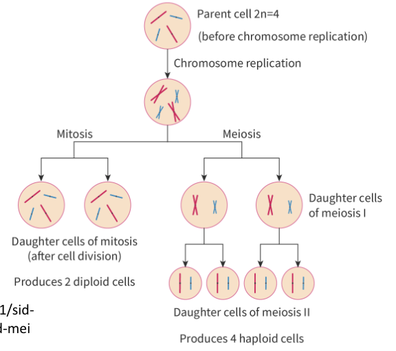
State the differences between mitosis and meiosis
Mitosis
Rounds of division: 1
No. of daughter cells produced:
2 genetically identical daughter cells from a single parent cell.
Purpose: growth, cell replacement and tissue repair of somatic cells
Meiosis
Rounds of division: 2
No. of daughter cells produced:
4 daughter cells that are genetically unique from the parent cell and contain only half as much DNA (haploid)
Purpose: Only occurs in the production of germ cells sperm and egg cells for sexual reproduction
State that DNA replication occurs in ___ phase of the cell cycle
Before mitosis or meiosis, DNA replication must occur.
DNA replication is the process by which a cell makes a copy of its DNA
DNA replication occurs during S-phase of interphase, a period in the cell cycle where cells are growing and are metabolically active.
Sister chromatids are formed when a chromosome is replicated
Sister chromatids are joined at the centromere

Explain how replicated DNA molecules are held together, with reference to chromatid, replicated chromosome, centromere and cohesin
Sister chromatids are held together in two ways:
1. At a region of the DNA called the centromere.
The centromere has two functions:
Adhere sister chromatids to each other
Site of kinetochore and microtubule attachment for movement of chromosomes
2. A protein complex called cohesin holds sister chromatids together so they remain connected to each other until anaphase.
Cohesin is established in interphase before both mitosis and meiosis.
The cohesin holding together the chromatids is removed by the start of anaphase. As a result, the sister chromatids are able to split apart and move to opposite poles of the cell, ensuring that each daughter cell will have a complete copy of the genetic material.
Explain how and WHY chromosomes condense during mitosis and meiosis (DNA packaging recap)
Condensation of DNA
DNA as a double helix made of two antiparallel strands of nucleotides with two strands linked by hydrogen bonding between complementary base pairs
Eukaryote cell DNA wraps around histone proteins to form a nucleosome.
Histone proteins have an overall positive charge, and so interact with negatively charged DNA.
The nucleosome is composed of eight histone proteins arranged in a core, around which DNA is coiled
The nucleosomes coil and stack together to form fibers called chromatin.
Because it needs to be accessible to enzymes, during interphase most of the DNA is in chromatin form and not tightly coiled.
Chromatin condenses during mitosis and meiosis to form chromosomes.
During prophase, replicated DNA in chromatin form, condenses to become a chromosome with two sister chromatids.
The DNA condenses by "supercoiling" so that it can more easily be moved to the poles of the cell without getting tangled and/or broken.
State the role of microtubules and kinetochore motor proteins
Movement of chromosomes:
Both mitosis and meiosis involve the movement of chromosomes.
Microtubules
long, thin, cylindrical fibrous proteins (tubulin)
they form the spindle apparatus (includes more than just spindle fibres) during cell division.
the shortening or lengthening of the spindle fibres is responsible for moving chromosomes during nuclear division
Kinetochore
is a protein complex that assembles at the centromere
each sister chromatid of a chromosome has it own kinetochore which faces opposite directions
the kinetochore links the chromatids to the microtubules
Microtubule motors
specialised proteins
bind to microtubules and hydrolyse ATP to provide the energy to move chromosomes to either pole (end) of the cell.
They literally "walk" along microtubules, which acts like tow trucks on tracks.
Microtubule organising centre
The MTOC is different in plant and animal cells
In animal cells, the MTOC are the centrioles
Plant cells (of multicellular plants) do not have centrioles but have different type of MTOC that is more spread out
Acts as the final destination for the chromatids
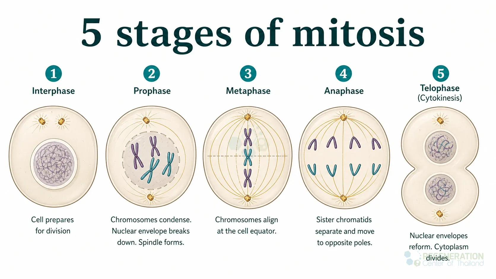
Draw typical eukaryotic cells as they would appear during the interphase and the four phases of mitosis.

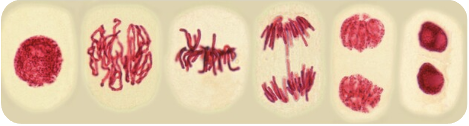
Outline Interphase
During interphase, the chromosomes are not supercoiled as the cells are undergoing processes like DNA replication and protein synthesis
Chromosome are not visible yet
TO spot:
Nuclei are rounded or oval, evenly distributed shading
Outline four events that occur during prophase
Replicated DNA, in chromatin form, condenses to become chromosome by supercoiling
Each replicated chromosome is a pair of sister chromatids joined at the centromere and held together by loops of cohesin
The kinetochore attaches to the centromere of the chromatids
Microtubules form to create the mitotic spindle
At the end of prophase, the nuclear membrane breaks apart
Look for clumps of DNA in real pictures

Outline the process of metaphase
Chromatids of the replicated chromosome are fully condensed and still attached to each other with cohesin
Microtubules continue to grow and attach to the centromeres on each chromosome.
The chromosomes are moved so they are equidistant from the two poles of the cell, along the equator
Outline the process of anaphase
The cohesin that has been holding the sister chromatids together are removed / centromere divides
allowing the sister chromatids to separate
creating two individual daughter chromosomes
Kinetochore removes tubulin subunits to shortening the length of the spindle microtubule to pull the genetically identical chromosomes (formerly sister chromatids) to opposite poles

Outline four events that occur during telophase
The chromosomes are pulled into a tight group near the MTOC
and a nuclear membrane reforms around them.
Chromosomes decondense to form chromatin
The microtubule spindle fibres are broken down
Occurs simultaneously as cytokinesis, division of the cytoplasm and organelles

Outline interphase following mitosis
chromosomes each consisting of a single DNA molecule are uncoiled and dispersed throughout the nucleus
cytokinesis completed
with the cytoplasm and plasma membrane pinched apart
cytoplasm is very active
Explain what it means for chromosomes to be “homologous”
Homologous chromosomes:
In many eukaryotic cells, chromosomes are found as a pair;
one inherited from the mother and one from the father
same size
same centromere location
same genes in the same order.
However, since the base sequence of a gene can change by mutation,
variations in the sequence of nucleotides within a specific gene.
Variations within a single gene are called alleles.
they are still considered the “same gene” because they still code for the same trait
The two chromosomes of a homologous pair might each have the
same allele of a gene (homozygous)
or might have different alleles of a gene (heterozygous)

Define diploid.
State the human cell diploid number.
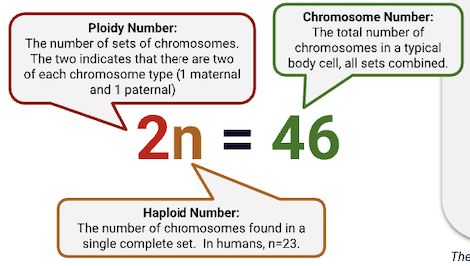
Cells and organisms that contain a homologous pair of each chromosome are called diploid (organisms).
with two complete sets of chromosomes in body cells
one copy of paternal and one copy of maternal chromosomes Diploid cells are described as 2n. In humans:
Define haploid.
State the human cell haploid number.
List example haploid cells, and its purpose.
A cell or organism with a single copy of each chromosome is called haploid.
Because there are no chromosome pairs, haploid cells do not contain homologous chromosomes.
Eggs and sperm are haploid cells with 23 chromosomes each. Haploid cells are described as "n".
In humans:

In sexual reproduction, the genetic information of two parents combine in an offspring.
In order to maintain the correct number of chromosomes in offspring, there must be a reduction in the number of chromosomes in the forming of eggs and sperm - halving from diploid to haploid.
At fertilisation the two haploid gametes combine to form a zygote
allowing it to have the full complement of 2n = 46 chromosomes.
If chromosome numbers are not halved, they will keep doubling / increasing every time fusion occurs
Polyploid can result and offspring are not viable (in the case of most animals)
Explain why meiosis I is a reductive division
Type of cell division happening in the process of gametogensis
Two consecutive nuclear divisions (instead of one in mitosis)
Meiosis I
the diploid (2n) germ cell that begins meiosis is split into two haploid (n) cells.
Meiosis II
the sister chromatids within the two daughter cells separate, forming four new haploid gametes.
Meiosis is known as the reduction division because
Results in 4 daughter cells with half the original number of chromosomes

Define nondisjunction and a named example
The process of meiosis is incredibly complex; mistakes can and do occur.
Nondisjunction is the failure of chromosomes to separate during either anaphase I or anaphase Il of meiosis.

Nondisjunction results in gamete cells that have an incorrect number of chromosomes.
Outcome:
E.g. Down Syndrome, which is caused by the presence of an extra chromosome 21. Results in developmental disorders.
Explain how meiosis leads to genetic variation in gametes
Genetic variation is promoted during meiosis due to
Crossing over during Prophase I
Random orientation of homologous chromosomes during Metaphase I due to “independent assortment”
Random orientation of chromosomes during Metaphase II

Define bivalent and synapsis
Synapsis occurs in prophase I
The pairing process of the homologous chromosomes is called synapsis.
In synapsis, the genes on the chromatids of the homologous chromosomes are aligned with each other.
Bivalent:
A pair of homologous chromosomes forms a "bivalent" on the equator of the cell during metaphase I of meiosis.
The pole to which each of the two chromosomes of the bivalent attaches to is random.

Because the two chromosomes can have different alleles of genes, the gametes that are formed at the end of meiosis will have different alleles from each other.
Describe the process and result of crossing over during prophase I of meiosis
During prophase I of meiosis, homologous chromosomes pair up align gene by gene (synapsis).
Because DNA replication has already occurred each chromosome consists of two genetically identical chromatids.
CROSSING OVER:
Crossing over is the process where corresponding sections of the chromatids are exchanged.
Two NON-sister chromatids of a pair homologous chromosomes are brought together at the precisely the same point along their gene sequence → mutual exchange of genes.
point at which they switch is known as chiasma (plu. chiasmata)
there can be multiple chiasmata along the chromosome
The DNA strands of the two non-sister chromatids is cut and rejoined to the other chromatid.
This results in an exchange of the DNA between the two chromatids.
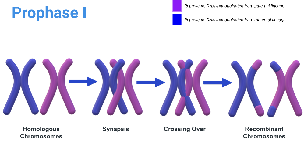
The produced recombinant chromosomes have new combinations of alleles that are not present in either parent, and the sister chromatids are no longer genetically identical
Define cell proliferation
Proliferation is the rapid increase in number of the cells by cell division
occurs in the meristematic tissues of plants and the early-stage embryos of animals
Outline cell proliferation during growth at plant meristems and early-stage animal embryos
Growth: Meristems
Meristems are regions of undifferentiated cells at the tips of roots and shoots that have the potential to become any cell type in the plant.
Although meristem cells are stem cells, they are usually not called that so as to avoid confusion with cells in the stem of a plant.
Growth: embryonic cells
During animal embryonic division, cells divide rapidly. During early human embryonic development, cell division occurs approximately once every 24 hours
This process is called cleavage and involves the division of the fertilised egg into a blastocyst.
Describe skin cell proliferation during cell replacement and tissue repair
Tissue Repair
Cell proliferation plays a critical role in wound healing, as it is responsible for the growth and repair of damaged tissues.
When a tissue is damaged, cells in the surrounding area are stimulated to divide and migrate to the site of injury.
After blood clotting, inflammation causes more blood to flow towards the wound
Hence fibroblasts and macrophages (white blood cells) travel towards the wound
Fibroblasts proliferate to heal the wound, breaking the clot and creates a collagen matrix to support other cells associated with wound healing
List the phases of the cell cycle
Sequence of events:
Interphase: G1(biochemical activity), S phase (DNA replication) and G2(prep for mitosis)
longest part of the cell cycle
In the S phase the cell replicates all the genetic material in its nucleus, so that after mitosis both the new cells have a complete set of genes.
Some do not progress beyond G1, because they are never going to divide so do not need to prepare for mitosis.
These cells enter a phase called G0 which may be temporary or permanent.
Mitosis: division of the nucleus
Cytokinesis: division of the rest of the cell (starting time varies for different cell types)
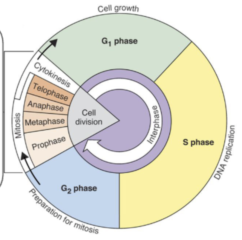
Outline events of the G1, S, and G2 phases of interphase
The longest part of the cell cycle
Cell is metabolically active during interphase
DNA replication in the nucleus and protein synthesis in the cytoplasm only happen during S-phase interphase (i.e. DNA replication and protein synthesis cannot occur during mitosis).
Other processes like cell respiration can occur during interphase and cell division (as the chromosomes are not directly involved)
During interphase the numbers of mitochondria in the cytoplasm increase (during G2).
Suggest why the cell would increase the number of mitochondria inside itself prior to cell division.
Need more ATP for cell division
More mitochondria are required to supply ATP to the two daughter cells that will be eventually produced
Some activities that take place (metabolically active)
Volume of cytoplasm increases
Number of organelles increases
Phospholipid bilayer area increases
More enzymes are synthesized
Respiration generates more energy for cell division to take place
What are cyclins and how does cyclin levels play a role in the cell cycle?
Are a group of proteins
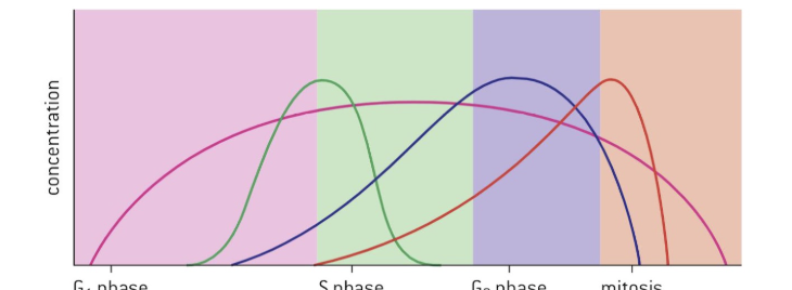
As cyclin concentrations increase, they bind to enzymes called cyclin-dependent kinases (CDKs) that form mitosis-promoting factors (MFP)
MFPs phosphorylate specific proteins to drive the cell cycle forward.
The levels of these cyclins rise and fall.
Unless these cyclins reach a threshold concentration, the cell does not progress to the next stage of the cell cycle.
Cyclins therefore control the cell cycle and ensure that cells divide when new cells are needed, but not at other times.
State the functions of cell cycle checkpoints
Three checkpoints are controlled by cyclin concentrations:
G1 checkpoint
Checks for cell size, nutrients, growth factors, and DNA damage before DNA synthesis begins.
G2 checkpoint
Ensures DNA replication is complete and undamaged before the cell enters mitosis.
M checkpoint
Confirms proper chromosome alignment and attachment to spindle fibers during metaphase
As cyclin concentrations increase, they combine with CDKs to form MFPs
MFPs triggers chromosome condensation, nuclear membrane fragmentation and spindle formation
Mitosis is switched on
Cyclins are broken down by anaphase but CDKs still persist
Explain how mutations leading to proto-oncogenes and tumor suppressor genes can lead to the development of cancer
Proto-oncogenes
are genes that code for proteins that help promote cell growth and division.
If a mutation in a proto-oncogene leads to these proteins becoming overexpressed, it can result in uncontrolled cell division. (Leads to cancer.)
The mutated proto-oncogene is called an oncogene.
Tumour suppressor genes
code for proteins that normally slow down or prevent cell division.
can also promote programmed cell death (apoptosis) to prevent the development of cancer.
Mutation of tumour suppressor genes
When these genes are mutated, the proteins they code for can no longer perform their protective function, leading to uncontrolled cell division.
Tumours
are abnormal groups of cells that develop at any stage of life in any part of the body.
Benign tumours:
In some cases the cells adhere to each other and do not invade nearby tissues or move to other parts of the body.
unlikely to cause much harm.
Malignant tumours:
The cells (of the primary tumour) can become detached and move elsewhere in the body
develop into secondary tumours
very likely to be life-threatening (carcinomas).
State the formula for calculation of a mitotic index
The mitotic index (MI) is the ratio of cells undergoing mitosis (cell division) to the total number of cells in a population

Outline the use of mitotic index calculations in diagnosis and treatment of cancer
Identifying malignancy: A significantly elevated mitotic index compared to healthy, tissue-specific normal tissue indicates rapid and uncontrolled cell proliferation, signaling a high likelihood of cancer.