Surgery - Derm (CMPP)
 Call Kai
 Learn
Learn Practice Test
 Spaced Repetition
 Match
Match Flashcards
 Knowt Play
1/51

There's no tags or description
Looks like no tags are added yet.
Name | Mastery | Learn | Test | Matching | Spaced | Call with Kai | Chat |
|---|
No analytics yet
Send a link to your students to track their progress
52 Terms
At what total body surface area does burn shock occur
>20%
Burn shock
significant metabolic changes occur, resulting in loss of intravascular volume, resulting in hemoconcentration
What are possible complications of fluid resuscitation of burns
Cerebral edema
Compartment syndrome
Pulmonary edema
1st Degree Burns
Affects the outer layer of skin (epidermis)
Skin looks red, dry, and painful with no blisters
2nd Degree Burns
Involves the epidermis and part of the dermis
Symptoms include intense pain, redness, swelling, and fluid-filled blisters
3rd Degree Burns
Destroys both the epidermis and dermis, reaching the fat layer underneath
The burn area looks white, leathery, brown, or charred and often causes little to no pain due to destroyed nerve endings
4th Degree Burns
Deepest burn destroying all skin layers and extending deep into underlying fascia, muscles, tendons, or bone.
Surface appears dry, blackened, charred and it painless
What is involved in the surgical evaluation and treatment of burns
Systematic wound assessment,
Early excision of dead tissue, and
Definitive wound closure using skin grafts or flaps
What is considered “early excision” for dead tissue from burns
2 days
Rule of 9s
A quick way to assess TBSA of a burn on an adult patient
Head = 9%
Left Arm = 9%
Right Arm = 9%
Chest = 9%
Abdomen = 9%
Upper Back = 9%
Lower Back = 9%
Anterior Left Leg = 9%
Posterior Left Leg = 9%
Anterior Right Leg = 9 %
Posterior Right Leg = 9%
Groin = 1%
Palmar Method
A burn estimation technique that considers the palm of the patient’s hand to be 1% of their TBSA
How is Rule of 9s modified for infants
Head → 9% to 18%
Legs → 18% for one leg to 14%
What fluid is used for burn fluid resuscitation
Lactated Ringer’s (LR) solution
Parkland Forumla
A calculation for the fluids for burns
Volume of Solution (LR) for 24 hrs = 4 mL x TBSA of burn (%) x weight (kg)
First half of the volume is given in 8 hours, then second half is given in 16 hours
Cleveland Clinic Burn Guide
A guide to assessing the TBSA of a burn
what secondary injuries should be checked for in burn patients
airway compromise,
carbon monoxide poisoning,
associated traumatic injuries requiring priority stabilization
tight, circumferential burns that restrict blood flow
What procedures can be performed for burns
Escharotomy/Fasciotomy:
Early Excision
Autografting and Closures:
Contracture Release:
Escharotomy
Make emergency incisions through tight, burned skin (eschar) to relieve compartment syndrome and restore perfusion
Autografting and Closures
Apply split-thickness skin grafts or temporary biological/synthetic dressings to close the wound bed and promote healing
Contracture Release
Perform later reconstructive procedures to release tight scar bands and recover functional range of motion
Cellulitis
Skin becomes infected with a bacterial pathogen
Presenation of Cellulitis
pain,
redness,
erythema of the affected skin
What pathogens typically responsible for cellulitis
Staphylococcus aureus,
Streptococcus pyogenes,
MRSA,
Pasteurella multocida
Patients with cellulitis also need to be assessed for
necrotizing soft tissue infections (NSTAs)
What are typical indicators to treat cellulitis inpatient
Fever
Leukocytosis
Outpatient Treatment for Cellulitis
Cephalexin or Augmentin x 5-10 days (up tp 14)
PCN Allergy → Clindamycin
MRSA Suspect → TMP-SMX or Doxycycline
Inpatient Treatment for Cellulitis
IV Cefazolin, Ceftriaxone, or Clindamycin x 5-10 days (up tp 14)
MRSA → IV Vancomycin or Clindamycin
Treatment for Cutaneous Abscess
Incision and drainage
Consider Abx
When should Abx be given after an I&D
immunocompromised,
DM,
fever,
tachycardia,
large abscess (over 2cm)
high-risk location (face, groin, perianal)
What condition is often called “flesh-eating bacteria”
Necrotizing soft tissue infections (NSTI)
Necrotizing soft tissue infections (NSTI)
Rapidly progressing, life-threatening bacterial infections that cause widespread destruction of the skin, subcutaneous fat, and deep fascia
Red Flags for Necrotizing soft tissue infections (NSTI)
Pain out of proportion
Redness, swelling, and warmth that spread visibly over the course of hours as compared to days
Development of a dusky, purplish, or bluish skin color, accompanied by bulla or areas of black skin necrosis
Cracking or popping sensation felt under the skin, caused by gas produced by invading bacteria
High fever,
tachycardia,
severe chills,
confusion,
hypotension (septic shock)
What are the types of NSTIs
Type I (Polymicrobial):
Type II (Monomicrobial):
Type III:
Anatomical Variants:
Most Common Type of Necrotizing soft tissue infections
Type 1
Cause of Type I Necrotizing soft tissue infections
a mix of aerobic and anaerobic bacteria but usually involves C. perfringens
Who typically gets Type I Necrotizing soft tissue infections
Olders Adults
DM
CKD
Obese
Cause of Type II Necrotizing soft tissue infections
by a single organism, most famously Group A Streptococcus or Staphylococcus aureus
Who is more at risk for Type II Necrotizing soft tissue infections
Trauma
Surgery
IVDA
Cause of Type III Necrotizing soft tissue infections
specific marine bacteria, most notably Vibrio vulnificus.
How are Type III Necrotizing soft tissue infections acquired
exposure of an open wound to warm, coastal seawater
handling raw or eating contaminated shellfish
What some anatomical variant necrotizing soft tissue infections
Fourneir’s Gangrene (Perineum and Genitals)
Ludwig’s Angina (Floor of Mouth and neck)
What should be done immediately for suspected necrotizing soft tissue infections
begin simultaneously in a hospital setting without waiting for laboratory or imaging results to confirm it
What is the general managment plan for necrotizing soft tissue infections
Immediate Surgical Debridement:
Broad-Spectrum Intravenous Antibiotics:
Aggressive Resuscitation
Adjuvant Therapies
What is typically the Abx regimn for empric necrotizing soft tissue infections
Piperacillin-Tazobactam (Zosyn) or Carbapenem (Broad Coverage)
Vancomycin (MRSA cover)
Clindamycin (Shuts down toxins by Strep and Staph)
What goes into the Aggressive Resuscitation for necrotizing soft tissue infection
substantial IV fluid resuscitation,
vasopressors to maintain blood pressure,
close monitoring in the ICU
What adjuvant therapies can be used for necrotizing soft tissue infections
hyperbaric oxygen therapy (HBOT) to inhibit anaerobic bacteria and promote healing,
Intravenous Immunoglobulin (IVIG) to help neutralize circulating bacterial toxins
What are the most important prognositc factors for necrotizing soft tissue infections
Early clinical recognition
immediate surgical consultation
Fournier’s gangrene
Type of necrotizing soft tissue infection occurring in the perineal, genital, and perianal region
Who is more likely to get Fournier’s gangrene
Men
What type of necrotizing soft tissue infection does Fournier’s tend to be
type 1 NSTI – almost always polymicrobial
Treatment of Fournier’s gangrene
Early wide debridement
broad-spectrum antibiotics
removal of the scrotum is not uncommon
May need penile degloving
Plastic surgery consultation for future reconstruction
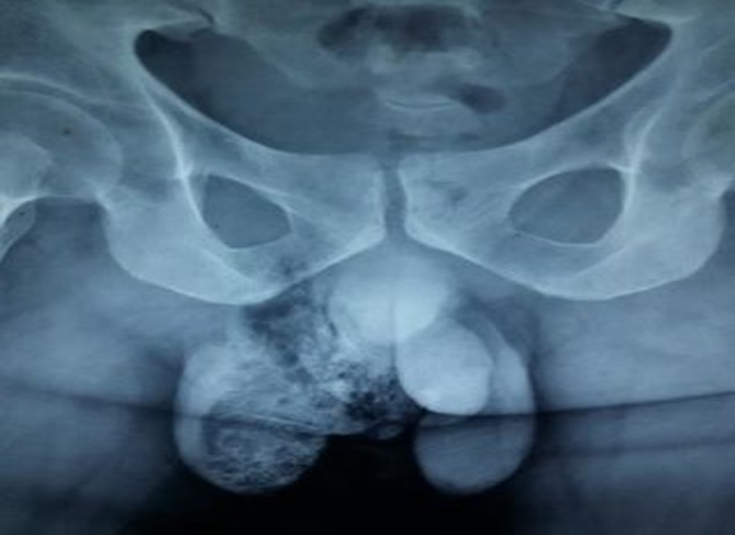
Fournier’s gangrene