Histo photo cards
 Call Kai
 Learn
Learn Practice Test
 Spaced Repetition
 Match
Match Flashcards
 Knowt Play
1/46

There's no tags or description
Looks like no tags are added yet.
Name | Mastery | Learn | Test | Matching | Spaced | Call with Kai | Chat |
|---|
No analytics yet
Send a link to your students to track their progress
47 Terms
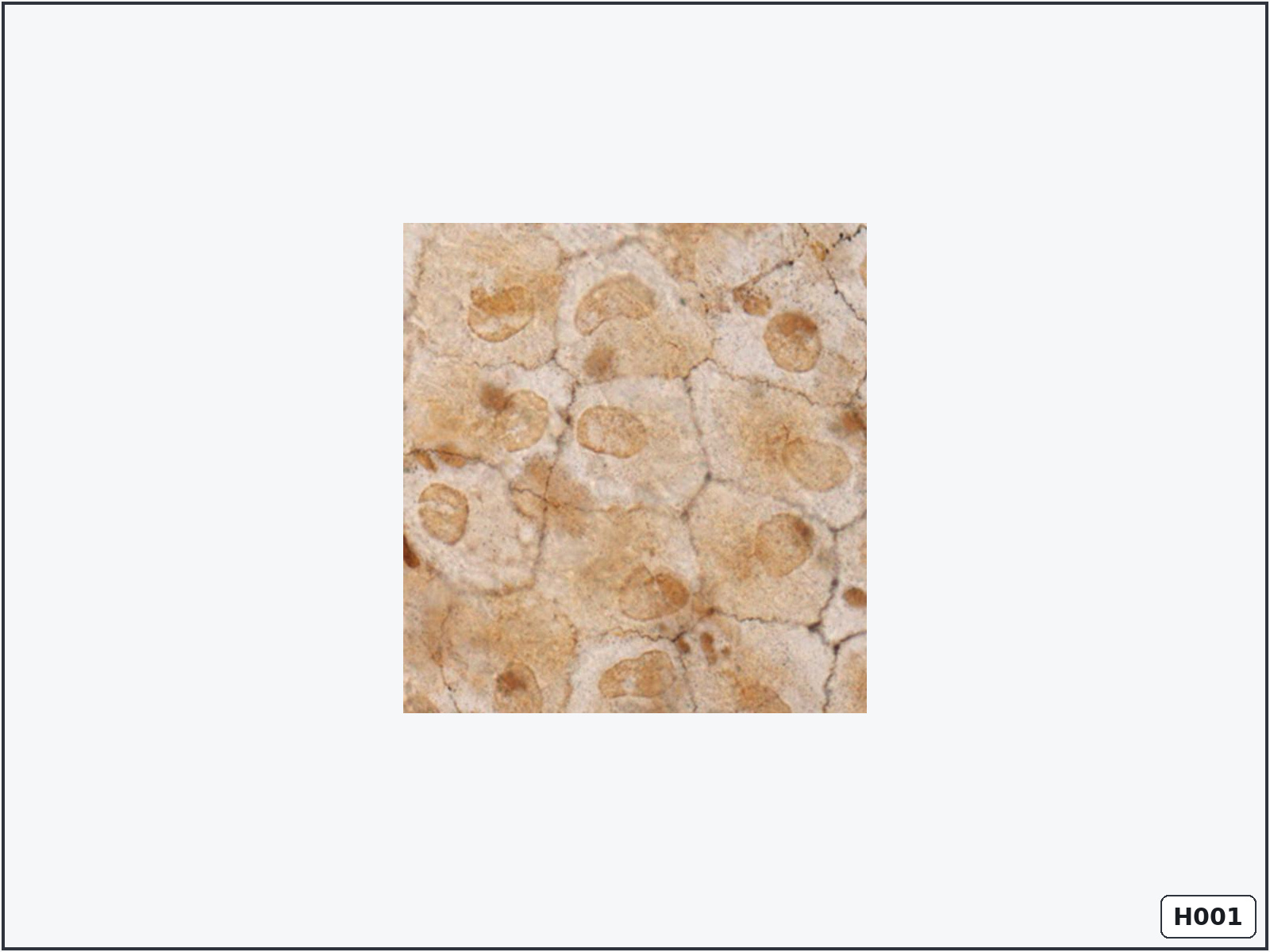
Squamous cells – mesothelium
Identify: flat polygonal cells with silver-outlined borders and central oval nuclei.
Important structures: cell membrane, nucleus and thin cytoplasm.
Know: mesothelium is simple squamous epithelium covering serous cavities and reducing friction.
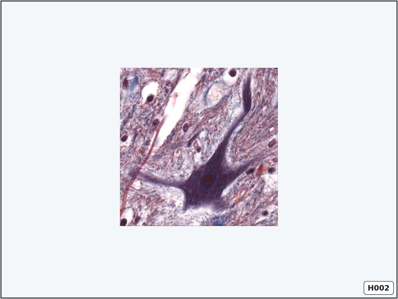
Multipolar neuron – spinal cord
Identify: a large stellate cell body with several processes and a prominent nucleus.
Important structures: perikaryon, nucleus, nucleolus, dendrites, axon and neurofibrils.
Know: dendrites receive signals, while the single axon carries signals away from the cell body.
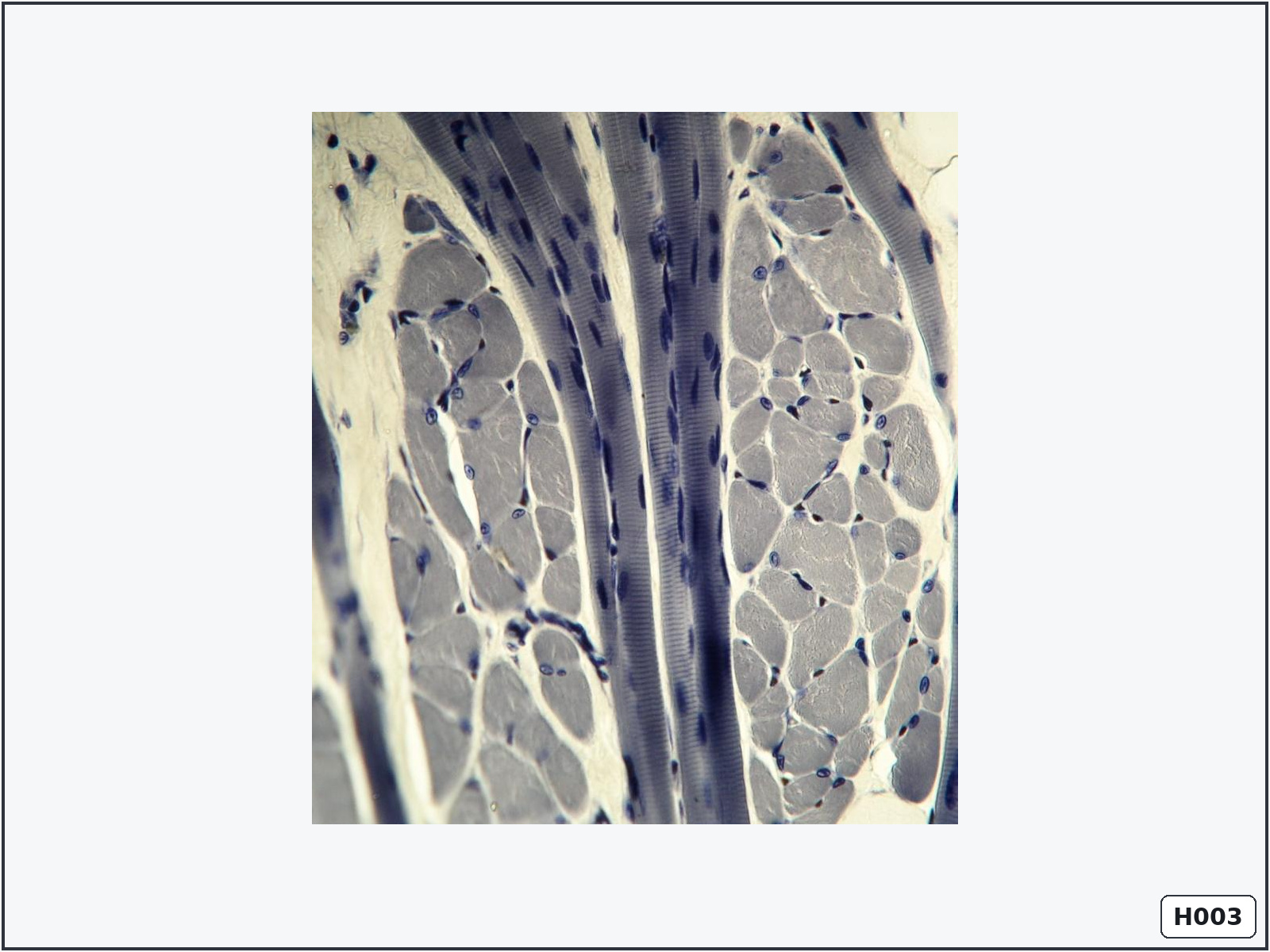
Striated skeletal muscle fibres – tongue
Identify: striated muscle fibres cut in longitudinal and transverse directions.
Important structures: muscle fibres, peripheral nuclei, sarcoplasm and endomysium.
Know: skeletal muscle fibres are long, unbranched, multinucleated and voluntarily controlled.
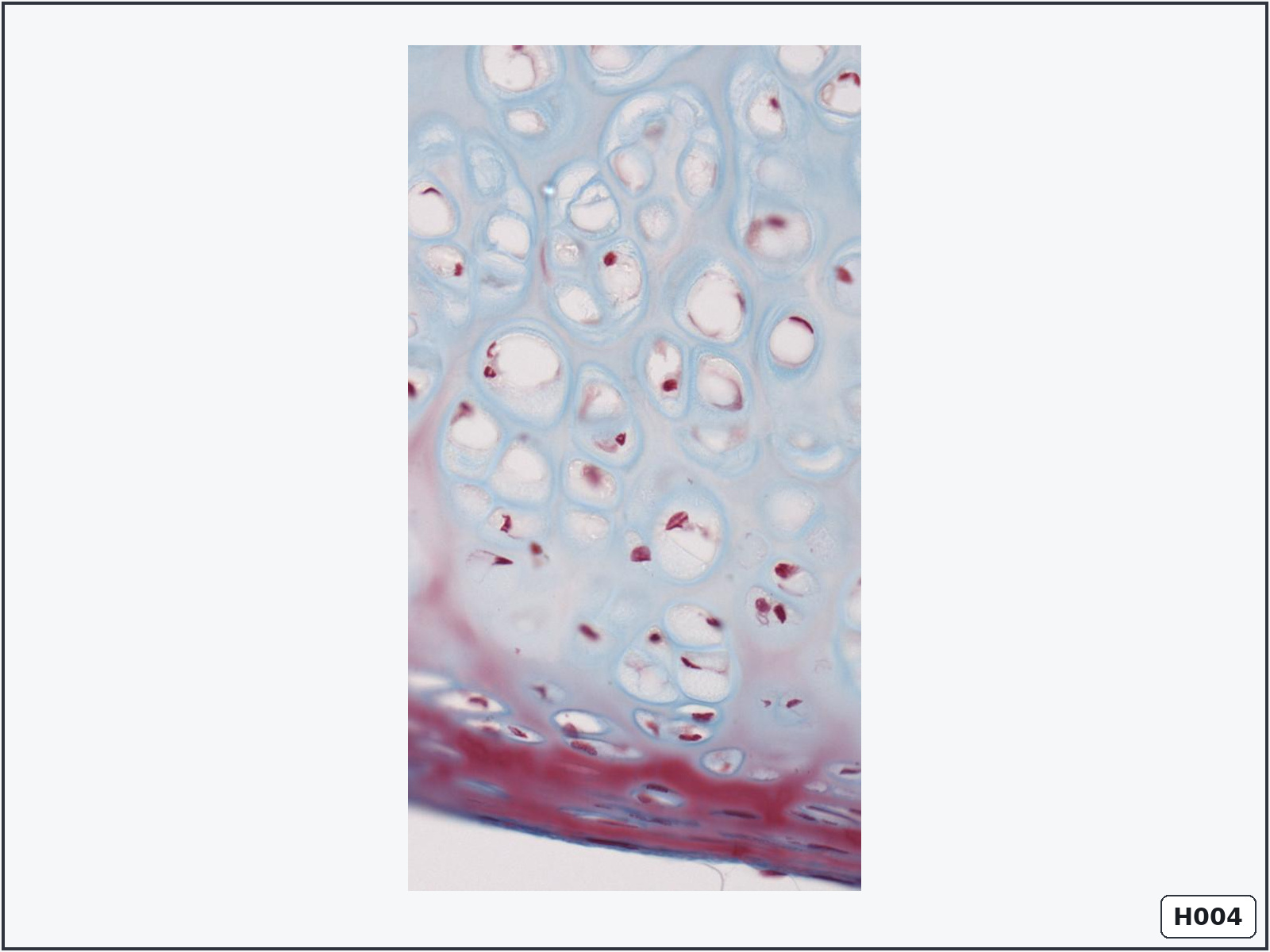
Hyaline cartilage – trachea
Identify: chondrocytes within lacunae surrounded by a smooth, glassy matrix.
Important structures: perichondrium, chondroblasts, chondrocytes, lacunae, isogenous groups and matrix.
Know: tracheal cartilage supports the airway. It is avascular and receives nutrients by diffusion.
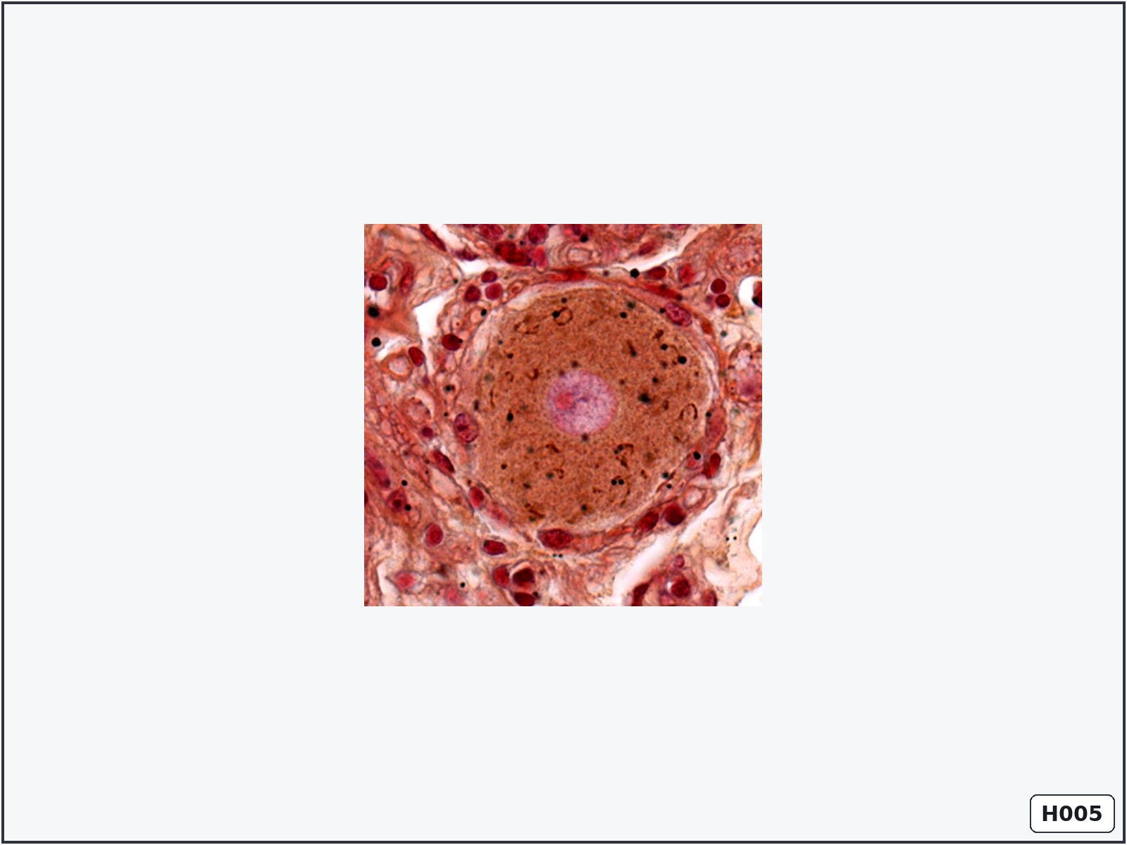
Pseudounipolar neurons – spinal ganglion
Identify: large round neuronal cell bodies surrounded by small satellite-cell nuclei.
Important structures: cell body, nucleus, nucleolus, cytoplasm and satellite cells.
Know: pseudounipolar neurons are sensory neurons. Satellite cells support and electrically isolate their cell bodies.
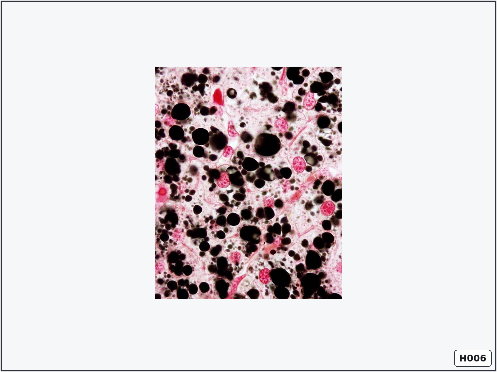
Lipid droplets in hepatocytes
Identify: numerous black lipid droplets inside hepatocytes.
Important structures: hepatocyte nuclei, cytoplasm and lipid droplets.
Know: osmium stains lipids black and preserves them during tissue preparation.
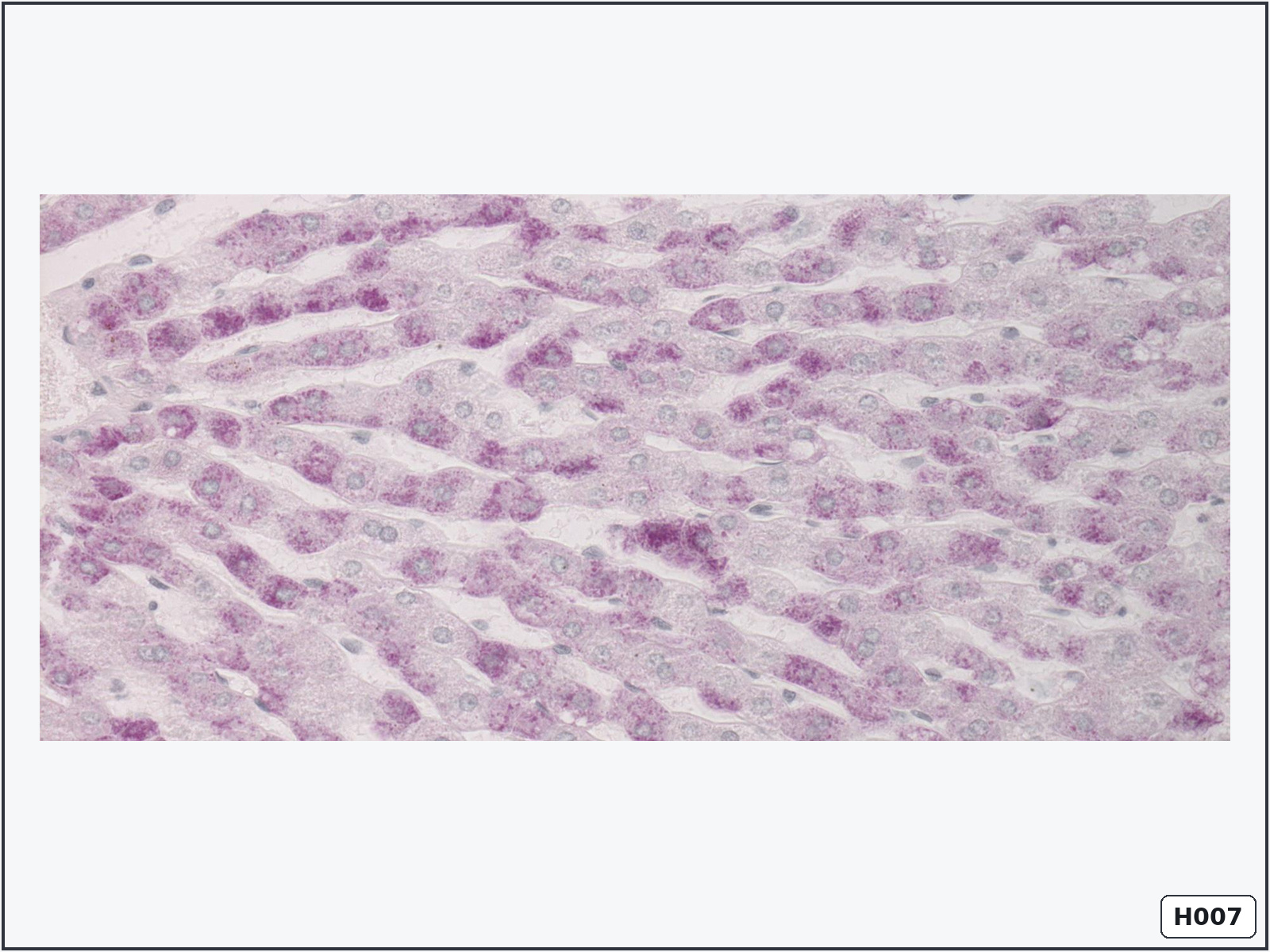
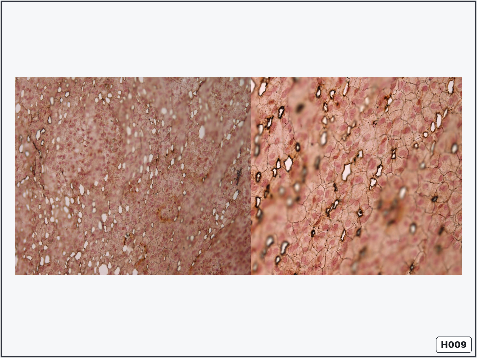
Glycogen granules in hepatocytes
Identify: magenta PAS-positive granules within polygonal hepatocytes.
Important structures: hepatocyte nucleus, cytoplasm, plasma membrane and glycogen granules.
Know: the PAS reaction demonstrates carbohydrate-rich substances such as glycogen.
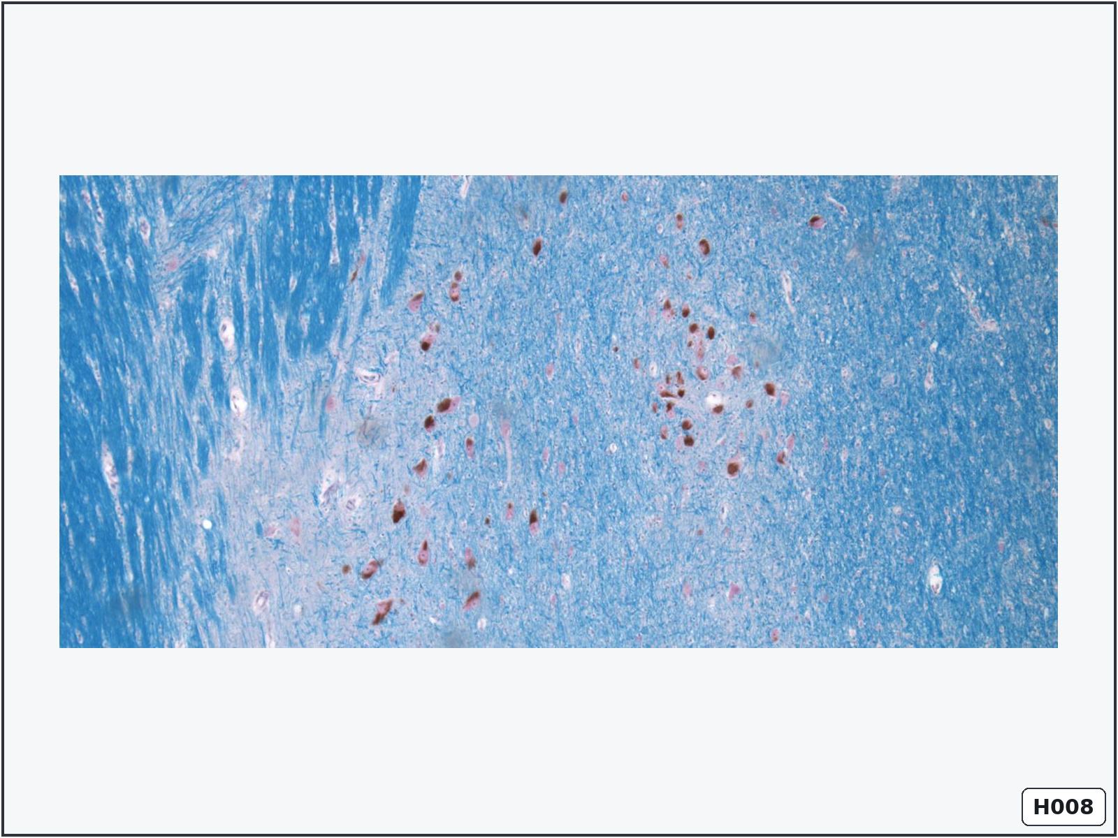
Melanin granules in multipolar neurons – mesencephalon
Identify: large multipolar neurons containing brown cytoplasmic pigment.
Important structures: perikaryon, nucleus, nucleolus, neuronal processes and pigment granules.
Know: pigmented dopaminergic neurons are characteristic of the substantia nigra.
Simple squamous epithelium – mesothelium
Identify: a surface mosaic of extremely thin polygonal cells with flattened nuclei.
Important structures: cell borders, nuclei and cytoplasm.
Know: all cells contact the basement membrane and form one thin layer specialised for exchange or low-friction lining.
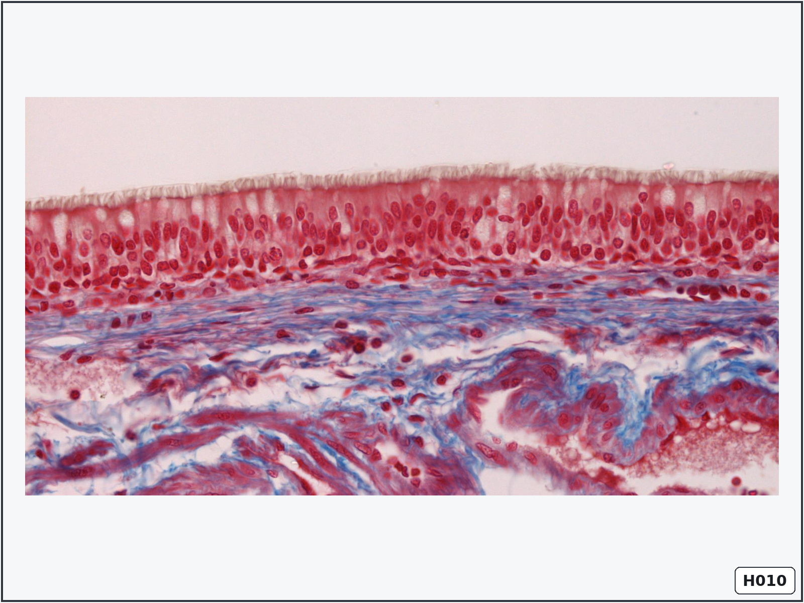
Pseudostratified ciliated columnar epithelium – trachea
Identify: nuclei at different heights, goblet cells and cilia on the apical surface.
Important structures: basement membrane, basal cells, columnar cells, goblet cells and cilia.
Know: every cell contacts the basement membrane, but not every cell reaches the surface. Cilia move mucus toward the pharynx.
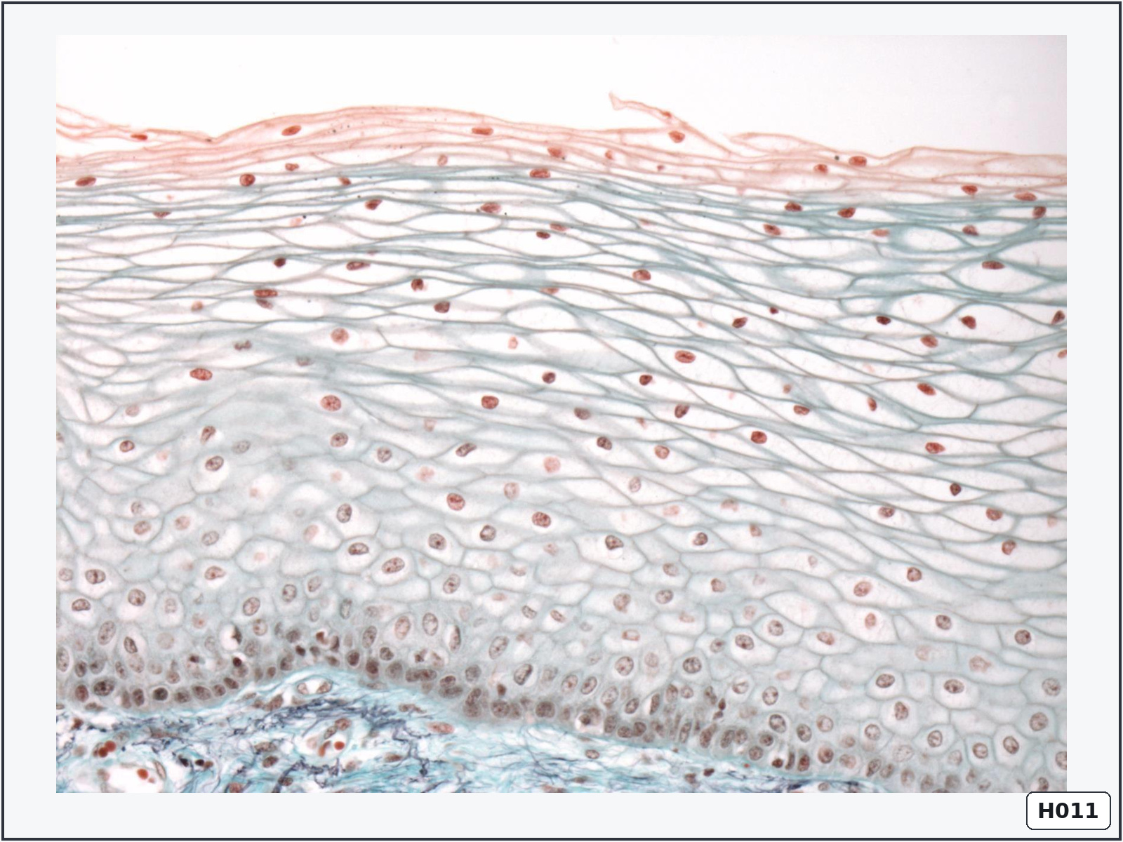
Stratified squamous non-keratinised epithelium – vagina
Identify: multiple cell layers with flattened superficial cells that retain their nuclei.
Important structures: basal layer, intermediate layers, superficial cells, basement membrane and lamina propria.
Know: it protects moist surfaces against abrasion and has no keratinised surface layer.
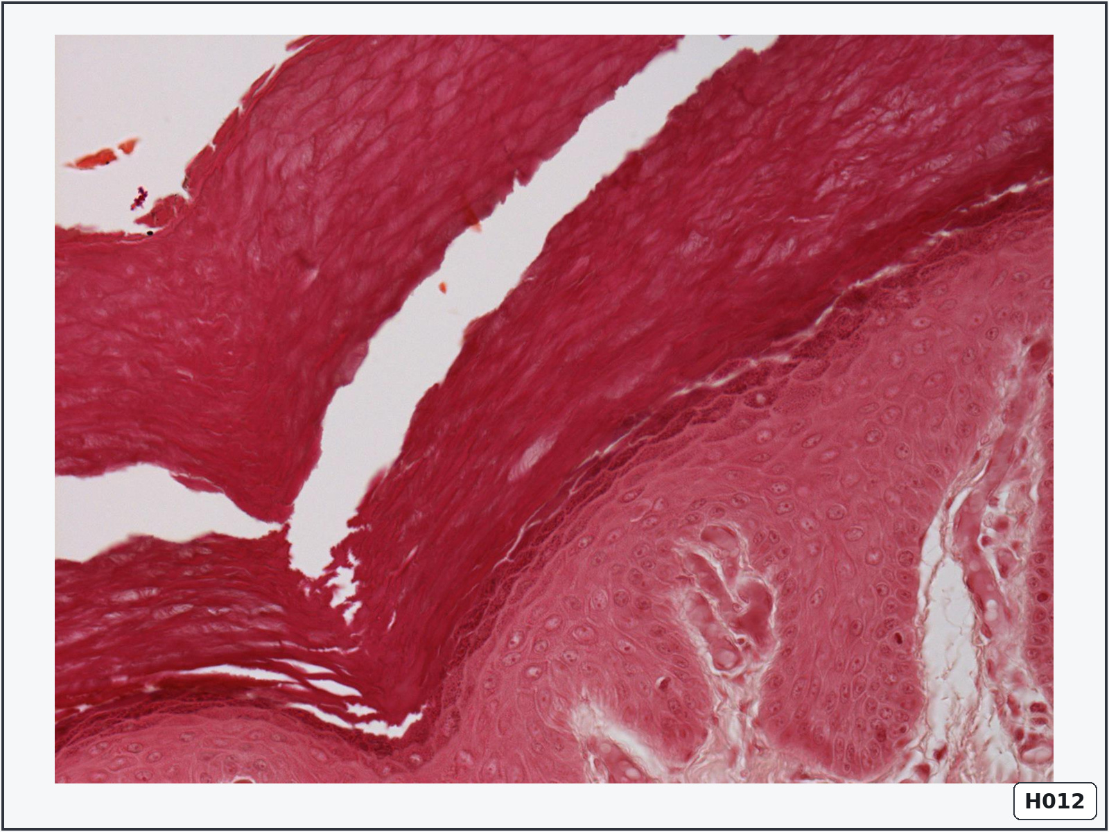
Stratified squamous keratinised epithelium – thick skin
Identify: a very thick, anuclear keratin layer over several epidermal layers.
Important structures: strata basale, spinosum, granulosum, lucidum and corneum.
Know: thick skin is found on palms and soles. The stratum lucidum is characteristic of thick skin.
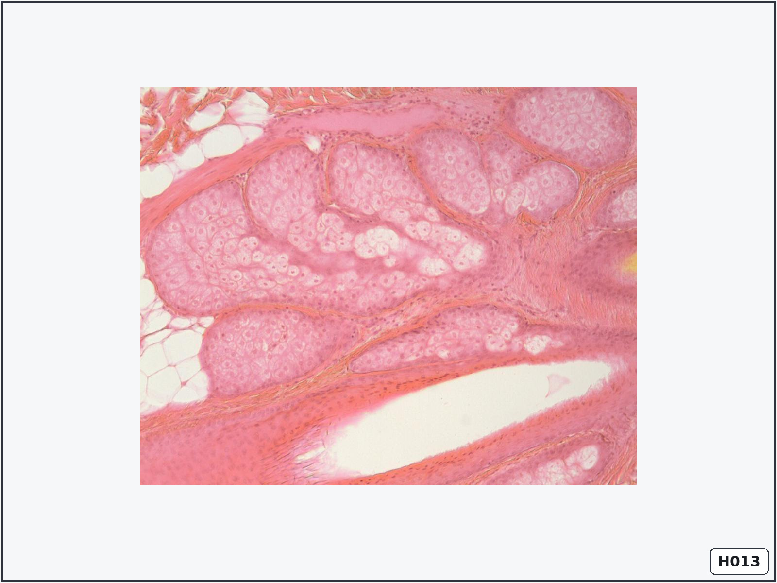
Holocrine sebaceous gland
Identify: lobules of pale lipid-filled cells associated with a duct.
Important structures: peripheral basal cells, sebocytes, glandular acini and duct.
Know: holocrine secretion releases the whole cell as sebum after the mature sebocyte disintegrates.
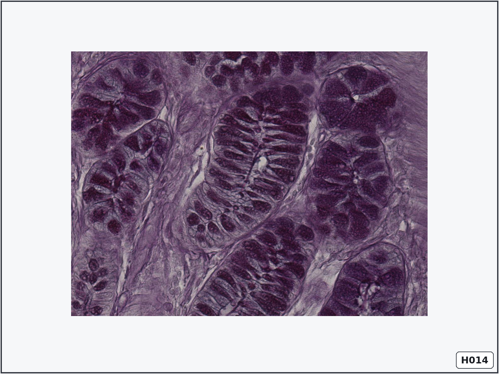
Goblet cells – large intestine
Identify: numerous pale goblet cells within straight intestinal crypts.
Important structures: simple columnar epithelium, goblet cells, absorptive cells, crypts and lamina propria.
Know: goblet cells are unicellular glands that secrete mucus to lubricate and protect the intestinal surface.
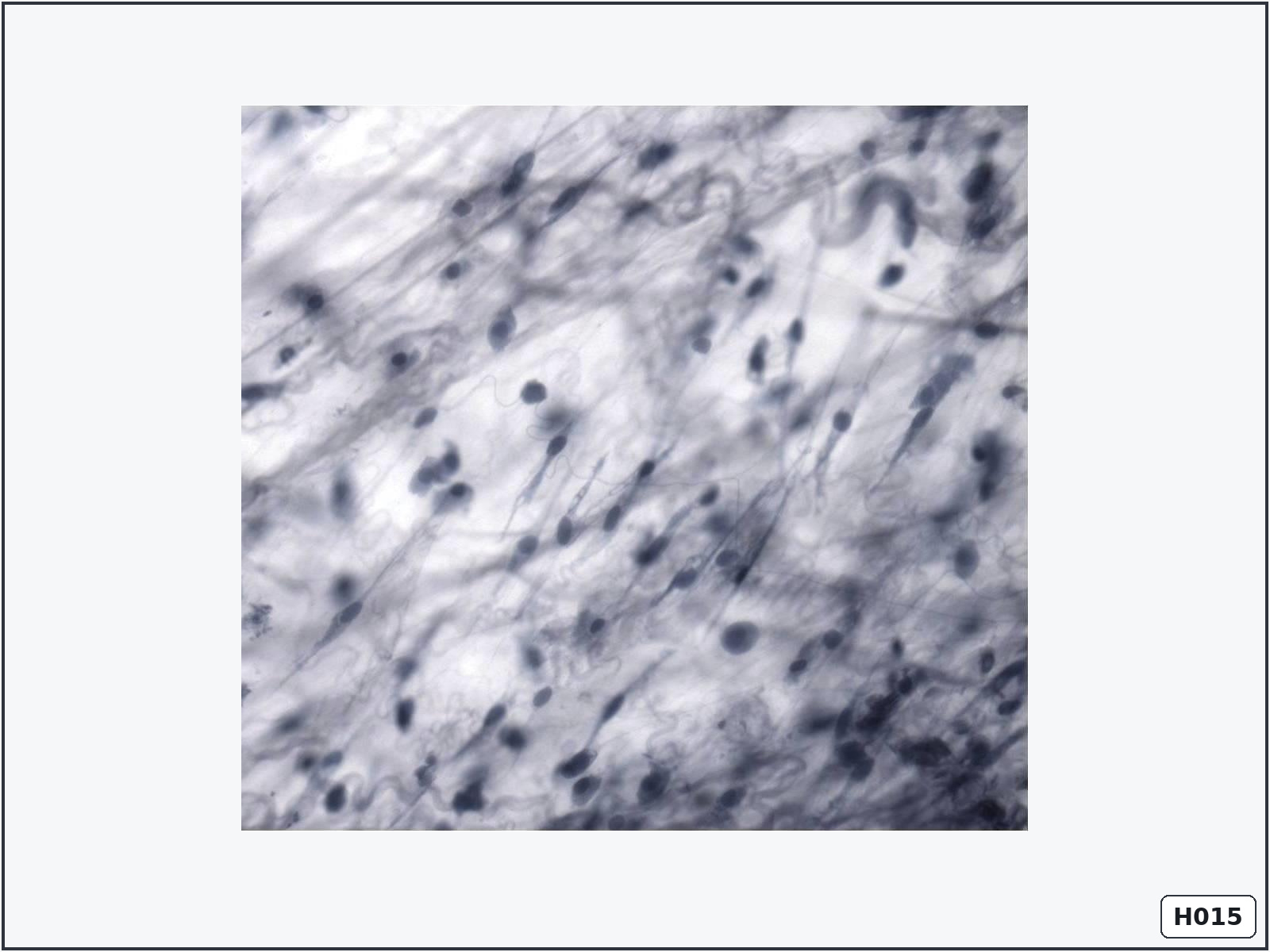
Loose connective tissue
Identify: loosely arranged fibres, abundant ground substance and several different cell types.
Important structures: collagen fibres, elastic fibres, fibroblasts, macrophages and ground substance.
Know: it supports epithelia, surrounds vessels and permits diffusion and immune-cell movement.
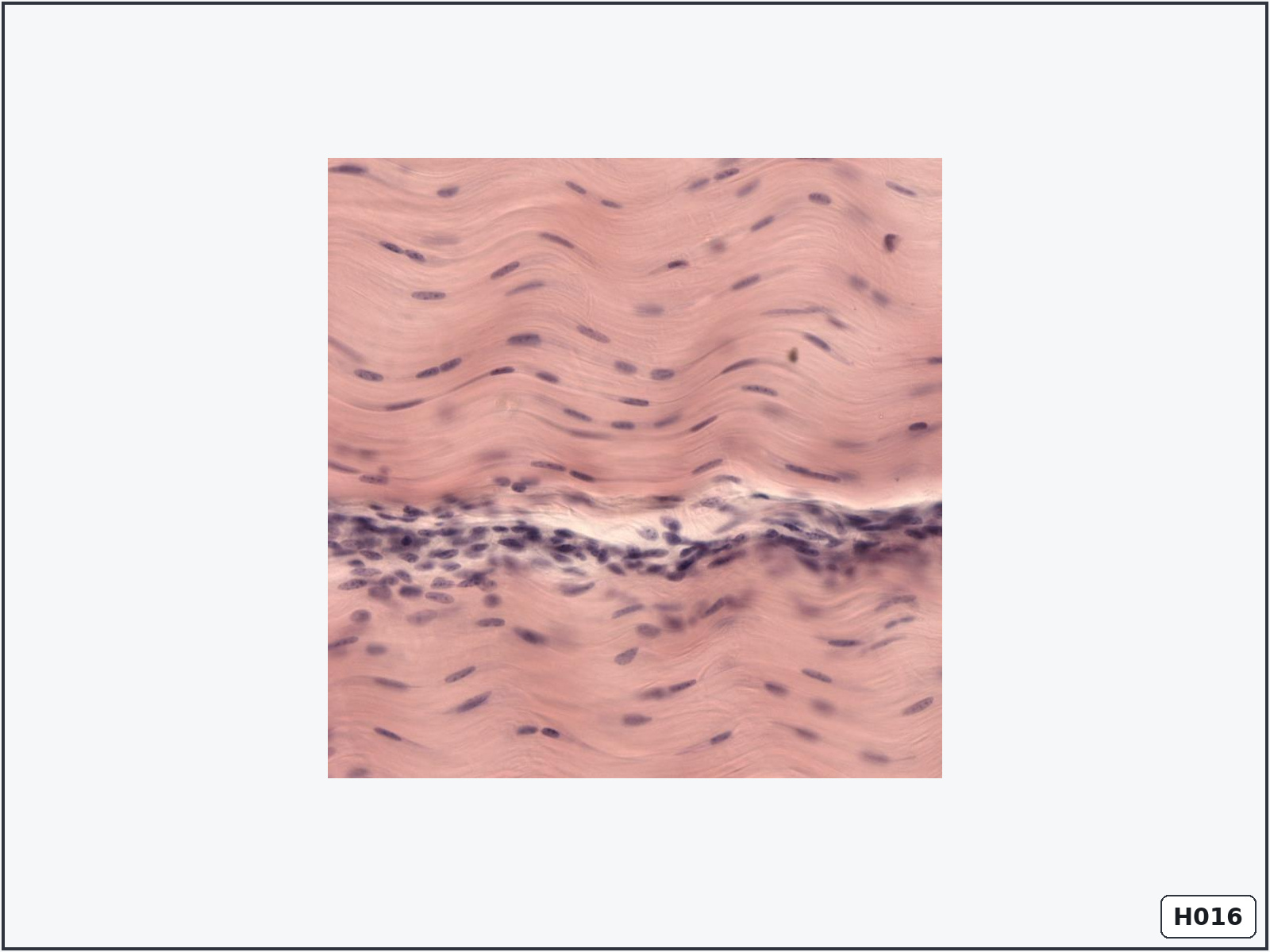
Dense regular connective tissue – tendon
Identify: densely packed parallel collagen bundles with rows of flattened nuclei.
Important structures: type I collagen bundles, tenocytes and scant ground substance.
Know: its parallel arrangement provides high tensile strength in one direction.
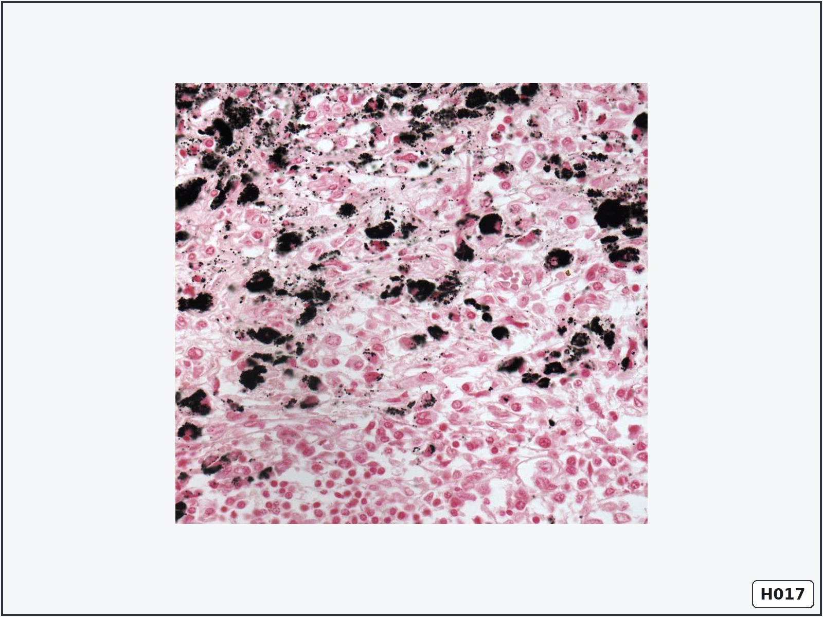
Macrophages in a lymph node
Identify: irregular cells containing coarse black carbon particles among lymphocytes.
Important structures: carbon-containing macrophages, macrophage nuclei, lymphocytes and reticular stroma.
Know: macrophages perform phagocytosis and present antigens to lymphocytes.
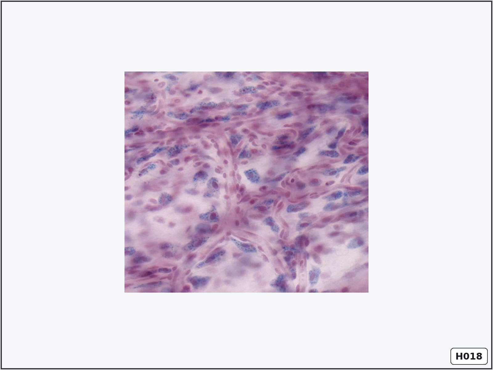
Macrophages in connective tissue
Identify: large irregular cells containing blue phagocytosed granules.
Important structures: macrophages, nuclei, dye granules, vessels and connective-tissue cells.
Know: macrophages originate from blood monocytes and remove foreign material and dead cells.
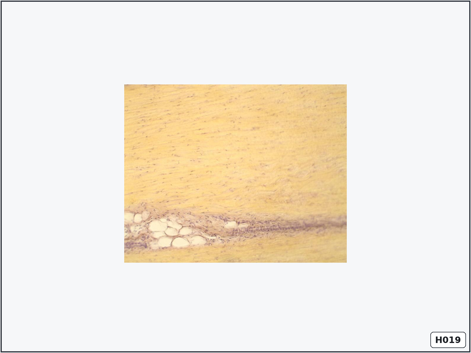
Elastic connective tissue
Identify: numerous dark, wavy and branching elastic fibres.
Important structures: elastic fibres, collagen fibres and fibroblast nuclei.
Know: elastic fibres contain elastin and allow tissues to stretch and return to their original shape.
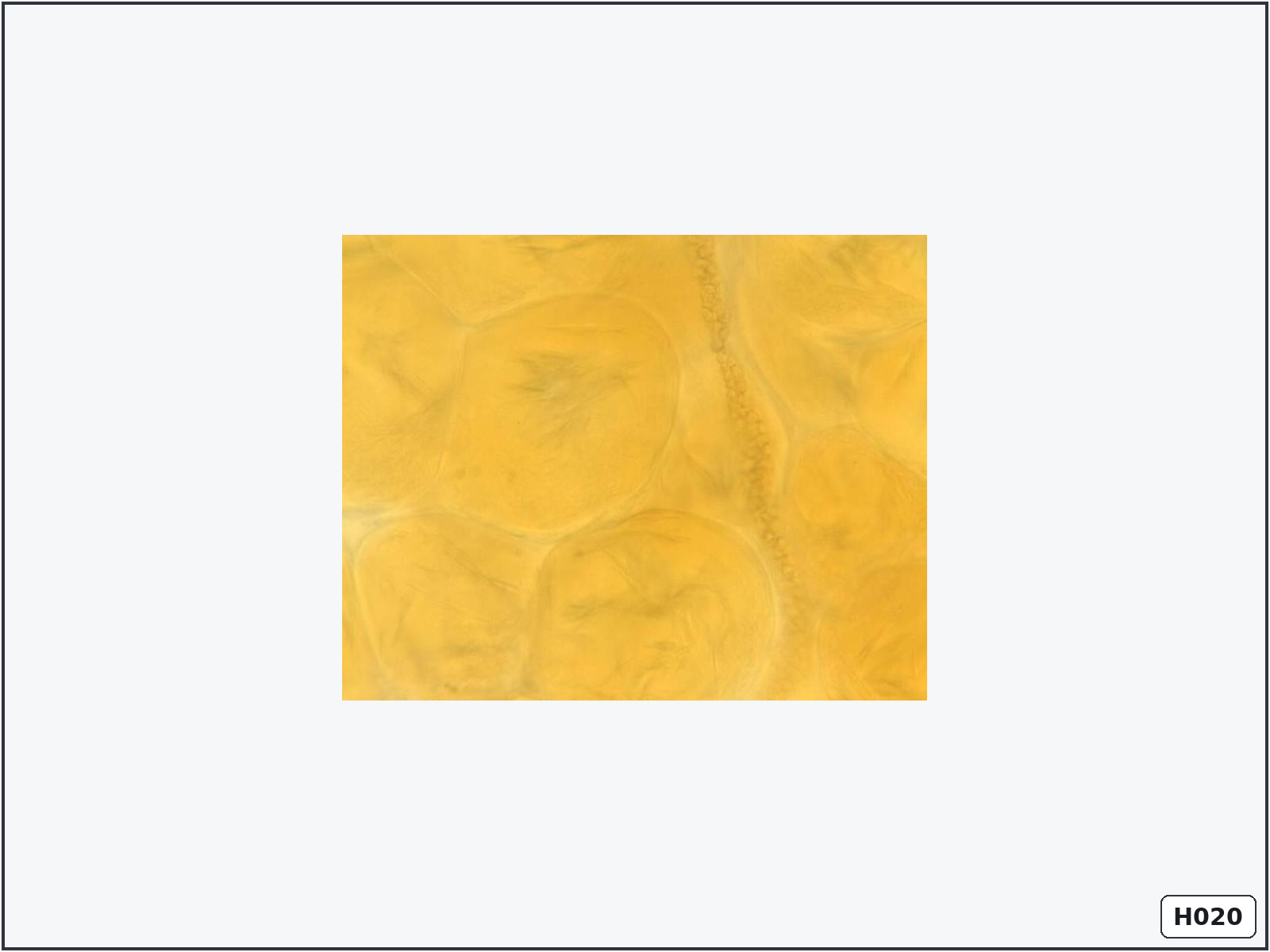
Adipose tissue – Sudan III preparation
Identify: large yellow-orange lipid droplets within adipocytes.
Important structures: adipocytes, lipid droplets, peripheral nuclei and capillaries.
Know: Sudan III demonstrates neutral fat because routine processing normally removes lipids.
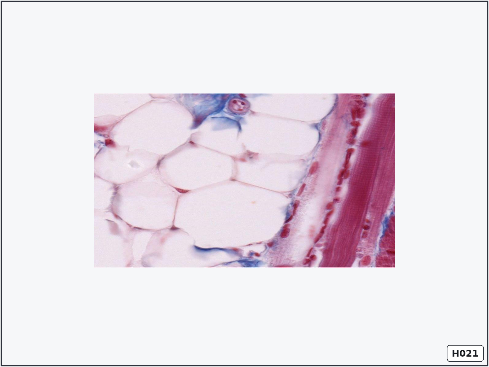
Unilocular adipose tissue – white fat
Identify: large empty signet-ring cells with a thin cytoplasmic rim and peripheral nuclei.
Important structures: one large lipid droplet, peripheral nucleus, cytoplasm, capillaries and connective-tissue septa.
Know: white adipose tissue stores energy, cushions organs, insulates the body and acts as an endocrine tissue.
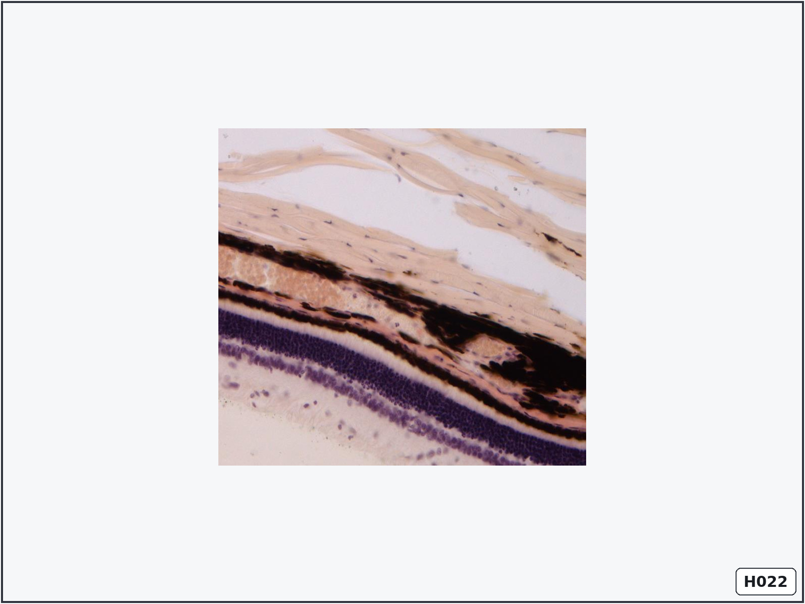
Pigmented connective tissue – choroid
Identify: numerous cells containing dark brown-black melanin near the retinal layers.
Important structures: melanocytes, melanin, connective tissue and blood vessels.
Know: choroidal melanin absorbs scattered light, while its vessels supply the outer retina.
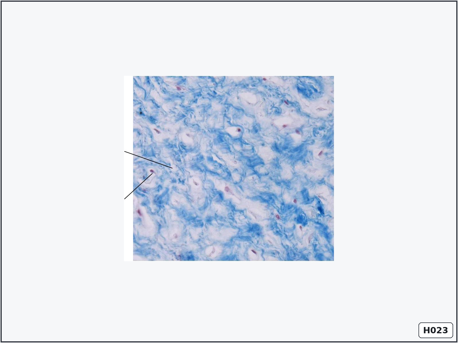
Mucous connective tissue – Wharton’s jelly
Identify: widely separated stellate cells in abundant pale gelatinous matrix.
Important structures: stellate fibroblasts, ground substance and fine collagen fibres.
Know: Wharton’s jelly surrounds and protects the umbilical vessels from compression.
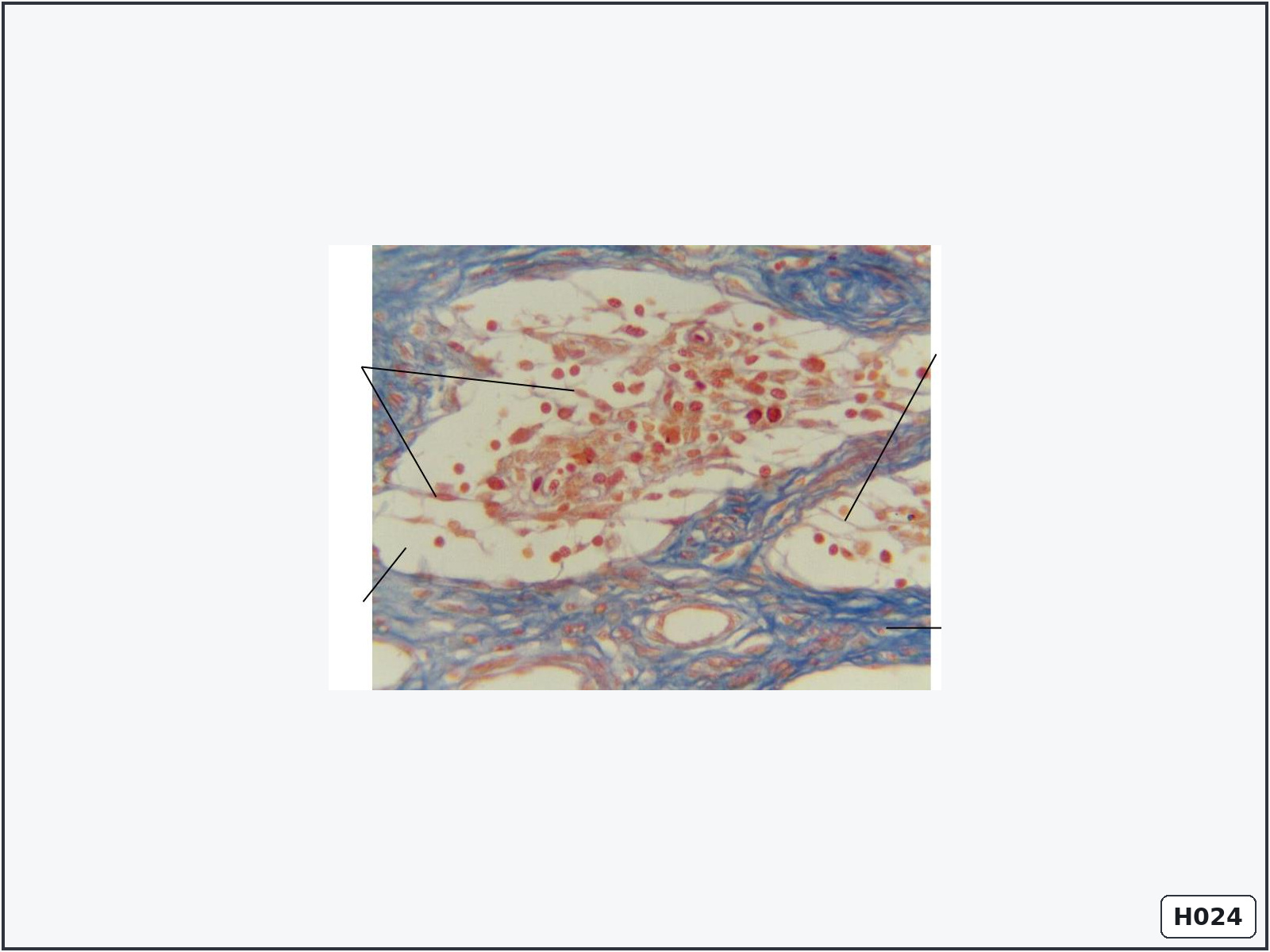
Reticular tissue – lymph node
Identify: a delicate branching fibre network supporting numerous free cells.
Important structures: reticular fibres, reticular cells, lymphocytes and macrophages.
Know: reticular fibres are mainly type III collagen and form the supporting stroma of lymphoid organs.
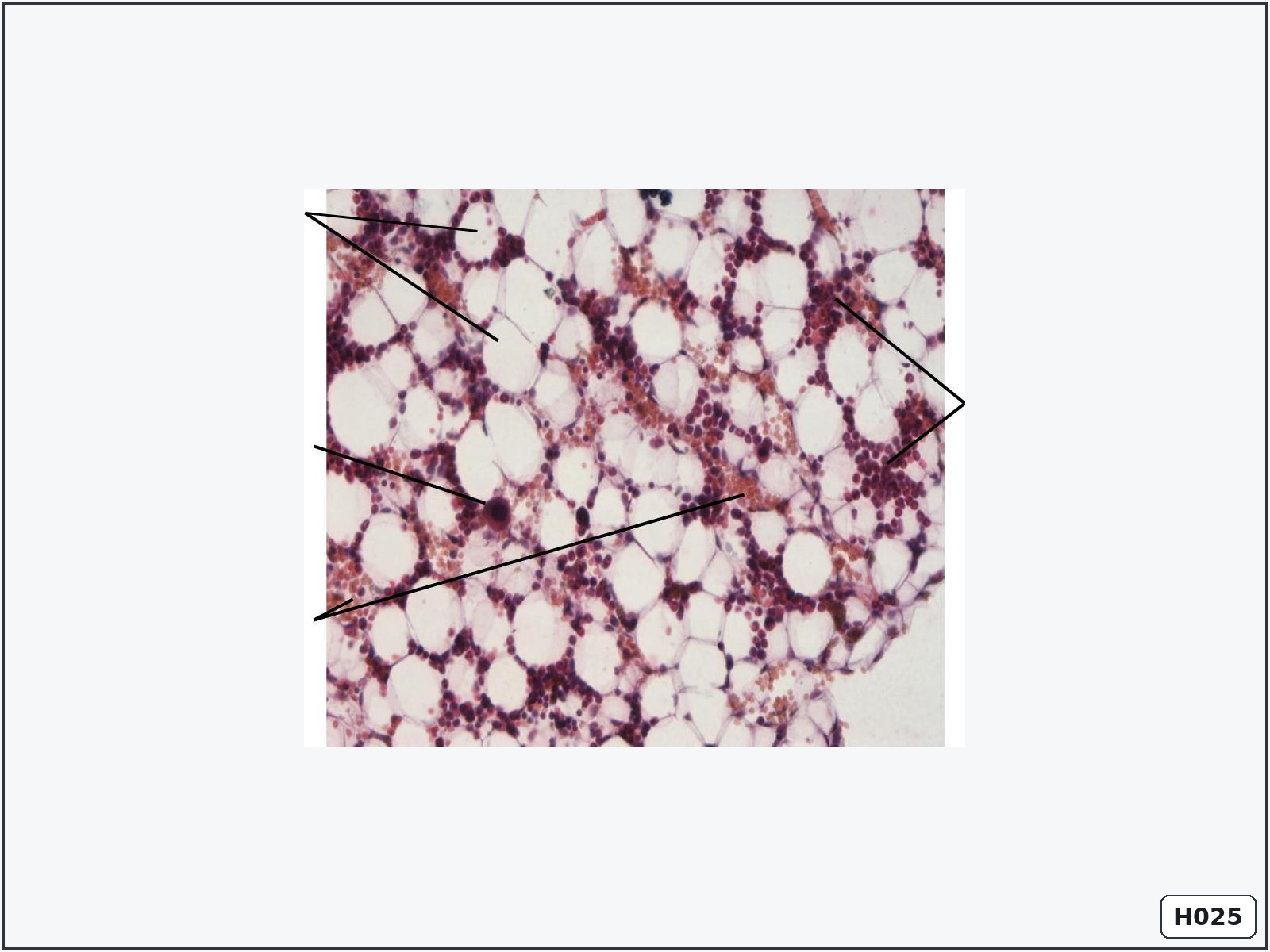
Bone marrow section
Identify: highly cellular tissue containing developing blood cells, adipocytes and giant megakaryocytes.
Important structures: haematopoietic cells, megakaryocytes, sinusoids, adipocytes and reticular stroma.
Know: red bone marrow produces erythrocytes, leukocytes and platelets.
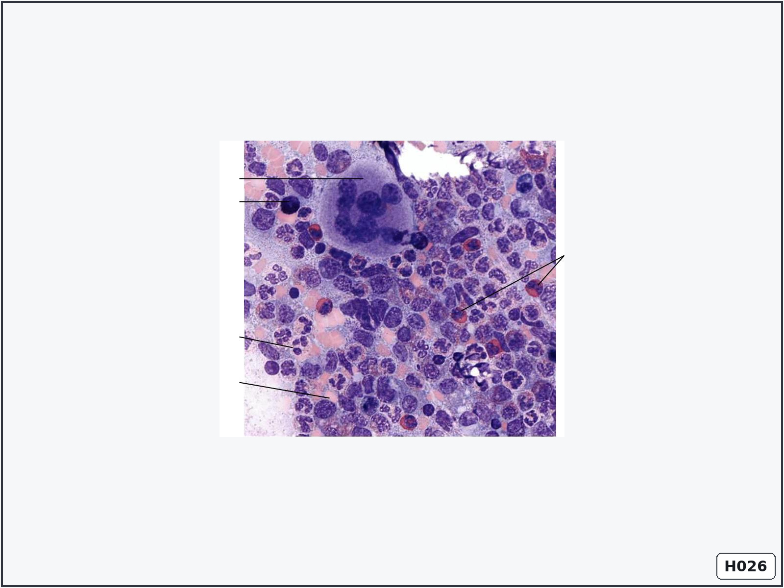
Bone marrow smear
Identify: blood-cell precursors at different developmental stages and a very large megakaryocyte.
Important structures: megakaryocyte, erythroid cells, granulocyte precursors and mature blood cells.
Know: megakaryocytes produce platelets by releasing cytoplasmic fragments.
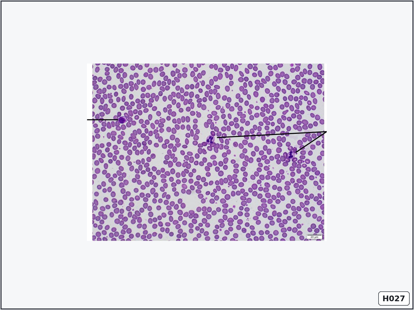
Peripheral blood smear
Identify: numerous pink erythrocytes with scattered leukocytes and small platelets.
Important structures: erythrocytes, neutrophils, lymphocytes and platelets.
Know: erythrocytes are anucleate biconcave cells; leukocytes contain nuclei; platelets are small cell fragments.
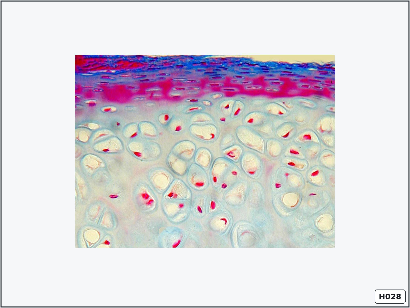
Hyaline cartilage
Identify: a glassy matrix containing chondrocytes in lacunae and isogenous groups.
Important structures: perichondrium, chondrocytes, lacunae, territorial matrix and interterritorial matrix.
Know: its matrix contains type II collagen, but individual fibres are usually not visible.
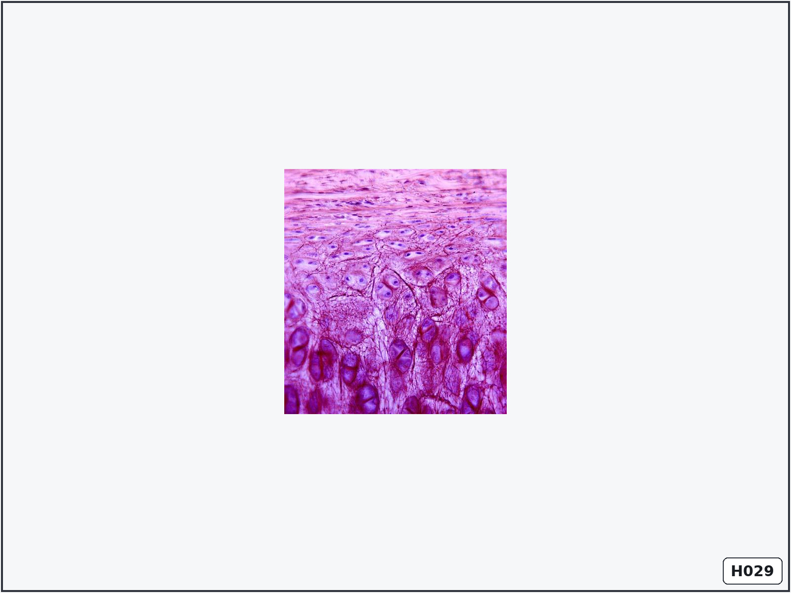
Elastic cartilage
Identify: cartilage containing a dense dark network of elastic fibres.
Important structures: perichondrium, chondrocytes, lacunae, isogenous groups and elastic fibres.
Know: elastic cartilage is flexible and is found in structures such as the external ear and epiglottis.
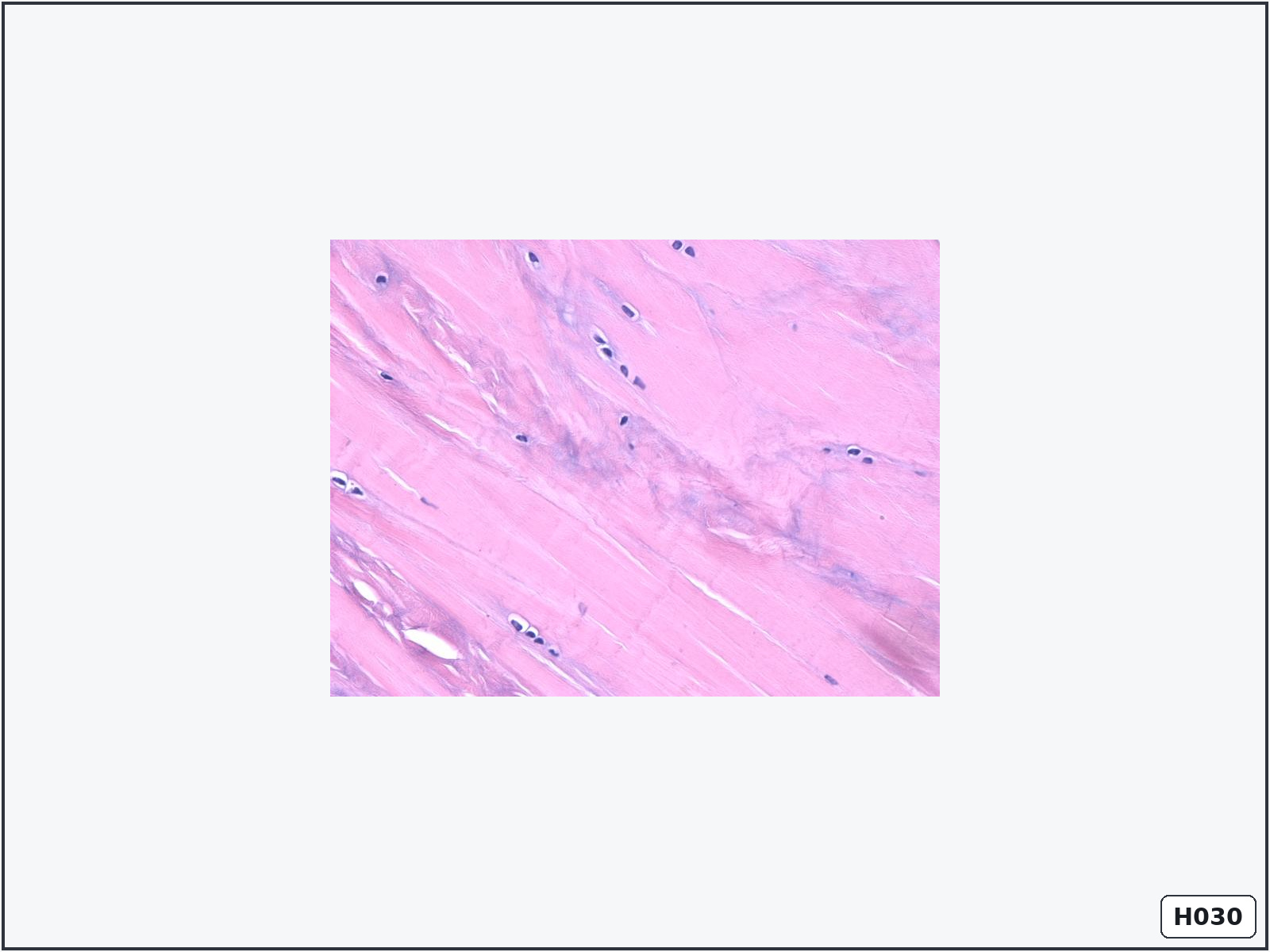
Fibrocartilage
Identify: rows of chondrocytes between thick parallel collagen bundles.
Important structures: chondrocytes, lacunae, type I collagen bundles and fibroblasts.
Know: fibrocartilage has no perichondrium and resists both compression and tension.
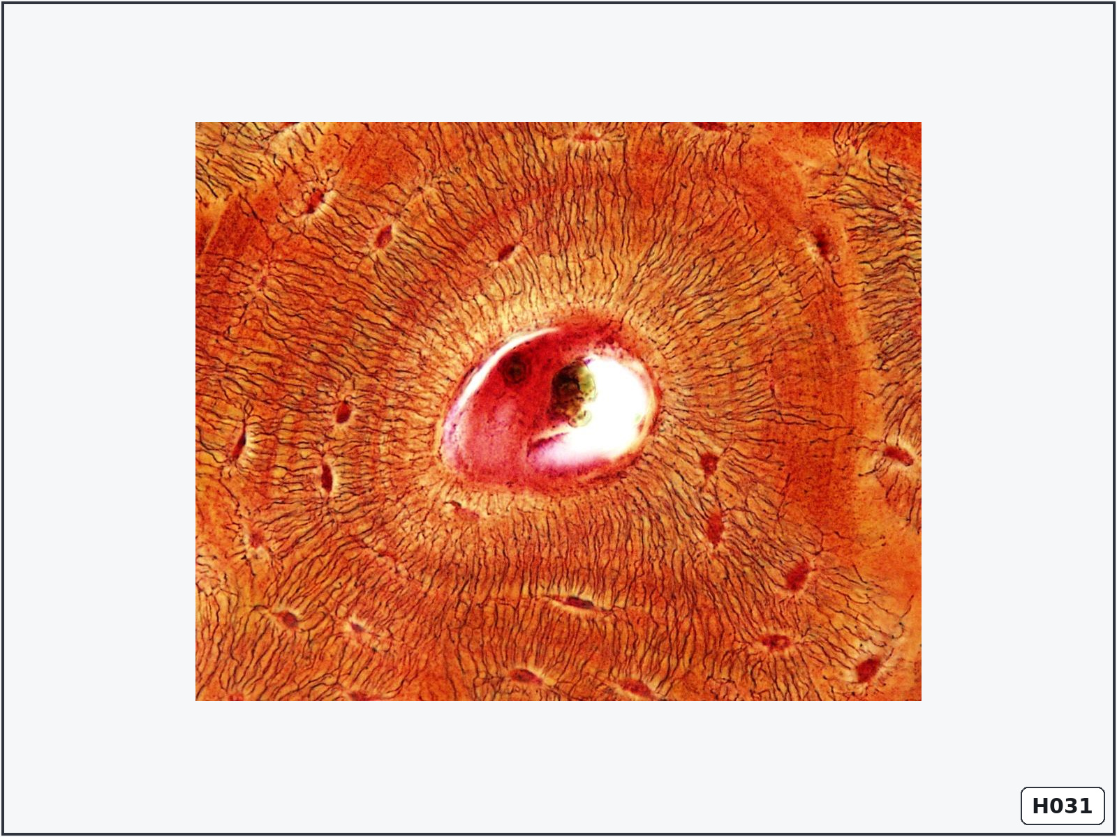
Compact lamellar bone – transverse section
Identify: circular osteons with central canals and concentric lamellae.
Important structures: osteons, Haversian canals, concentric lamellae, osteocyte lacunae, canaliculi and interstitial lamellae.
Know: Haversian canals contain blood vessels and nerves. Canaliculi connect osteocytes.
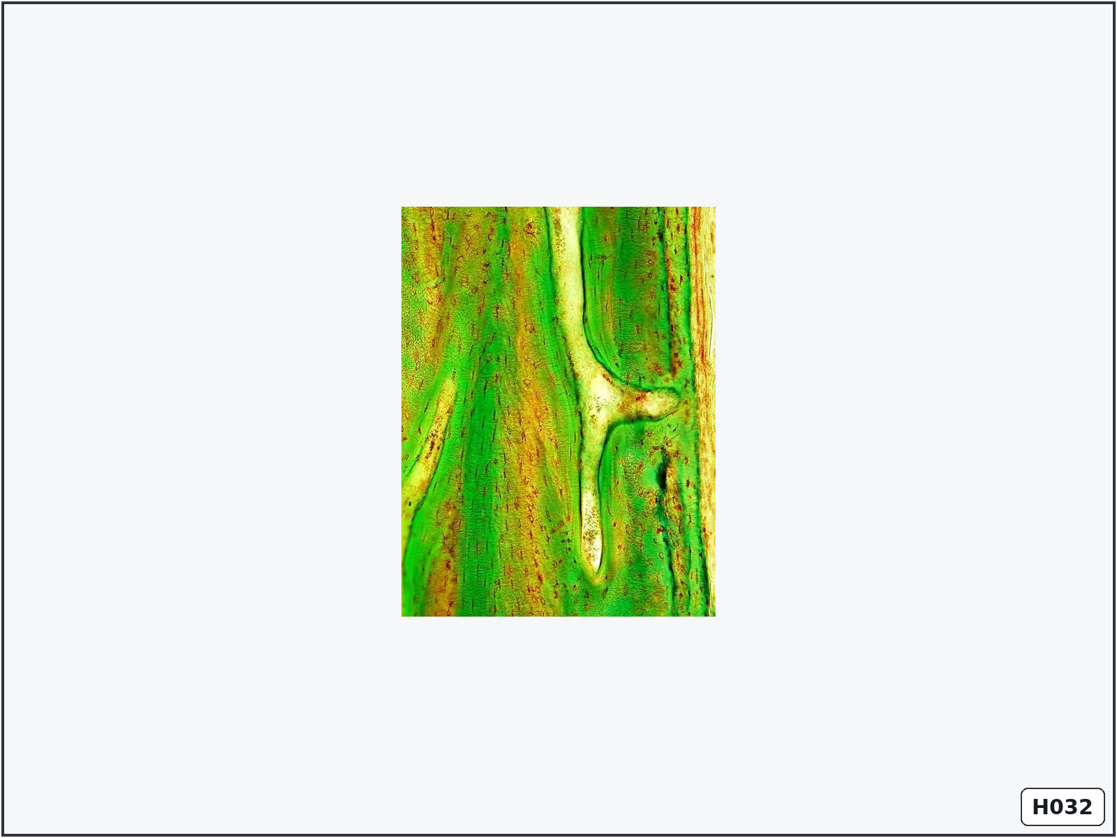
Compact lamellar bone – longitudinal section
Identify: long parallel Haversian canals connected by transverse Volkmann canals.
Important structures: Haversian canals, Volkmann canals, lamellae, lacunae and canaliculi.
Know: Volkmann canals connect neighbouring Haversian canals and communicate with the periosteum and marrow cavity.
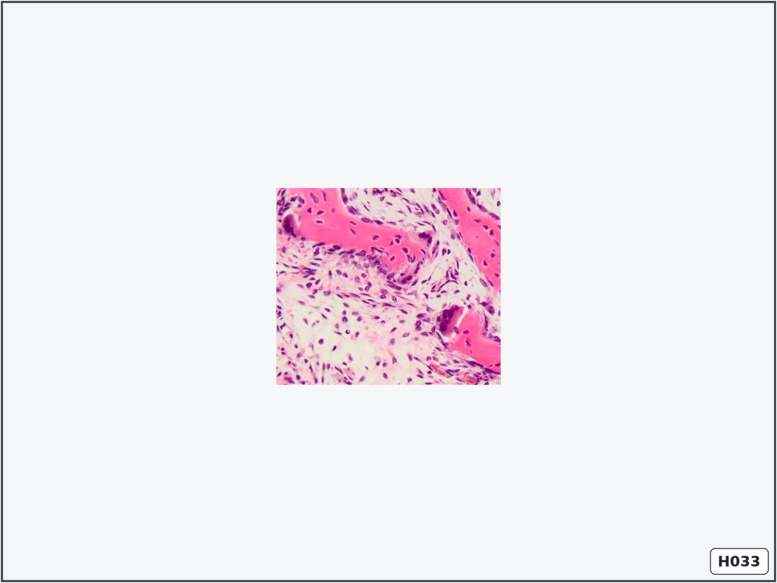
Intramembranous ossification
Identify: irregular woven-bone trabeculae forming directly within vascular mesenchyme.
Important structures: mesenchymal cells, osteoblasts, osteocytes, osteoclasts, bone trabeculae and vessels.
Know: bone forms directly from mesenchyme without a cartilage model. This produces many flat bones of the skull.
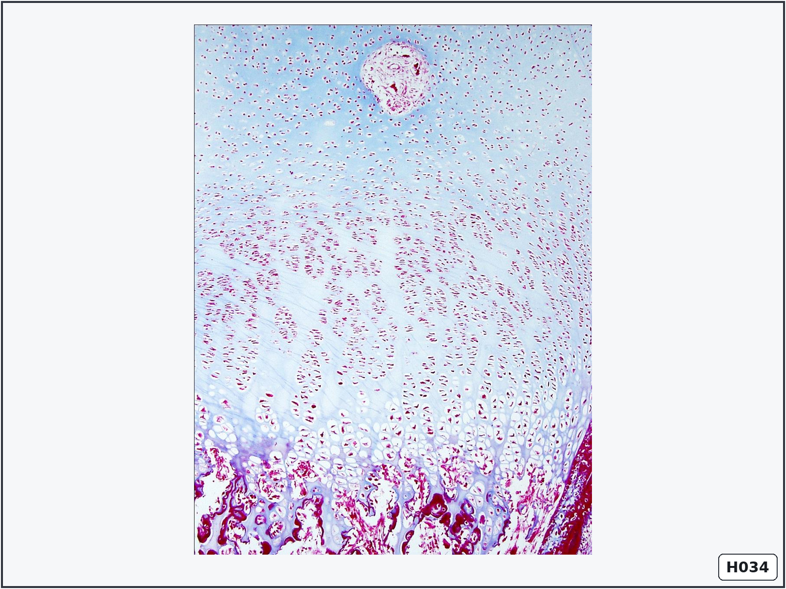
Endochondral ossification – epiphyseal plate
Identify: orderly columns of chondrocytes progressing toward newly formed bone.
Important structures: reserve, proliferation, hypertrophy, calcification and ossification zones.
Know: endochondral ossification replaces a cartilage model and is responsible for longitudinal growth of long bones.
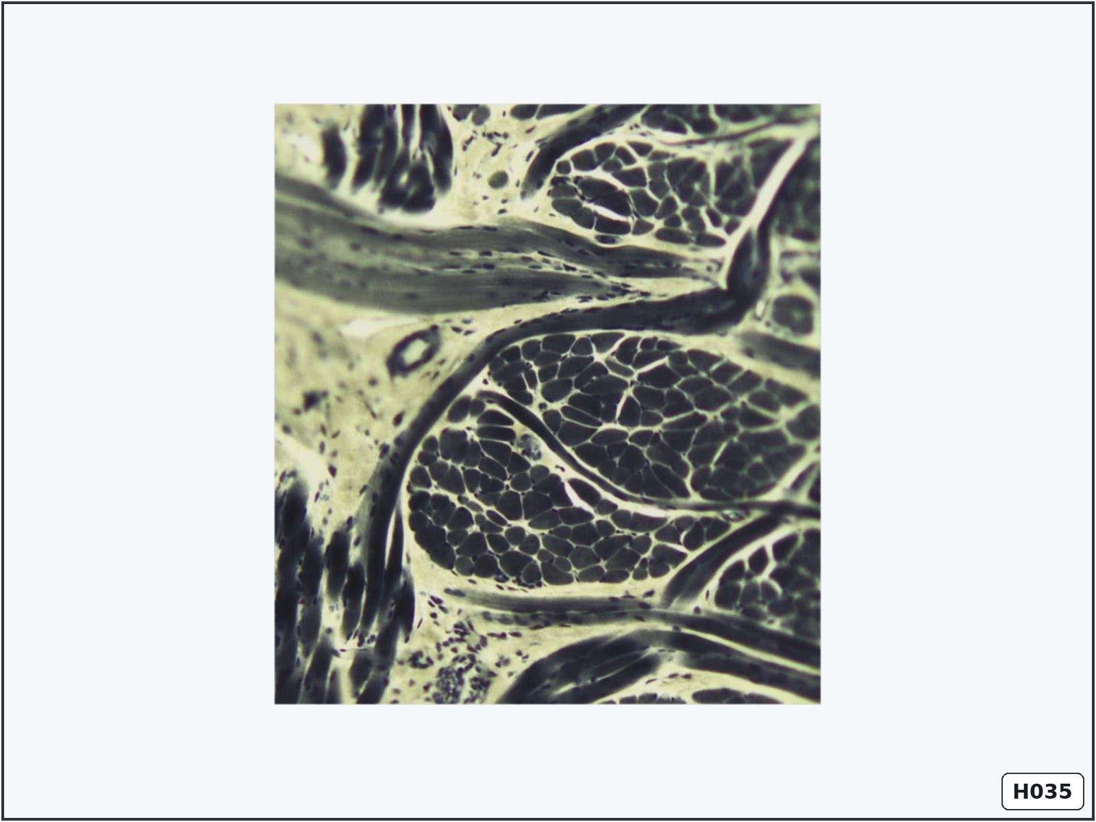
Striated skeletal muscle – transverse section
Identify: large polygonal muscle fibres with peripheral nuclei arranged into fascicles.
Important structures: muscle fibres, peripheral nuclei, endomysium, perimysium and blood vessels.
Know: endomysium surrounds individual fibres; perimysium surrounds fascicles.
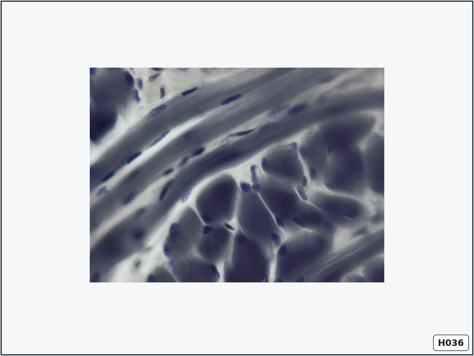
Striated skeletal muscle – longitudinal and transverse sections
Identify: long striated fibres together with polygonal cross-sections and peripheral nuclei.
Important structures: longitudinal fibres, transverse fibres, cross-striations, endomysium and perimysium.
Know: skeletal muscle fibres are multinucleated syncytia with nuclei positioned beneath the sarcolemma.
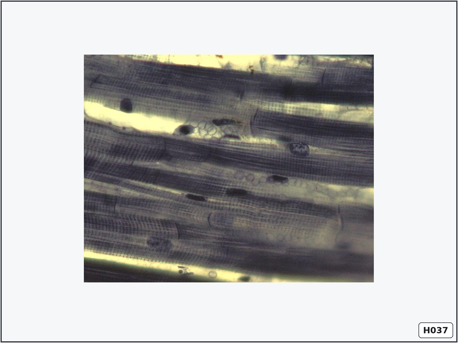
Cardiac muscle – longitudinal section
Identify: branching striated cells with central nuclei and dark intercalated discs.
Important structures: cardiomyocytes, central nuclei, striations, intercalated discs and endomysium.
Know: intercalated discs mechanically and electrically connect cardiomyocytes, allowing coordinated contraction.
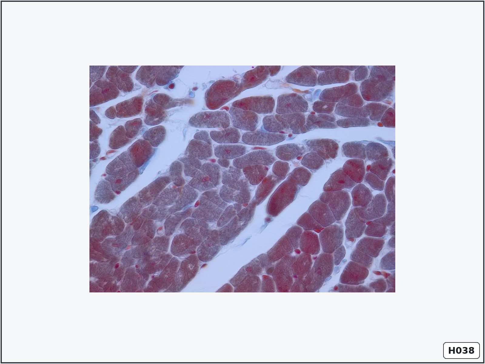
Cardiac muscle – transverse section
Identify: irregular round or polygonal cell profiles, often with central nuclei.
Important structures: cardiomyocytes, central nuclei, perinuclear sarcoplasm, endomysium and capillaries.
Know: central nuclei and abundant capillaries help distinguish cardiac muscle from skeletal muscle.
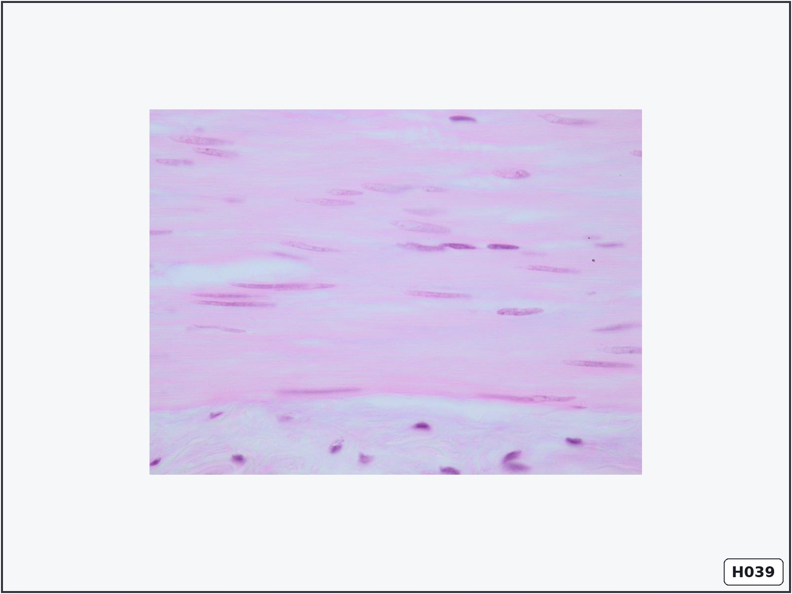
Smooth muscle – longitudinal section
Identify: closely packed spindle-shaped cells with elongated central nuclei and no striations.
Important structures: smooth-muscle cells, cigar-shaped nuclei, sarcoplasm and indistinct cell borders.
Know: smooth muscle is involuntary and contains actin and myosin but no sarcomeres.
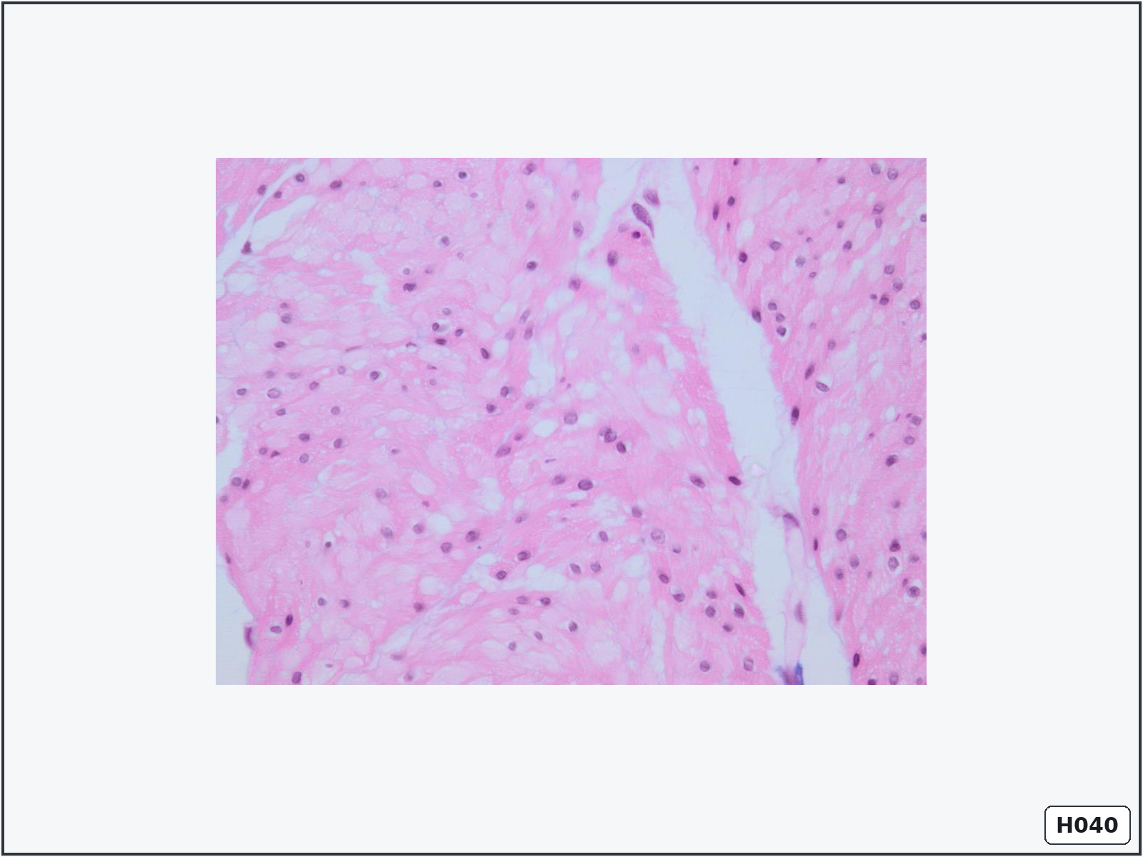
Smooth muscle – transverse section
Identify: small circular profiles of different sizes; only some contain nuclei.
Important structures: cell profiles, central nuclei, sarcoplasm and surrounding connective tissue.
Know: profiles differ in diameter because spindle-shaped cells are cut at different levels.
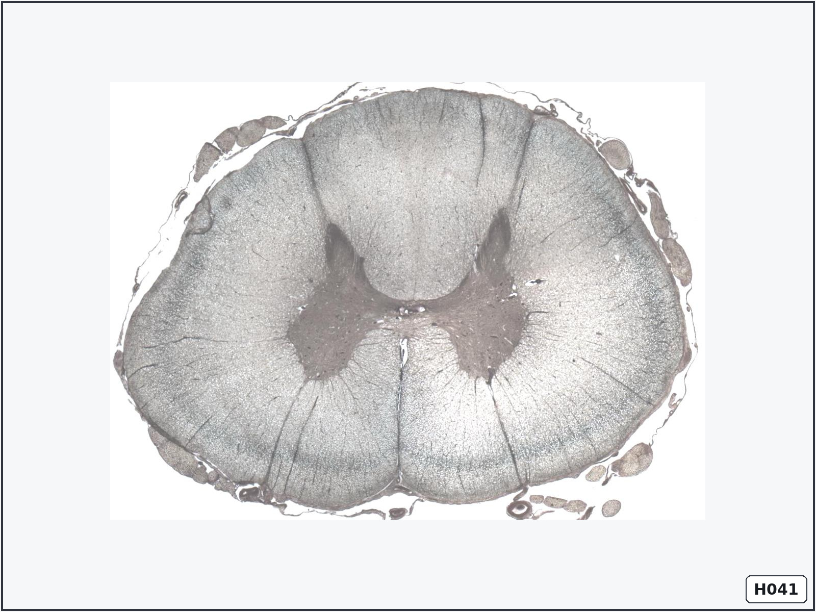
Spinal cord – transverse section
Identify: butterfly-shaped central grey matter surrounded by peripheral white matter.
Important structures: anterior horns, posterior horns, grey matter, white matter, central canal and anterior median fissure.
Know: grey matter contains neuronal cell bodies; white matter mainly contains myelinated axons.
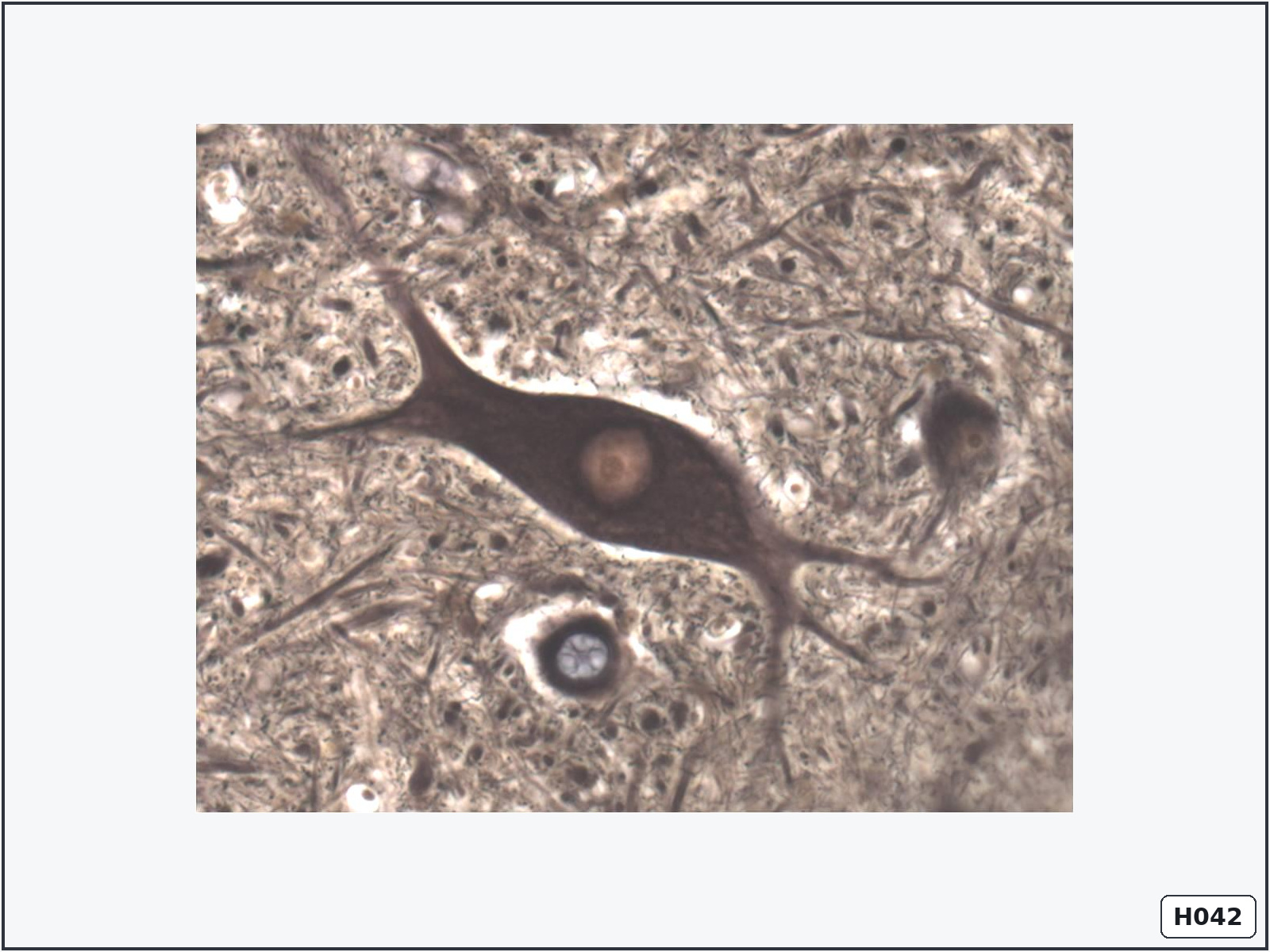
Multipolar neuron with neurofibrils
Identify: a large stellate neuron with several processes and dark fibrillar material.
Important structures: cell body, nucleus, nucleolus, dendrites, axon and neurofibrils.
Know: neurofibrils are cytoskeletal elements that support the neuron and assist intracellular transport.
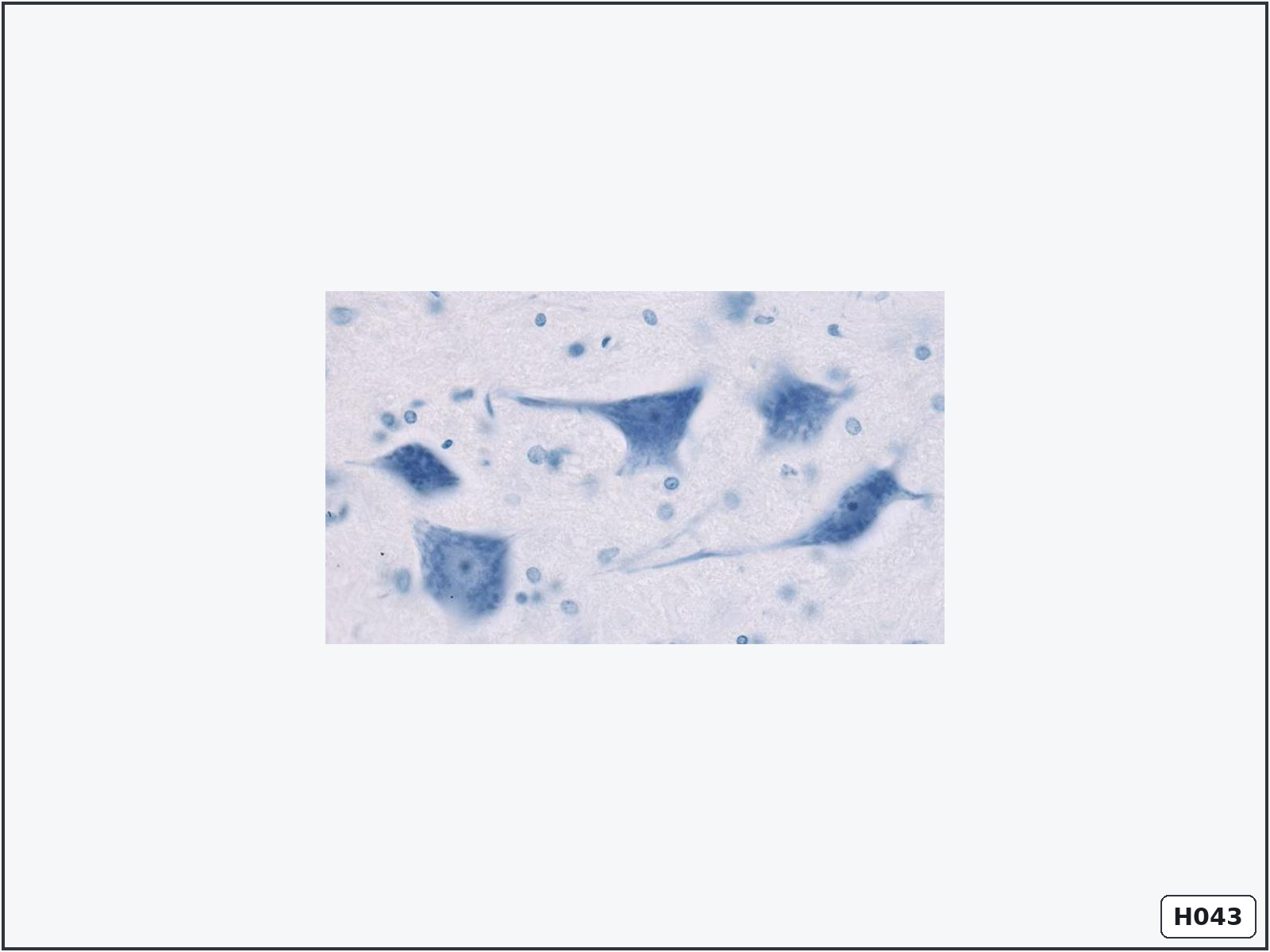
Neurons with Nissl bodies – spinal cord
Identify: large neurons containing coarse basophilic material in the soma and dendrites.
Important structures: cell body, nucleus, nucleolus, Nissl bodies, dendrites and glial nuclei.
Know: Nissl bodies consist of rough endoplasmic reticulum and ribosomes. They are absent from the axon hillock and axon.
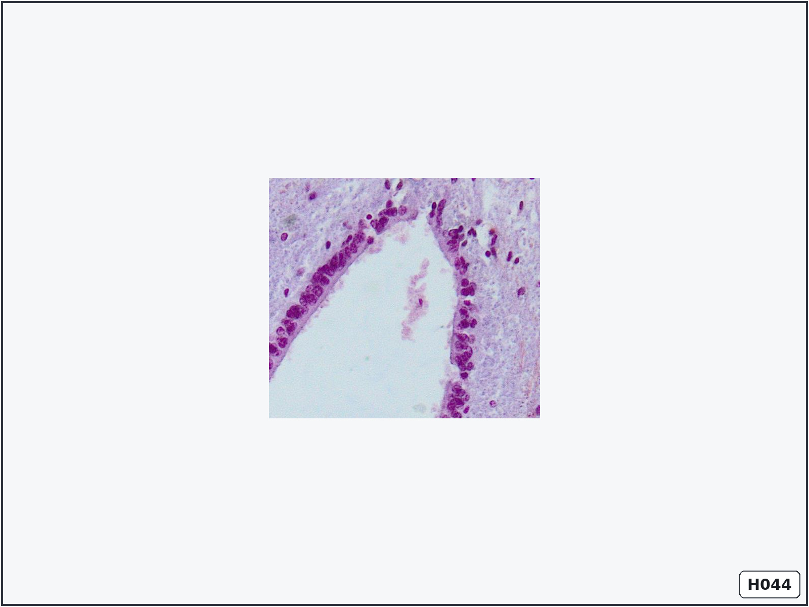
Ependymal cells lining the central canal
Identify: a single layer of cuboidal or columnar cells surrounding the central canal.
Important structures: central canal, ependymal cells, ependymal nuclei, glial nuclei and grey matter.
Know: ependymal cells line the brain ventricles and spinal central canal and help circulate cerebrospinal fluid.
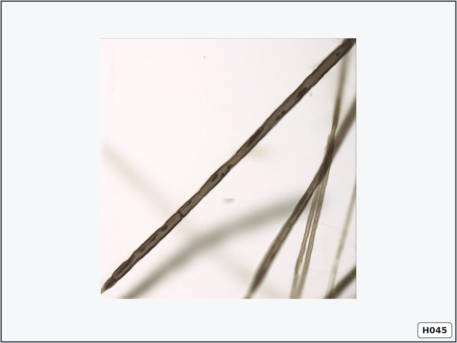
Myelinated peripheral nerve fibre
Identify: a dark segmented myelin sheath surrounding a central axon, interrupted by nodes.
Important structures: axon, myelin sheath, Schwann-cell nucleus, internode and node of Ranvier.
Know: one Schwann cell forms one myelin internode in the PNS. Nodes allow saltatory conduction.
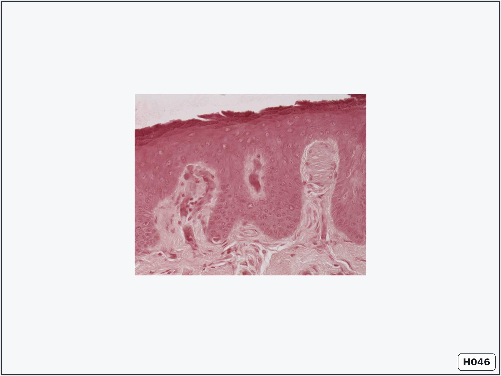
Meissner tactile corpuscle – thick skin
Identify: a small oval receptor located superficially inside a dermal papilla.
Important structures: capsule, flattened tactile cells and afferent nerve fibre.
Know: it is a rapidly adapting mechanoreceptor for light touch and low-frequency vibration.
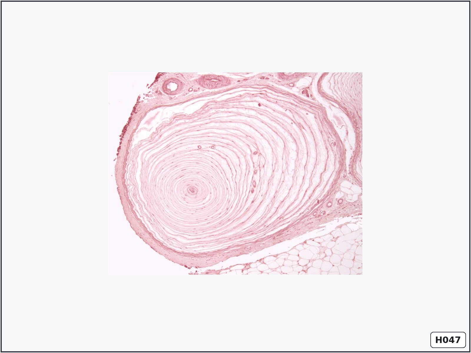
Pacinian corpuscle
Identify: a large onion-like receptor with concentric lamellae around a central axon.
Important structures: capsule, concentric lamellae, fibrocyte nuclei, central core and afferent nerve fibre.
Know: it is a rapidly adapting receptor for deep pressure and high-frequency vibration.