Looks like no one added any tags here yet for you.
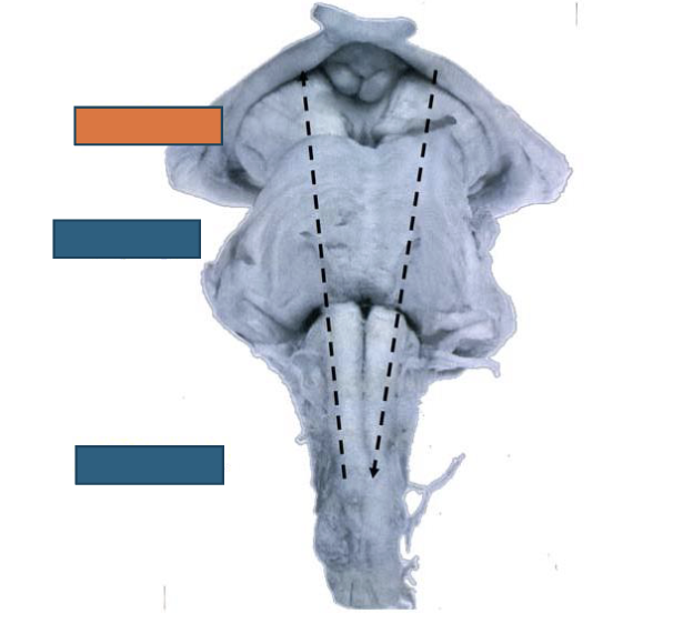
Name the structure:
Midbrain
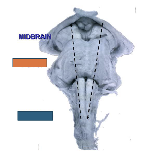
Name the structure:
Pons
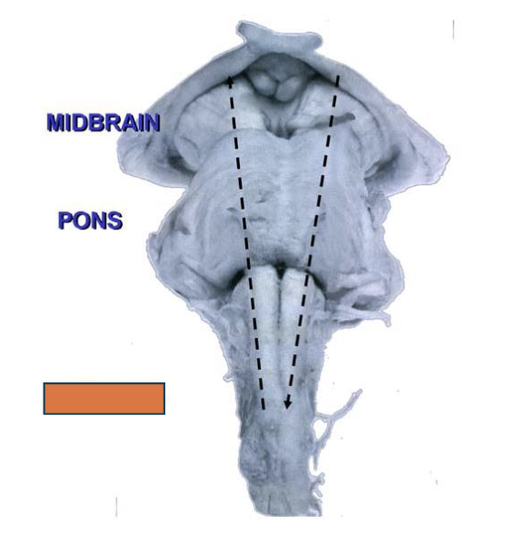
Name the structure:
Medulla
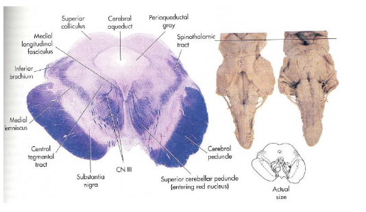
Midbrain
contains cerebral peduncles
sleep/wake cycles
alertness
temperature rgulation
Pons
contains cerebellar peduncles
connects with cerebellum
Medulla
vital functioning
breathing, heart rate, swallowing
Name this structure:
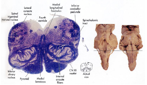
Caudal medulla
Name this structure:
Rostral medulla
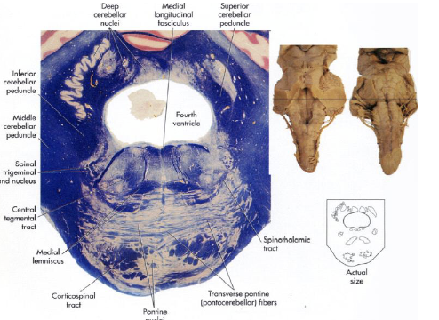
Name this structure:
Caudal pons
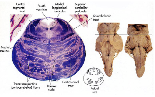
Name this structure:
Rostral pons
Name this structure:
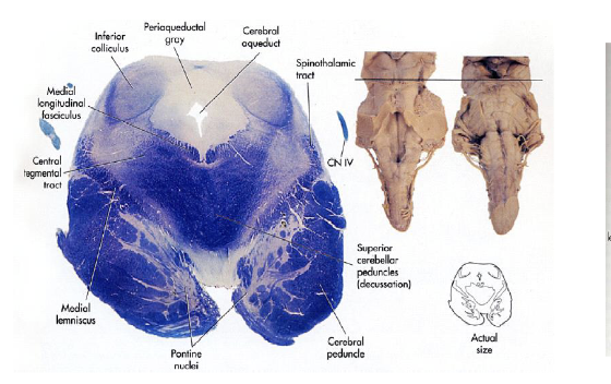
Caudal midbrain
Name this structure:
Rostral midbrain
Afferent
Ascending - SENSORY (body → brain)
Efferent
Descending - MOTOR (brain → body)
Midbrain CNs
CN III → CN IV
Pons CNs
CN V → CN VIII
Medulla CNs
CN IX → CN XII
CN I
Olfactory
smell
sensory
CN II
Optic
visual
sensory
CN III
CN IV
CN V
CN VI
CN VII
CN VIII
CN IX
CN X
CN XI
CN XII
Apex of cochlea
LOW frequency
Base of cochlea
HIGH frequency
Inner Hair Cells
Afferents (SENSORY)
responsible for hearing
SPECIFIC - each AN fiber receives signal from ONE IHC
Outer Hair Cells
Efferents
responsible for helping to better align IHC to tectorial membrane
amplify signal
SENSITIVE - MANY OHC to one nerve fiber
if ONE OHC goes off → nerve fires
auditory pathway
cochlear nucleus
superior olive
inferior colliculus
ITD pathway
ILD Pathway
medial geniculate body
cochlear nucleus
MONAURAL hearing
receives information from IPSILATERAL cochlear nerve fibers
superior olive
BINAURAL hearing
picks up BILATERAL cues
inferior colliculus
where sound localization occurs
ITD pathway (inferior colliculus)
sound localization at LOW frequencies where there is a TIMING difference
ILD pathway (inferior colliculus)
sound localization at HIGH frequencies where there is a LEVEL difference
medial geniculate body
located in the thalamus
anatomy of the eye
divided into:
small anterior cavity
large and round posterior cavity
anterior cavity of the eye contains:
iris
cornea
lens
aqueous humor - fluid similar to CSF
Where in the brainstem can you see the cerebral peduncles most prominently?
rostral midbrain
posterior cavity of the eye is the area between the _____.
lens & retina
what fills the posterior cavity of the eye?
vitreous fluid - jellylike substance
maintains normal intraocular pressure
prevents eyeball from collapsing
outer ear
pinna - funnels sound into ear canal
middle ear
amplifies sound by changing it from large → small diameter
ossicles - change diameter of sound vibration
inner ear
cochlea - translates sound from mechanical energy into electrical information
fluid in scala vestibuli
perilymph
fluid in scala media
endolymph
fluid in scala tympani
perilymph
where is the organ of corti located
scala media
organ of corti
primarily responsible for sound transduction
where are hair cells located?
on the basilar membrane
filaments on top of hair cells
stereocilia - get deformed by sound waves
all connected
Ca 2+ and K+ enter hair cell → action potential
cellular transduction
photos move POSTERIORLY (towards back of eye)
signal moves ANTERIORLY (towards front of eye)
rods
night/low light vision
mostly in PERIPHERY
cones
color & acuity (most accurate/crisp vision)
mostly concentrated in fovea
streams of association processing:
dorsal stream
ventral stream
dorsal stream
motion processing
“where”
projects to PARIETAL lobe
ventral stream
objective recognition
“what”
projects to TEMPORAL lobe
limbic system
learning, emotions, & homeostasis
the limbic system contains:
thalamus
hypothalamus
amygdala
hippocampus
fornix
thalamus
sensory relay station
medial geniculate body
lateral geniculate body
hypothalamus
homeostasis, hunger, thirst, vital functioning
amygdala
emotions (fear pleasure, reward); how one sees themselves
hippocampus
learning & memory
fornix
“C” shape that connects many structures
where in the auditory pathway is hearing binaural?
superior olive
inferior colliculus
thalamus - medial geniculate body
reward/punishment functions of limbic system:
help us “learn'“ and motivates behavior (or lack of behavior)
impaired by drugs and psychiatric disorders
reward centers:
hypothalamus
septum
amygdala
thalamus
basal ganglia
punishment centers:
periaqueductal grey
hypothalamus
thalamus
what can take precedence over pleasure & reward?
punishment & fear
sensorimotor
a task is NEVER purely motor
always getting sensory feedback and correcting
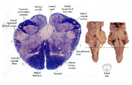
anatomy of spinal cord:
DORSAL - afferent (SENSORY in)
VENTRAL - efferent (MOTOR out)
sensory fiber tracts
dorsal column pathway
spinothalamic tract
dorsal column pathway
touch, proprioception, vibration
optic chaism
point where PARTIAL crossing of optic nerves occurs
optic tract
AFTER crossing; white mater tract
optic radiations
AFTER crossing & thalamus; white matter tract
send signals to primary visual cortex
spinothalamic tract
slower, unmyelinated, pain, & temperature
important MOTOR spinal tracts
corticospinal tract
corticobulbar tract
corticospinal tract
fibers originated from cortex & descend to spinal cord
cross midline in the pyramids of medulla (brainstem)
control CONTRALATERAL muscles throughout the entire body
corticobulbar tract
fibers originated from cerebral cortex & descend…
through the internal capsule
in the midbrain through cerebral peduncle
cross midline in brainstem at level of the CN nuclei they innervate
BILATERAL projections
visual system pathway:
retina
optic nerve
optic chiasm
optic tracy
lateral geniculate body
optic radiation
primary visual cortex (V1)
Damage to the left optic tract would cause:
loss of vision in the right visual field
Upper motor neurons
located in CNS
within motor cortex of brain & certain brainstem nuclei'
responsible for conveying motor signal from the brain to the LMNs in the spinal cord or brainstem
play crucial role in the initiation, planning, & modulation of VOLUNTARY movements
Lower motor neurons
located in PNS
within anterior horn of spinal cord & certain brainstem nuclei
innervate skeletal muscles
transmit motor commands initiated by UMNs
responsible for the execution of VOLUNTARY muscle movemtns
* releases ACh *
types of movement:
reflex
central pathway
volitional
pyramidal (DIRECT) pathway
corticospinal tract
corticobulbar tract
originated from primary motor cortex
extrapyramidal (INDIRECT) pathway
tectospinal tract
rubrospinal tract
vestibulospinal tract
involved with the basal gangli and cerebellum
takes LONGER
basal ganglia
striatum - major INPUT (from motor cortex)
globus pallidus - major OUTPUT (thalamus)
substantia nigra - major OUTPUT (brainstem)
CONTRALATERAL deficits in body if there is damage to basal ganglia
damage to basal ganglia causes:
parkinson’s disease
huntington’s disease
parkinson’s disease
HYPOkinesia - lack of movement
depleted dopamine (substantia nigra)
rigidity
resting tremor
huntington’s disease
HYPERkinesia - excessive movement
uncontrolled movements
cognitive & psychological deficits
cerebellum
input:
sensory systems (throughout body)
sensorimotor systems (cortex & basal ganglia)
output:
brainstem, extrapyramidal system, thalamus → cerebral motor cortices
damage causes:
ataxia - poorly coordinated movements
posture/gait problems
IPSILATERAL effects on body
What is the primary nucleus in the basal ganglia with inputs from the cortex?
striatum
What kind of deficits are caused by damage to the basal ganglia?
contralateral
What kind of deficits are caused by damage to the cerebellum?
ipsilateral
A patient has increased muscle tone, spasticity, & exaggerated reflexes. Where do you expect there to be damage?
Upper motor neuron