2 - Urinary System
 Call Kai
 Learn
Learn Practice Test
 Spaced Repetition
 Match
Match Flashcards
 Knowt Play
1/68

There's no tags or description
Looks like no tags are added yet.
Name | Mastery | Learn | Test | Matching | Spaced | Call with Kai | Chat |
|---|
No analytics yet
Send a link to your students to track their progress
69 Terms
The kidney maintains homeostasis through
1. filtration,
2. selective reabsorption,
3. secretion,
4. concentration and dilution of urine,
5. electrolyte and
6. acid–base regulation, and endocrine functions.
The nephron consists of
renal corpuscle and tubular system.
Nephrons differ in location and
loop length, contributing to
urine-concentrating ability.
Renal blood flow is _ relative to _
because filtration and tubular processing depend
on an extensive vascular network.
high; organ mass
The filtration barrier consists
principally of
fenestrated endothelium, glomerular basement membrane, and podocyte slit
diaphragms. It restricts cells and most large plasma proteins.
Glomerular barrier injury most characteristically produces
proteinuria.
Severe diffuse glomerular damage can reduce _ and contribute _
filtration and contribute to azotemia and chronic renal dysfunction.
•Persistent protein loss may contribute to
progressive renal injury.
reflect proliferation of intrinsic cells or accumulation of inflammatory cells.
Hypercellularity
when identifying patterns of glomerular injury, what should we remember
•Assess capillary loops, mesangium, urinary space, and basement membranes.
•Record whether the lesion is focal, segmental, diffuse, or global.
•Look for necrosis, fibrin, crescents, synechiae, and sclerosis when present
Inflammatory or immune-mediated glomerular
injury may involve capillary walls, mesangium,
endothelial cells, epithelial cells, and podocytes.
Morphologic pattern is more useful than
prematurely assigning etiology.
GLOMERULONEPHRITIS
represents increased extracellular
matrix with loss or obliteration of functional
capillary loops. It is commonly a marker of
chronic glomerular injury and nephron loss.
GLOMERULOSCLEROSIS
Extracellular amyloid deposition may expand
the glomerular tuft, compress capillary lumina,
and impair filtration. H&E is supportive but not
definitive; Congo red is used for confirmation.
GLOMERULAR AMYLOIDOSIS
have high metabolic
requirements and are exposed to
substances concentrated in tubular fluid.
Tubular epithelial cells
commonly cause tubular injury.
Ischemia and nephrotoxins
ranges from reversible epithelial damage to frank
necrosis. Lesions may be patchy even when renal dysfunction is severe.
ACUTE TUBULAR NECROSIS
Surviving tubular cells can re-enter the cell cycle after injury.
Regenerating cells may
have basophilic cytoplasm, enlarged nuclei,
prominent nucleoli, and mitotic figures.
TUBULAR REGENERATION
FUNCTIONAL CONSEQUENCES OF TUBULAR INJURY
•Reduced sodium and water reabsorption.
•Impaired urine concentration.
•Altered potassium and acid–base handling.
•Potential back-leak or obstruction of tubular fluid
impairs reabsorption and secretion and may reduce urine concentrating ability.
Tubular damage
can contribute to acute kidney injury and azotemia
Severe acute tubular injury
are centered in the renal interstitium and commonly involve tubules
secondarily.
Tubulointerstitial lesions
•Acute lesions of TUBULOINTERSTITIAL DISEASE may include
edema and neutrophils.
•Chronic lesions of TUBULOINTERSTITIAL DISEASE commonly contain
lymphocytes/plasma cells, fibrosis, and tubular atrophy.
may accompany interstitial inflammation during TUBULOINTERSTITIAL DISEASE
•Tubular degeneration or necrosis
commonly produces interstitial fibrosis, tubular atrophy, chronic
inflammation, and progressive nephron loss. In advanced
disease, the original cause may no longer be identifiable.
CHRONIC INTERSTITIAL NEPHRITIS
involves the renal pelvis and renal parenchyma.
Infection may ascend from the lower urinary tract or reach the
kidney hematogenously.
PYELONEPHRITIS
Vascular compromise can produce glomerular and tubular ischemic injury.
Distribution often reflects the affected vascular territory.
•Reduced perfusion can cause tubular injury.
•Severe interruption of blood supply can cause infarction.
•Chronic vascular injury can contribute to fibrosis and nephron loss.
RENAL VASCULAR INJURY
An infarct is tissue necrosis caused by interruption of blood supply.
Grossly it may be wedge-shaped;
microscopically early lesions typically show
ischemic/coagulative necrosis with
preservation of basic architecture.
RENAL INFARCTION
A useful conceptual sequence is loss of functional nephrons followed by adaptive changes and progressive injury.
•Primary injury → nephron loss.
•Surviving nephrons undergo compensatory hypertrophy/hyperfiltration.
•Persistent increased workload can promote further glomerular/tubular injury.
•Interstitial fibrosis and tubular atrophy reduce functional renal mass.
•Compensation eventually becomes inadequate and clinical renal failure develops.
CHRONIC KIDNEY DISEASE
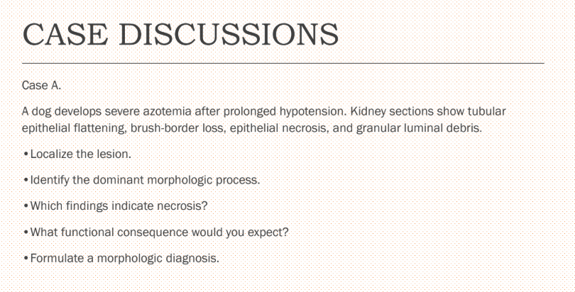
1. Localize the lesion
Renal tubules (specifically the proximal convoluted tubules).
Indicated by the loss of the brush border, which is unique to proximal tubular epithelium.
2. Identify the dominant morphologic process
Acute tubular injury (ATI) / Acute tubular necrosis (ATN).
Specifically, this is an ischemic injury triggered by prolonged systemic hypotension.
3. Which findings indicate necrosis?
Epithelial necrosis (direct mention).
Granular luminal debris (representing sloughed, dead epithelial cell fragments casting into the tubule lumens).
4. What functional consequence would you expect?
Acute renal failure (ARF) / Acute kidney injury (AKI).
Mechanistically, this causes an abrupt drop in glomerular filtration rate (GFR), clinical oliguria or anuria, and progressive uremia/azotemia.
5. Formulate a morphologic diagnosis
Kidney: Tubular necrosis (or degeneration and necrosis), acute, diffuse, severe.
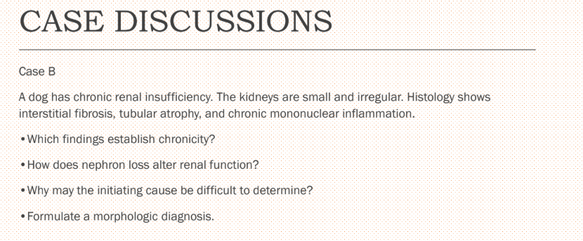
1. Which findings establish chronicity?
Gross findings: Small and irregular kidneys (indicating parenchymal contraction and scarring over time).
Histological findings: Interstitial fibrosis (scar tissue formation), tubular atrophy (wasting away of nephron components), and chronic mononuclear inflammation (infiltration of lymphocytes and macrophages rather than neutrophils).
2. How does nephron loss alter renal function?
Reduced GFR: Loss of functional nephrons directly lowers the total Glomerular Filtration Rate.
Compensatory hypertrophy: Remaining intact nephrons undergo hyperfiltration and hypertrophy to handle the increased load.
Loss of concentrating ability: Damaged tubules cannot maintain the medullary osmotic gradient, leading to polyuria (increased urination) and polydipsia (increased thirst).
Azotemia: The kidneys fail to adequately excrete metabolic waste products, leading to a buildup of blood urea nitrogen (BUN) and creatinine.
Systemic effects: Secondary consequences often develop, including non-regenerative anemia (due to decreased erythropoietin production) and altered calcium-phosphorus homeostasis (secondary hyperparathyroidism).
3. Why may the initiating cause be difficult to determine?
End-stage convergence: Many distinct primary renal diseases (e.g., chronic glomerulonephritis, chronic interstitial nephritis, chronic pyelonephritis) result in the same identical, nonspecific end-stage picture of widespread fibrosis and atrophy.
Chronological delay: By the time clinical signs appear and a biopsy or necropsy is performed, the original inciting agent (such as a previous bacterial infection, toxin, or immune complex) is typically no longer present.
4. Morphologic Diagnosis
Diagnosis: Kidneys: Nephritis, tubulointerstitial, chronic, diffuse, severe, with interstitial fibrosis and tubular atrophy (commonly referred to as "End-stage kidney" or "Chronic kidney disease").
transport urine from the kidneys to the bladder, where it is stored until voluntary or reflex emptying occurs.
ureters
provides the final pathway for urine elimination.
urethra
lined by urothelium, a specialized transitional epithelium.
urinary bladder
contribute to the permeability barrier and allow the mucosa to
change its apparent thickness as the bladder fills and empties.
superficial umbrella cells of the urinary bladder
Beneath the urothelium is the _, followed by the _
lamina propria, muscular wall.
Inflammation of the urinary bladder.
Acute Cystitis
May result from infection, chemical irritation, trauma, urolithiasis, urinary stasis,
or other causes.
Grossly, the mucosa may be congested, reddened, edematous, friable, or
hemorrhagic.
Histologically, neutrophilic inflammation is often prominent.
Acute Cystitis
Develops when irritation or inflammation persists or recurs.
Lymphocytes and plasma cells commonly predominate, although mixed inflammatory populations may be present.
The bladder mucosa can become thickened, irregular and fibrotic.
Chronic Cystitis
common in chronic cystitis and can create substantial epithelial
thickening.
Reactive urothelial hyperplasia
An inflammatory lesion that can resemble a urinary bladder neoplasm grossly and microscopically.
It is characterized by polypoid mucosal projections supported by edematous, inflamed, and often fibrovascular stroma.
Reactive urothelial hyperplasia may be prominent.
Polypoid Cystitis
May occur at any point from the renal pelvis to the urethral opening.
Causes include uroliths, strictures, congenital abnormalities, inflammatory lesions, blood clots, and neoplasia.
The pathologic consequences depend on the location, completeness, duration, and unilateral or bilateral nature of the obstruction.
Urinary Obstruction
dilation of the ureter caused by impaired urine outflow.
Hydroureter
refers to dilation of the renal pelvis
and associated progressive atrophy of renal
parenchyma.
Hydronephrosis
Formation of mineralized or otherwise organized
concretions within the urinary tract.
Urolithiasis
favored by supersaturation of urine,
appropriate physicochemical conditions for crystal
formation, retention of crystals, and failure of normal
inhibitory mechanisms.
Stone formation
can influence stone formation depending
on composition.
Diet, urine pH, infection, metabolism, genetics, and urinary stasis
A bladder containing uroliths often develops chronic mucosal irritation.
Histologically, there may be epithelial erosion, ulceration, hemorrhage, edema, neutrophilic or mixed inflammation, and reactive epithelial hyperplasia.
Chronic irritation can produce fibrosis and polypoid mucosal lesions
Cystitis Associated with Urolithiasis
clinically important because they can produce persistent hematuria,
irritative lower urinary tract signs, obstruction, and local invasion.
Urinary bladder tumors
a major diagnostic consideration in dogs with a bladder mass, but
inflammation and reactive lesions can produce similar clinical and gross findings.
Urothelial tumors
may be papillary, solid, or infiltrative.
Urothelial carcinoma
may show loss of orderly maturation, increased nuclear-to- cytoplasmic ratios, nuclear enlargement, anisokaryosis, prominent nucleoli, and increased mitotic activity.
neoplastic epithelium
multiple papillary, exophytic, frond-like, or polypoid mass
projecting into the lumen from the mucosal surface
Irregular nests and sheets of malignant cells breaching
the lamina propria and muscularis propria.
Urothelial carcinoma
basahin mo lang, come up with a trend or story line
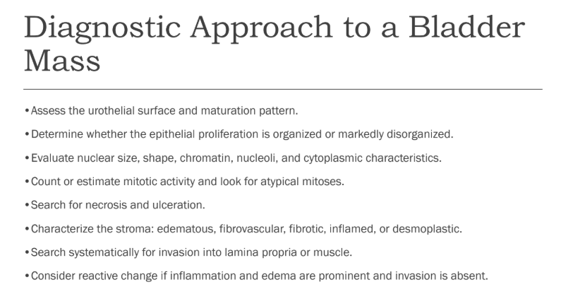
consider inflammation, urolithiasis, trauma, vascular injury, and neoplasia.
•Hematuria
consider cystitis, urethral obstruction, and painful urolithiasis.
•Dysuria/stranguria
commonly reflects lower urinary tract irritation.
•Pollakiuria
investigate obstruction and its level.
•Hydronephrosis
distinguish polypoid/reactive lesions from urothelial and other neoplasms
•Bladder mass
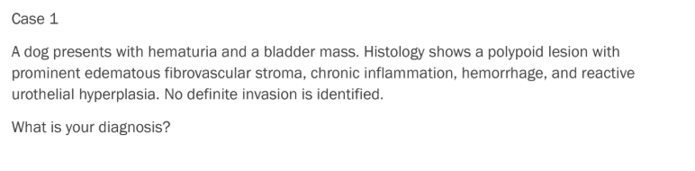
polypoid cystitis
cystitis assocaited with uroliths















