ANT 3331 - Frontal and Parietal bone terms
1/26
There's no tags or description
Looks like no tags are added yet.
Name | Mastery | Learn | Test | Matching | Spaced | Call with Kai |
|---|
No analytics yet
Send a link to your students to track their progress
27 Terms
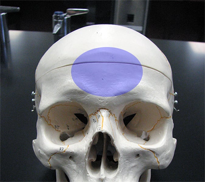
frontal squama
vertical forehead
temporal lines
lateral sides of bone; attachment of temporalis muscle (chewing)
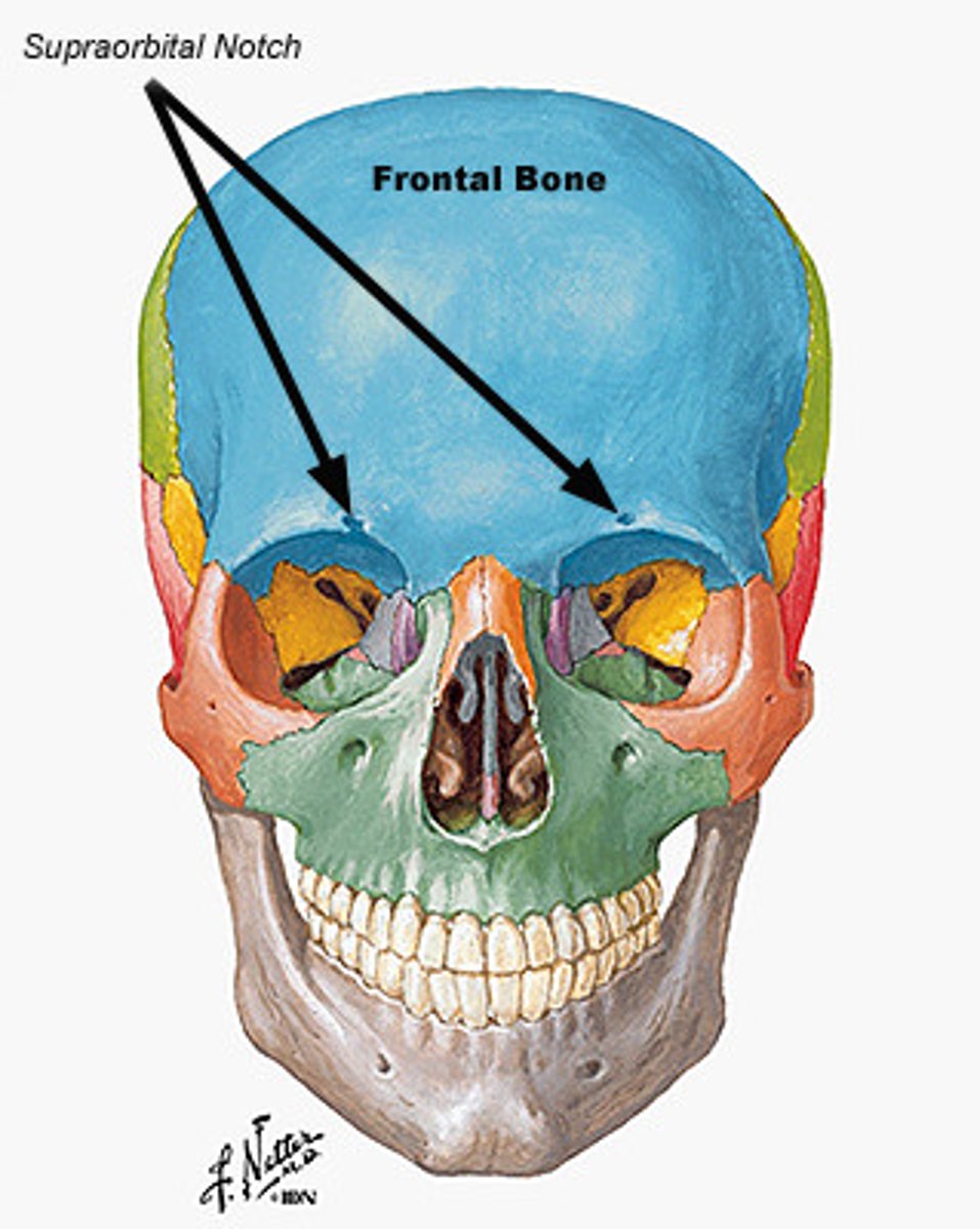
supraorbital margins
upper edges of orbits; notched or pierced by supraorbital notch or foramen
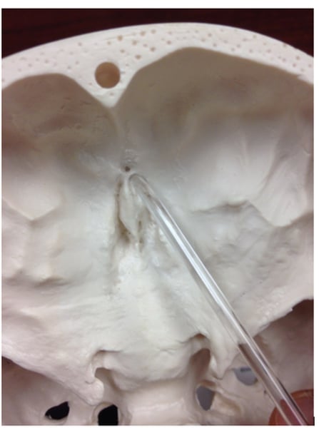
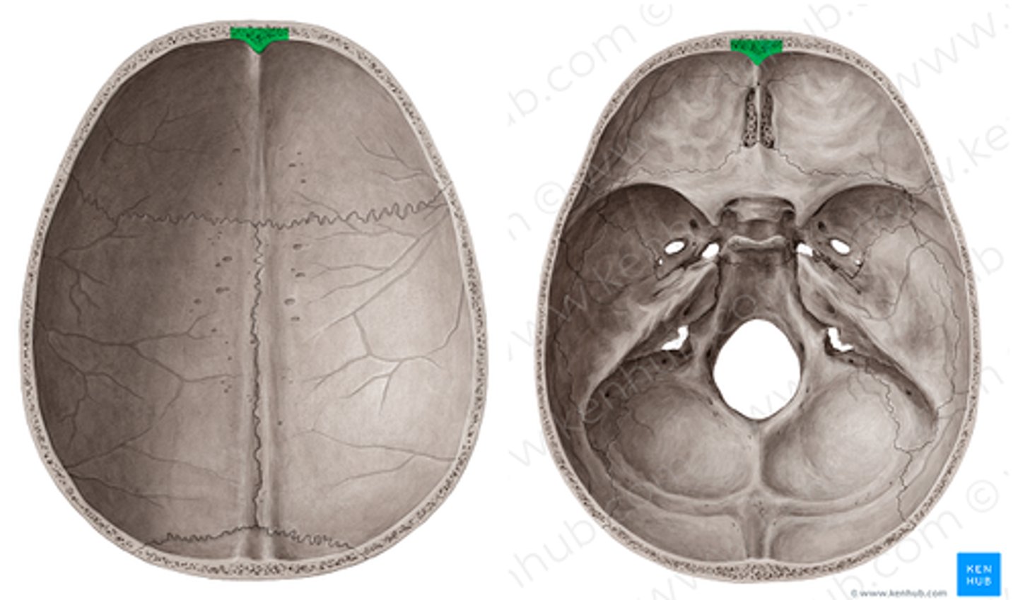
foramen secum
hole at base of frontal crest
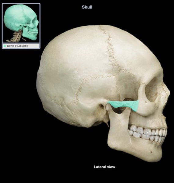
lacrimal fossae
lateral, inferior part of orbital surface; for lacrimal gland
sides and roof of neurocranium
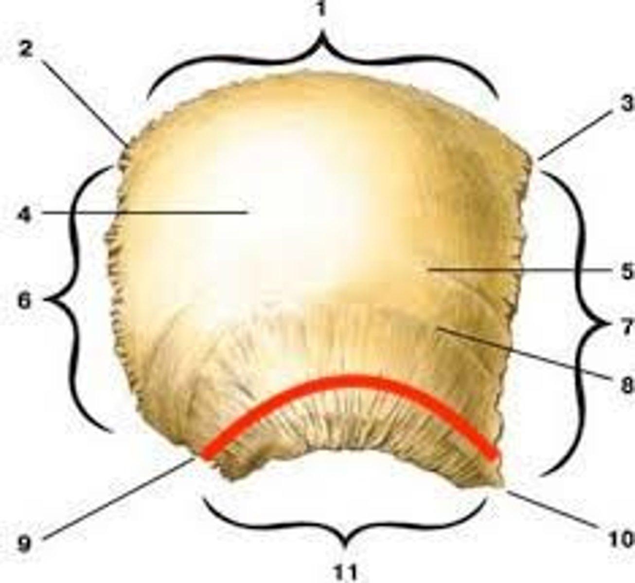
articulates with other parietal, frontal, temporal, occipital and sphenoid; one of largest bones in neurocranium; ossifies in membrane from center near parietal boss
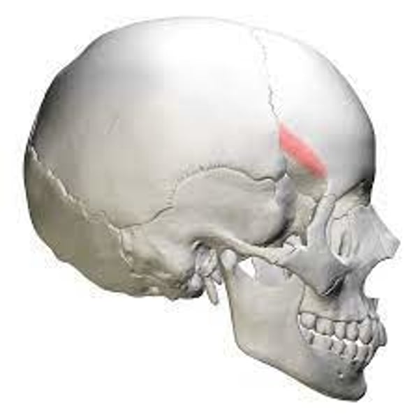
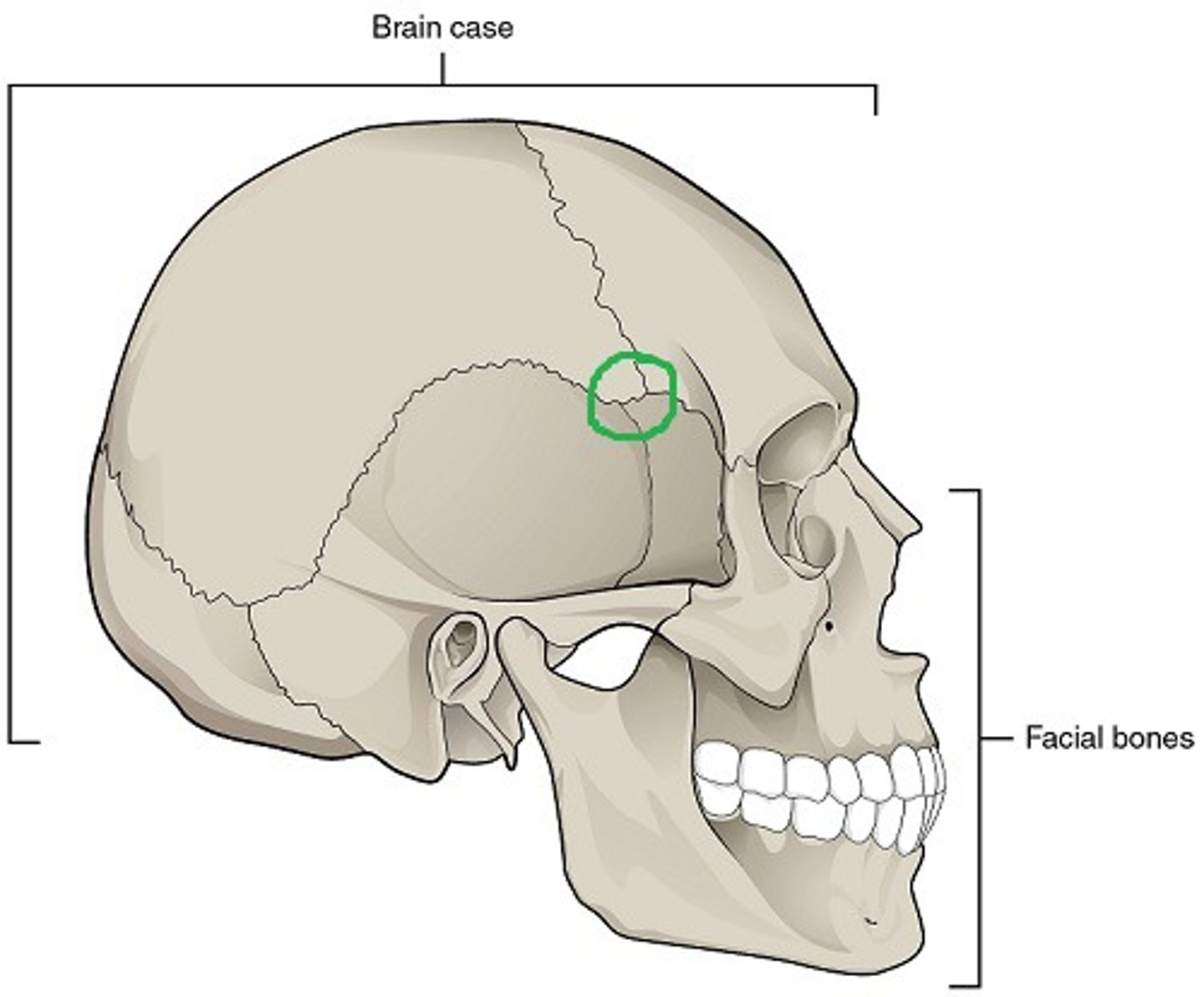
sphenoidal angle
located at pterion
mastoid angle
located at asterion
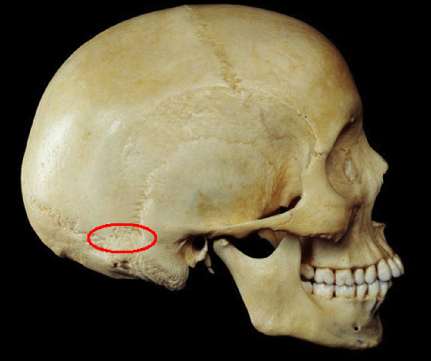
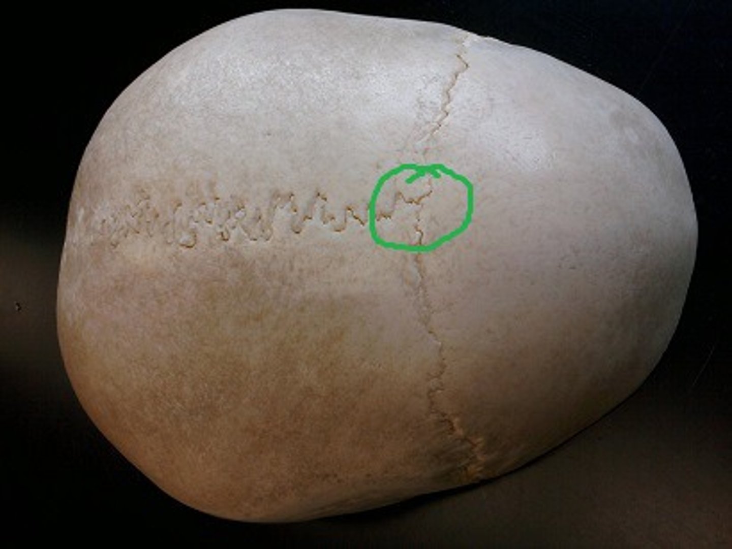
parietal striae
ectocranial striations near squamosal suture
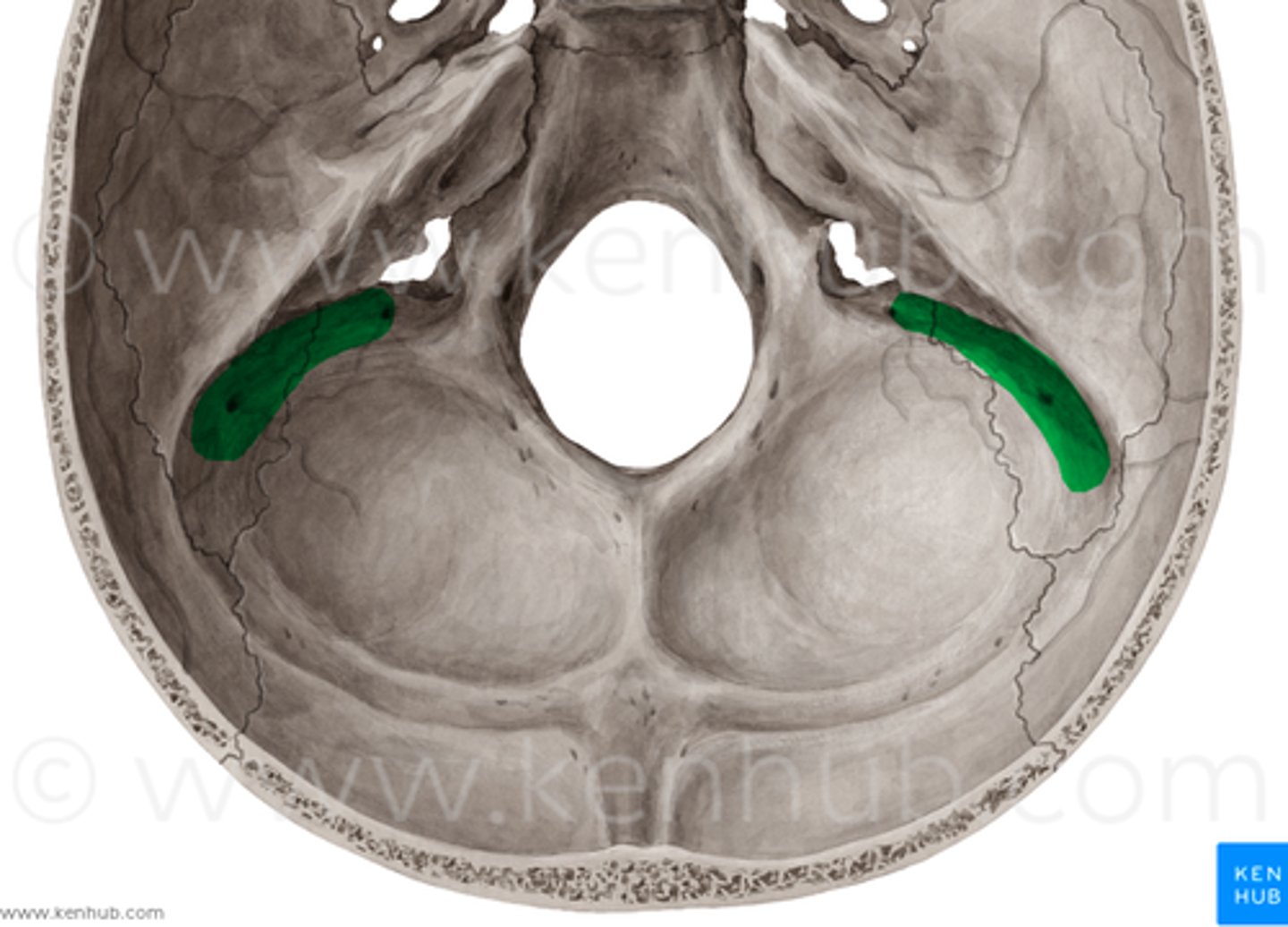
sigmoid sulcus
groove crosses mastoid angle of endocranial surface; for sigmoid sinus (drains blood from brain)
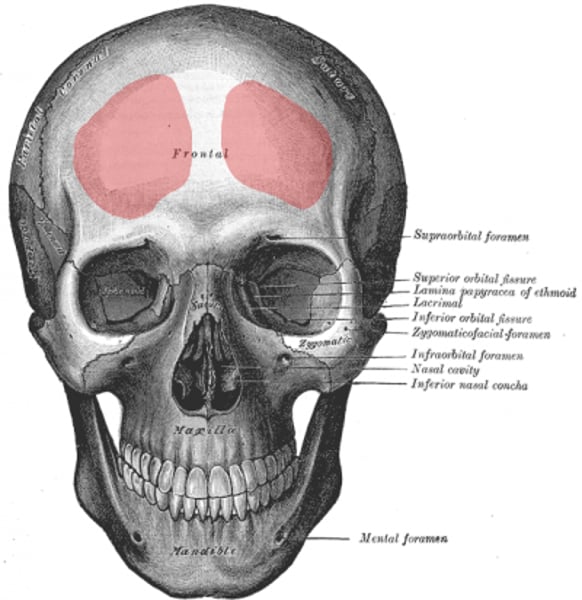
frontal bone
- front of neurocranium
- articulates with parietals, nasals, maxillae, sphenoid, ethmoid, lacrimals, and zygomatics
- ossifies on membrane from 2 primary centers (fuse along metopic suture)
orbital plates
- horizontal portions of frontal
- orbital side on smooth and concave, the endocranial side is rugose
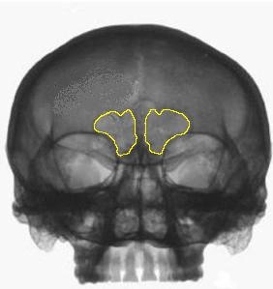
frontal bosses
2 eminences marking original centers of ossification
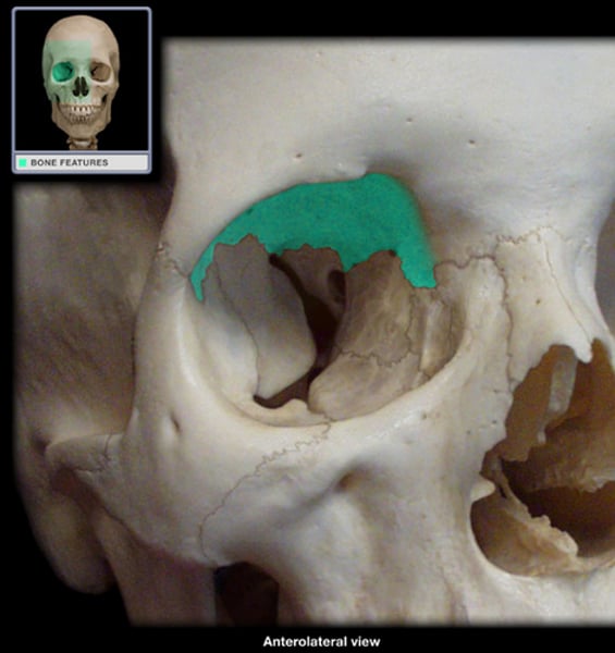
zygomatic processes
lateral, anterior corners of frontal
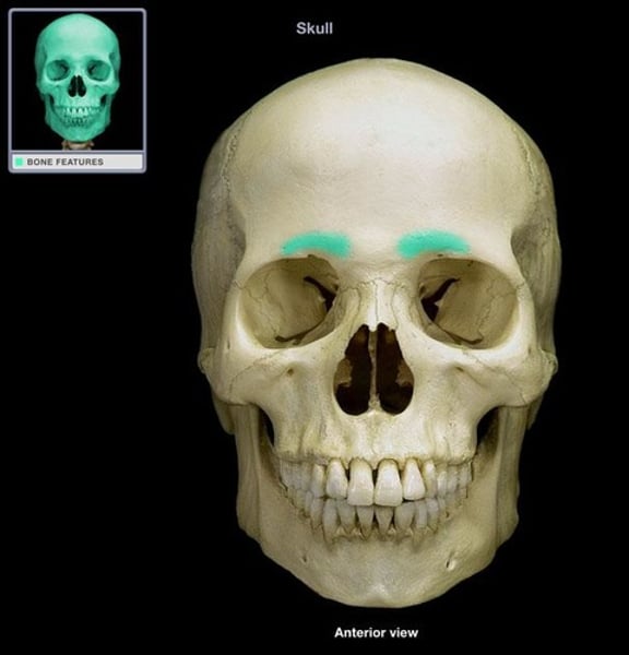
supercilliary arches
brow ridges above orbits; prominent in males; can be joined by prominent glabellar region
supraorbital notch or foramen
on medial half of superior orbital rim; transmit supraorbital nerve and vessels
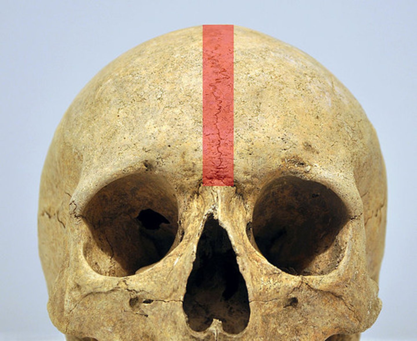
metopic suture
vertical suture between right and left frontal halves; rare in adults, but may be found at glabella
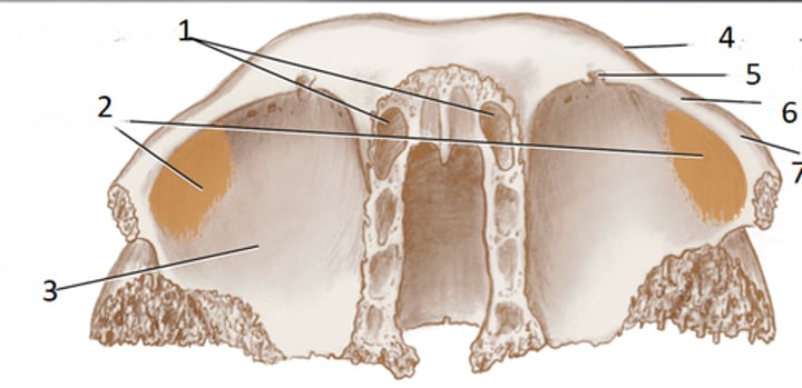
meningeal grooves
endocranial surface of frontal squama; for middle meningeal arteries (supply dura mater)
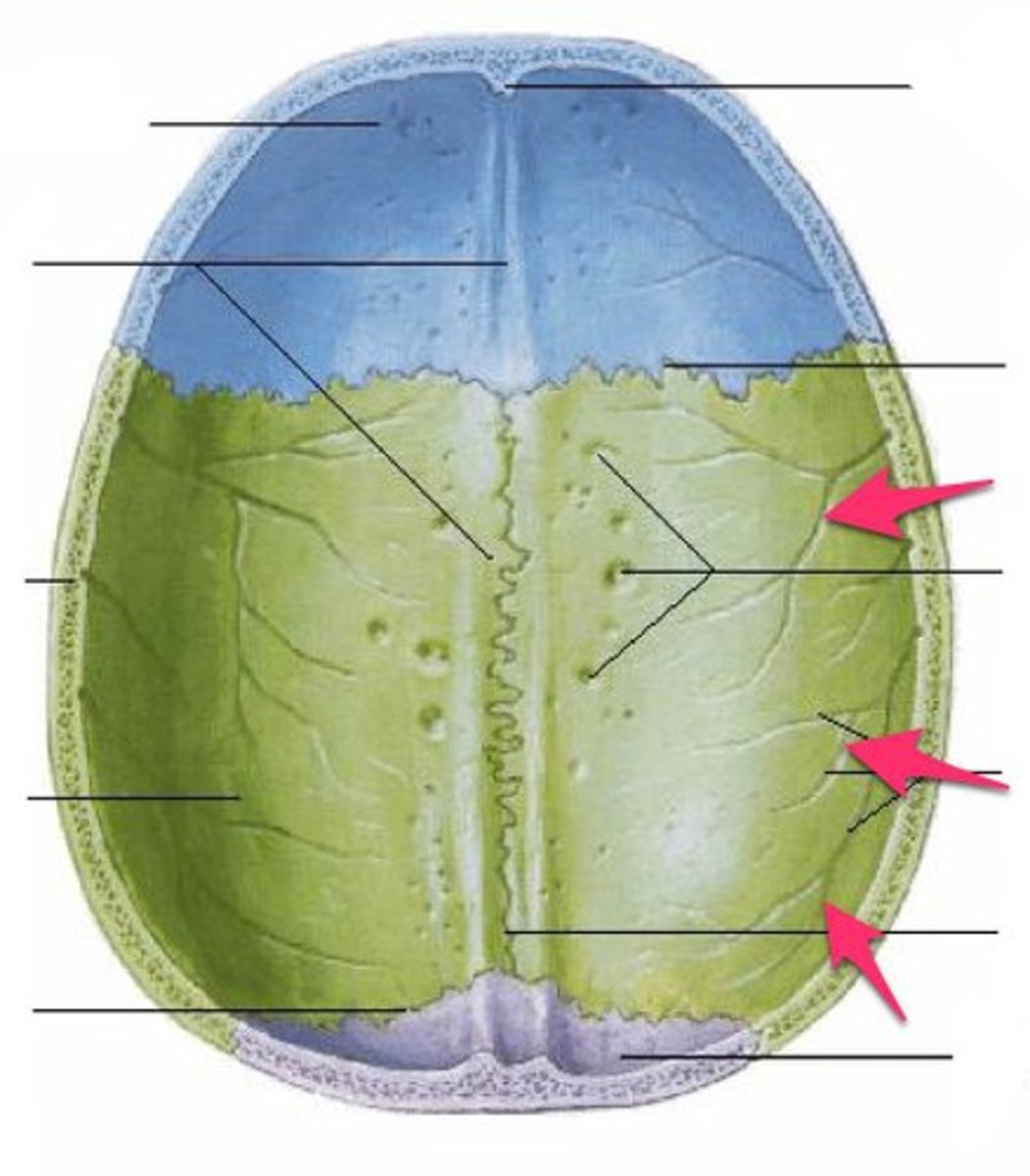
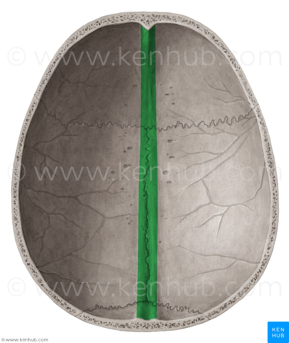
sagittal sulcus
vertical groove at sagittal edge (midline) of endocranial surface; for superior sagittal sinus (drains blood from brain)
frontal crest
endocranial midline crest; falx cerebri attaches here
ethmoidal notch
gap separating 2 orbital plates of frontal, ethmoid bone fills this in an articulated cranium
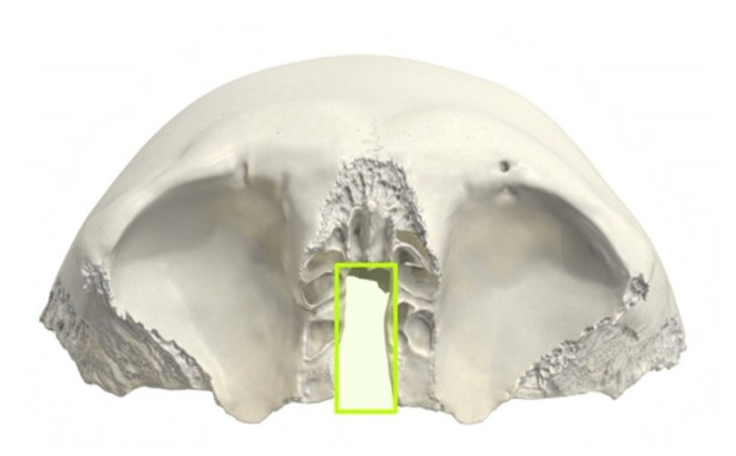
frontal sinus
anterior to ethmoidal notch, a hollow chamber
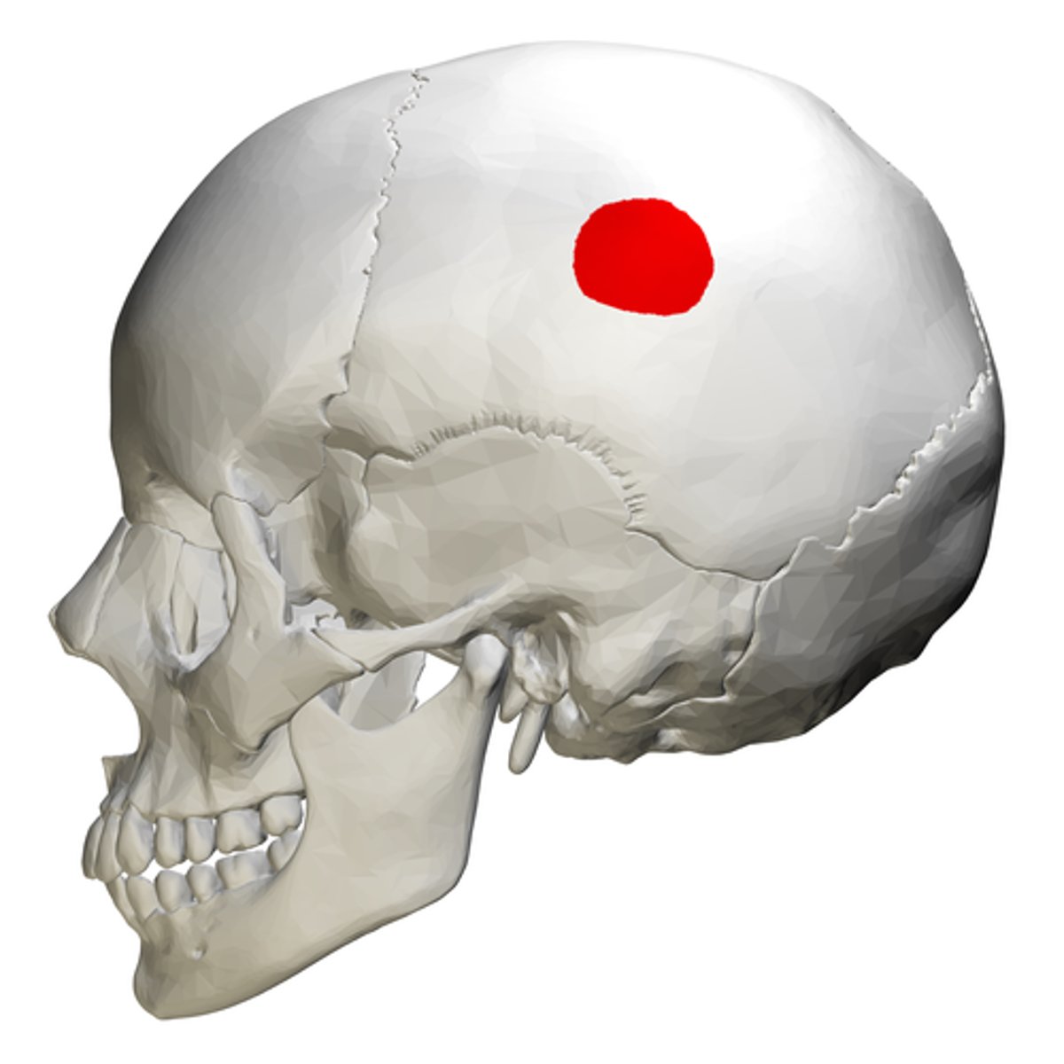
parietal boss
large eminence marking original center of ossification
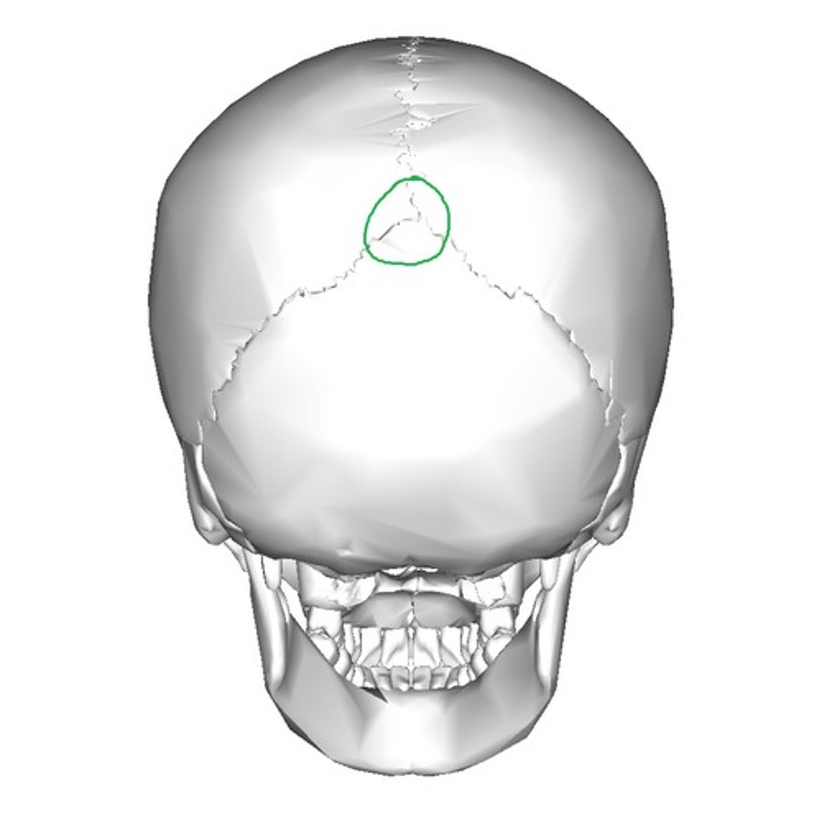
frontal angle
located at bregma
occipital angle
located at lambda
superior temporal line
attachment of temporal fascia (covering) (1)
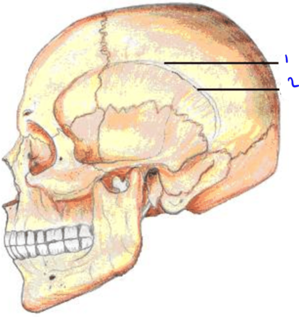
inferior temporal line
attachments of temporalis muscle (chewing) (2)
