Quiz 2 L3(end stuff) - L8
 Call Kai
 Learn
Learn Practice Test
 Spaced Repetition
 Match
Match Flashcards
 Knowt Play
1/55

There's no tags or description
Looks like no tags are added yet.
Name | Mastery | Learn | Test | Matching | Spaced | Call with Kai | Chat |
|---|
No analytics yet
Send a link to your students to track their progress
56 Terms
Role of Hemoglobin(Hb) vs Myoglobin(Mb) and their globin fold
Hemoglobin - transports oxygen throughout our bloodstream + deliver it to all tissues in our body that are low in pH/metabolically active
Myoglobin - Stores oxygen in muscle tissues + release to different tissues when oxygen is needed.
Both have same globin fold (8-helix with 2 His residues)
How many subunits do Hemoglobin vs Myoglobin have
Hemoglobin = 4 subunits(tetramer) so can bind 4 oxygen molecule
Myoglobin = 1 subunits(monomer) so can bind 1 oxygen molecule

How does Myoglobin increase the storage of O2 in the cells
Myoglobin is at high conc(100mM) in the cytoplasm & free O2 in aq solution/blood solubility is low (0.3mM) so Mb increases the solubility by 333-fold
Formation of MbO2 increase/decrease the total amount of O2 in the cytosol by 300-fold
increases

What is the heme group in Mb & what are the 2 reasons we dont use free heme to store O2
Fe2+ (only form that can be used to accept oxygen)
Free heme can form dimers that react with O2 forming Fe3+ that bind with H2O and not O2
How many coordination is the Fe2+ in Mb and how many are ocupied by nitrogen
6 coordinations for Fe2+
4 occupied by nitrogen
5th group occupied by proximal HIS(has a nitrogen from his) which holds the heme in the F helix Mb
6th group occupied by distal HIS which allows binding to O2 in the heme pocket of the E helix of Mb

Without Mb, Water binds to Fe2+ heme which causes
Fe2+ heme will become Fe3+ heme
Oxygen reacts with heme but doesn’t bind with heme
What is the function of Mb polypeptide
Shield the heme from bulk water inside a hydrophobic pocket, while keeping the protein soluble in the aqueous cytosol via its hydrophilic exterior
Keep the heme Fe2+ away from contact with all other hemes, because it is actually stacks of hemes(into a 4 heme complex) that chemically react with the oxygen.
Enable hundreds of times higher concentration of oxygen than could be achieved in water, since Mb is so soluble in the water inside a cell and free O2 is not.
Is the Mb binding of Oxygen a reversible process
Yes since no covalent bond is formed so the reaction can run right to left but its favored the right
What is a Prosthetic group, Holoprotein and Apoprotein
Prosthetic Group - Non AA(heme in the case of Mb) structure covalently bonded in the protein
Holoprotein - entire protein + prossthetic group
Apoprotein - protein w/o its prosthetic group
What is Hemoglobin and Myoglobin as a Holoprotein vs Apoprotein
Holoprotien
Hb = binded to heme
Mb = binded to heme
Apoprotein
Hb = not binded to heme
Mb = not binded to heme
How many methods are there to find amino acid sequence(primary structure) and what are they
2 methods
Protein-based (older method via Edman degradation or mass spec) - each amino acid is cleaved and identified
DNA-based (newer method) - obtain a small bit of the AA sequence via method 1(i.e mass spec) and identify the sequence as nucleotides
What are Isozymes = isoenzyme = isoforms
Enzymes found in the same organism that are structurally (3D fold) and catalytically similar, but differ slightly in A.A sequence
i.e hexokinase
Why do we have different Hexokinase in different cell types
HK IV binds to glucose weakly but when glucose conc is high then HK IV does bind glucose and catalyze rxn to form glucose phosphate
What are Homologs and the 2 types
Homologs = shared common ancestor
Paralogs = in same speices
Orthologs = in different speices
What can AA sequence do if not 3D structure yet
1) Enzyme activity
Comparison of an unknown AA sequence with a known sequence(similar sequences)
2) Building Domains
Proteins or parts of proteins that are not catalysts (so no chemical reactions)
What are the short sequence motifs.
Serine’s role: Target of “phosphorylation” (many proteins in cytosol and in biomembranes).
Serine in amino acid sequence, found only in glycogen phosphorylase, is also a target of “phosphorylation”. (due to the OH) group
Understand why comparison of primary structures of orthologs provides evolutionary relationships among organisms.
Orthologs provide evolutionary relationships through phylogenetic trees. The similarity in protein sequence can help us identify common ancestors that the mutations in amino acid sequence came from. Homology models can be built based on related amino acid sequences between the investigated sequence and the “databank”.

What is AlphaFold 2 (by deepmind,london, UK: Computational Method)
Predicts 3D protein structure given the AA sequence
What is Bioinformatics
Computational/technological methods to understand protein/gene function/evolution
Protein sequence analysis
Genome annotation/information to DNA sequence
Evolutionary biology w/progression
Gene/protein expression
Interactions in biology system
Cancer and other mutations related to disease
What are the 3 main ways atomic 3D structure protein can be determined(Experimental method)
X-Ray Crystallography: Protein crystals
NMR Spectroscopy: Protein dissolved in aqueous buffer
Cryo Electron Microscopy: Newest technology to get atomic level microscopy at ~3 Angstrom
Who is Dorothy Crowfoot Hodgkin & Kendrew & Perutz
Hodgkin: 1964 Nobel Prize Winner for proving how x-ray crystallography could directly map the atomic positions of complex biological molecules
Kendrew & Perutz: Nearly all amino acid positions located in 3D for myoglobin

Describe Light
Packs of photons
has no mass
has distinct energy determined by its frequency
Wave and particle properties through X-Ray beam (high energy light) with an oscillating electromagnetic wave
Range of X-Ray wavelengths possible
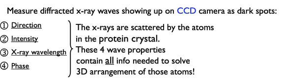
What is the Phase problem in X-Ray Crystallography
X-rays are shot at protein crystals, leading to scattering in all directions by every atom. However, during the process, only information about the intensity of the light was found, which yielded missing information on the phase of the light.
It took them 30 years to find the phase information, suggesting that formation of good crystals was a limiting factor.
What are the 4 ways to show protein strucutre
Sticks
Spheres
Ribbon
Cartoon

What are the 6 categories of non-covalent bonds important for construction of protein structure
1) Hydrogen Bonds - H with FON
2) Electrostatic - every positive and negative must have an equal opposite charge
3) Disulfide - weak for covalent but stronger than H-Bond
4) Weak and weakly directional forces - dipoles, London dispersion
5) Ring Stacking (Phe, Tyr & Trp)
6) Hydrophobic interactions - favorable water entropy
What does it mean that a protein crystal is 50% water
Salts when in crystal have no empty space whereas proteins have empty spaced filled by fluid
What does it mean that alpha helix(8 alpha helices) 70% Mb
Mb has one of the higest alpha helix content and some protein have no alpha helix
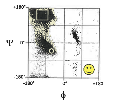
(think about the Ramachandran Map
What does it mean by Heme in a crevice for Mb
Protein makes precise fit , Fe2+ kept away from water
What are the 6 atoms in the peptide plan
1) Calpha of residue i
2) C (carbonyl carbon) of residue i
3) O (carbonyl carbon) of residue i
4) N (amide nitrogen) of residue i+1
5) H (amide hydrogen) of residue i+1
6) Calpha of residue i+1

Why are the 6 atoms in the peptide plane constrained in a plane
Resonance between C-O and C-N, each having partial double bond character.

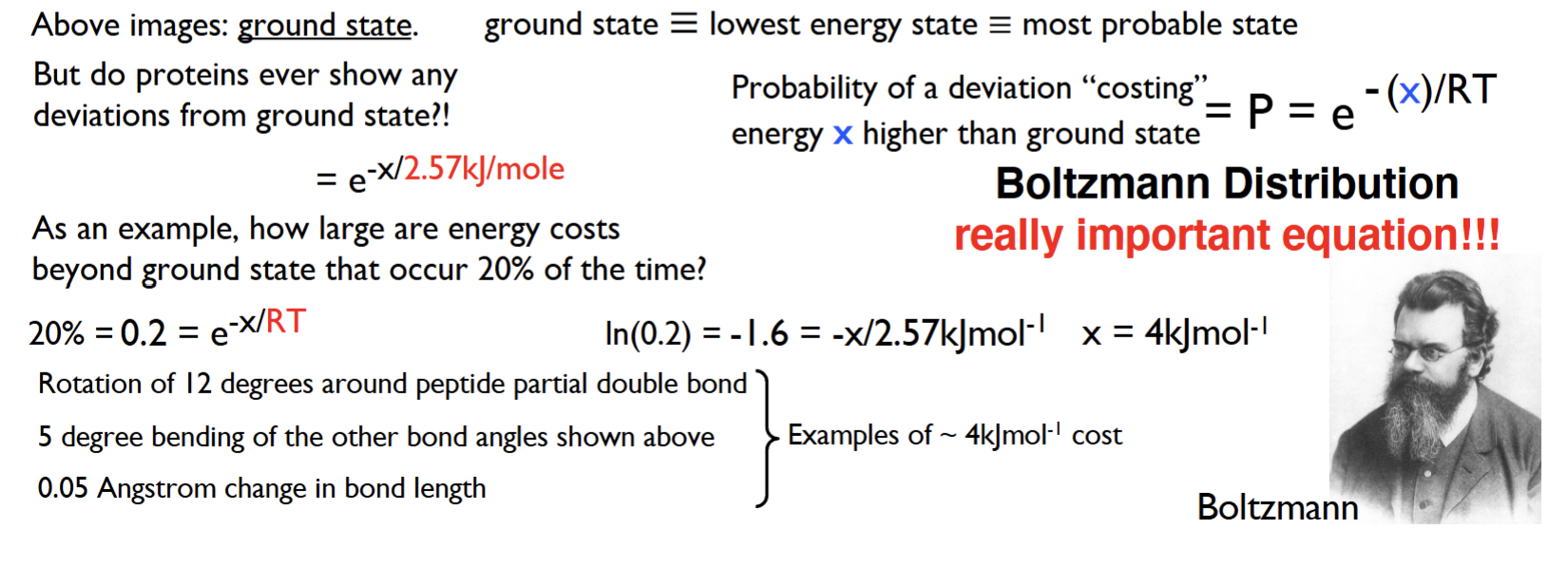
What is the Boltzmann Distribution equation and what does it tell you
Boltzmann Distribution equation: P = e-x/RT
P = the probability of the certain energy cost being sufficient to reach a certain deviation from the ground state
x = the amount of energy needed to reach a certain deviation from the ground state (in kJ/mol)
R = the gas constant, which is equal to 8.31 J/mol * K
T = the temperature (in Kelvin)
RT = 2.57kj/mole
The equation tells you the conformation of known energy relative to ground state
What are the bonds corresponding to backbone torsion angles phi, psi and omega
Phi (ɸ) → bond between Cɑ and N → 180 degrees
Psi (Ѱ) → bond between Cɑ and C → 180 degrees
Omega (Ѡ) → bond between C and N (peptide bond) → 0 degrees

Explain the info displayed on a Ramachandran Map
The info shows the possible combination of torsion angle values (ɸ and Ѱ) in a secondary structure of polypeptide(all the secondary structure) due to steric hindrance
Each point on the plot shows a single amino acid’s torsion angles, rather than the entire protein
We cannot tell the 3D structure information of protein from the graph as we are missing the primary amino acid sequence information.
Note: White regions are allowed regions = no steric hindrance

What are the allowed regions for Gly and Pro vs all other AA

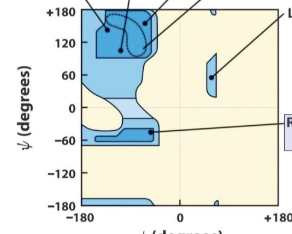
Where are the general locations of the alpha helix and beta sheets

What is the meaning of calculated vs.“real protein” Ramachandran Plot
Calculated Ramachandran Plot provides an estimate for the allowed regions (theoretical modeling based on steric constraints) while “real protein” Ramachandran Plot provides the actual, empirical database plotting of ɸ, Ѱ value combinations for a few 100 proteins.
Calculated: shaded region is forbidden
Real data: shaded region is where you find the protein
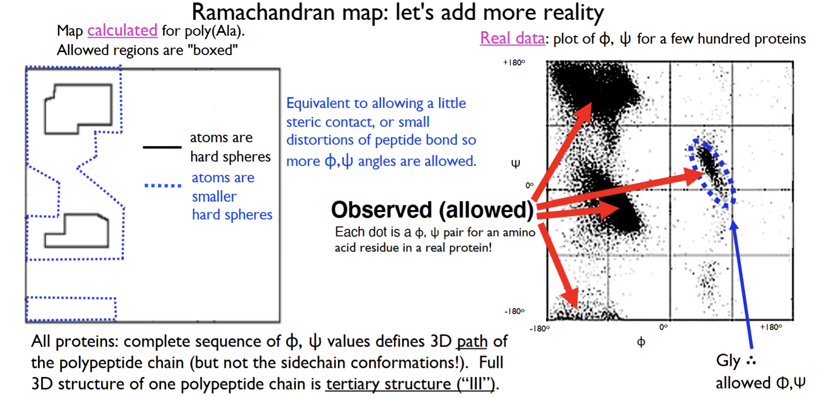
Explain why the Ramachandran Plots for hundreds of folded and unfolded proteins look similar
What are the secondary structures found on polyproline type II helix
Both folded and unfolded proteins are subject to same physical constraint of peptide backbone, so certain combinations of ɸ and Ѱ torsion angles are energetically unfavorable and not always sterically possible.
Alpha helix, beta sheet, turns, collagen are the four secondary structures found -> polyproline type II helix
Identify the Cis and Trans isomer of the peptide bond
Red = Oxygen
Green = Carbon
Blue = Nitrogen

In nature is cis/trans the dominant form and why.
Trans is the dominant form and it’s because of steric hinderance that occurs in cis
Why is Proline special in the cis/trans idea
What are the phi and psi values
Proline (Pro), 5-50% of the Proline exists in cis form.
Proline has a fixed ɸ(phi) value at -65 degrees
Proline cannot have a Ѱ(psi) value at near -40 degrees due to steric hindrance with ring carbons

Draw and recognize the backbone hydrogen bonding and key features of the alpha-helix
phi and psi values
average length: amino acid residue
which handed
R group pointed
Which AA is almost never found & which unfavored
AA residue per 360 degree
Orientation
ɸ = -60 degrees and Ѱ = -40 degrees
Average length: 10-11 amino acid residues
Alpha helices are always right-handed
All R groups are pointed away from the helix axis (spiral motion)
Proline almost never(if found then has to be at end terminals) & runs of positive or negative are unfavorable
For every 360 degree turn of the helix, there are 3.6 amino acid residues
Peptide plane orientation: All carbonyl groups (C-terminus) and N-H groups (N-terminus) point in the same direction(plane is relative to helix axis. The C-terminus and N-terminus point in opposite directions.


Draw and recognize the backbone hydrogen bonding and key features of the beta-sheet
phi and psi values
Average length: amino acid residue
R group are pointed
Orientation
ɸ = ranging from -60 to -150 degrees (-120 degrees) and Ѱ = 90 to 180 degrees (120 degrees)
Average strand length of 6 amino acids
R-groups located above or below the plane of the strand(alternate)
Peptide plane orientation: All of the carbonyl groups (C-terminus) and the N-H groups (N-terminus) point in opposite/alternate directions. The C-terminus and N-terminus point in opposite directions.
Can have either antiparallel or parallel strands in terms of orientation
All possible H-bonds are made between strands


Describe how beta turns are defined (4 amino acid residues, i+1 and i+2 phi,psi torsion angles & between which are H bonds
Beta turns: defined by the (i+1) and (i+2) ɸ, Ѱ torsion angles
4 amino acid residues = minimum for a chain to turn 180 degrees (completed rotation of polypeptide chain). However, note that these 4 amino acid residues do not have repeating ɸ, Ѱ values
Hydrogen bonds between i and i+3

Identify beta turn positions where Pro is favored or where Gly is required
Proline (Pro) is favored only in the (i+1) turn for both Type I and II
Glycine (Gly) is required in the (i+2) turn for only Type II

Describe the structure of Collagen (primary, secondary, tertiary, quaternary)
Primary: (Gly-Pro-Pro)n
Secondary: Poly-Proline Type II helix, repeating ɸ, Ѱ torsion angles (ɸ = -60 degrees, Ѱ = 140 degrees) no alpha helices
Tertiary: Not usually distinguished
Quaternary: cross linked form (fibrils)
Describe collagen synthesis (what occurs inside vs. outside the cell)
Synthesis on ribosomes(signal sequence for lumen of ER)
Vitamin C-mediated hydroxylation in RER
Sugar(glucose or galactose) added via glycosylation in SER
Transfer vesicle into lumen of Golgi, forming triple helix(procollagen) with ends not in triple helix(and not trimmed)
Secretory vesicle transfers procollagen out of cell where lysyl oxidase (enzyme converting lysine residues to carbonyl group) and aldol condensation (takes place between lysine and lysine)

Explain link between Vitamin C deficiency and Scurvy
Vitamin C deficiency leads to increased risk of Scurvy.
Problem occurs in the hydroxlyation via vitamin C
Identify what amino acid sidechains undergo post-translational modification (hydroxylation, cross-linking) and where (inside vs. outside the cell) these modifications happen.
Lysine undergoes both hydroxylation and crosslinking, which are examples of post-translational modifications.
Proline undergoes only hydroxylation
Hydroxylation happens inside the cell in the ER, through Vitamin C mediation (ascorbic acid).
Crosslinking happens outside the cell through aldol condensation.
List and recognize the main categories of folds (e.g., all alpha, all beta, etc.)
All alpha
All beta
Mixed alpha and beta
Give reasons for the limited number of unique protein folds.
Why does intrinsically disordered protein not fold
Due to the limited ways to get stabilized folds that can fold quickly.
Intrinsically disordered proteins have a large fraction of polar/charged residues, which means it doesn’t fold.
Describe what a topology diagram shows
Conformation/connection of beta-pleated sheet (drawn in 2D)
Define fold, superfamily, family
Fold: The characteristic arrangement of the major secondary structures
Superfamily: Similar protein structure + function (same fold) that may prove an evolutionary relationship, but varying amino acid sequence. A superfamily can contain only one or many families.
Family: common evolutionary origin, with either high sequence identity or lower sequence identity that has greater similarity in structure and function
Describe the purpose of SCOP/SCOPe, what they do and what they provide
A database/software that provides the structural + evolutionary relationship between all known protein structures
AKA Structural Classification of Protein (extended)
True or False, In a folded protein, all or nearly all hydrogen bonds must be formed
True
Which amino acid has the greatest amount of conformational freedom of the 20 AA
Glycine