Respiratory Phys
 Call Kai
 Learn
Learn Practice Test
 Spaced Repetition
 Match
Match Flashcards
 Knowt Play
1/101

There's no tags or description
Looks like no tags are added yet.
Name | Mastery | Learn | Test | Matching | Spaced | Call with Kai | Chat |
|---|
No analytics yet
Send a link to your students to track their progress
102 Terms
functions of the respiratory system
oxygen provision
CO2 elimination
microbial infection protection
blood pH regulation
respiratory system contributes to phonation, […], and acts a blood reservoir
olfaction
Components of resp system (6)
upper airways
trachea
lungs
resp muscles
rib cage
CNS
upper airways includes
nostril, oral cavity, pharynx, larynx
Trachea and primary bronchi arrangment
anterior c-shape cartilage
posterior smooth muscle
bronchi structure
cartilage and smooth muscle
bronchioles structure
smooth muscle
2 zones of larynx
conducting zone
respiratory zone
conducting zone
leads gas to gas exchange region of lungs
no alveoli
thus, no gas exchange
conducting zone components
bronchi
bronchioles
terminal bronchioles
respiratory zone
where gas exchange occurs
respiratory zone components
respiratory bronchioles
alveolar ducts
alveolar sacs
Terminal bronchioles
smallest airway w/o alveoli
respiratory bronchioles
contains occasional alveoli
where does gas exchange occur?
alveoli
alveoli structure + function
small, thin-walled, capillary rich sac in lungs where the gas exchange occurs
Type I alveolar cell
most cells
lined by continuous mono layer of flat epithelial cell
don’t divide
Type II alveolar cell
produce surfactant
act as progenitor cells (can differentiate into type I)
Transfer of O2 and CO2 occurs by diffusion through the […]
respiratory membrane
respiratory membrane
incredibly thin tissue barrier inside the lungs where gas exchange occurs
Steps of respiration (general)
ventilation via bulk flow
exchange via diffusion
transport bulk flow
exchange via diffusion
cellular utilization of O2 and CO2 prod
How is resp airflow produced (general)
CNS sends rhythmic drive to resp muscle
resp muscles contract rhythmically
change in vol and pressure
air flows in/out
resp muscle categories
pump muscles
airway muscles
accessory muscles
Diaphragm (3)
dome-shaped muscles which flattens during contraction (INS)
abdominal contents forced down + forward
widened rib cage
External intercostal muscles
contract and pull ribs upward increasing the lateral volume of the thorax
bucket handle motion
Parasternal intercostal muscles
contract and pull sternum forward, increasing anterior posterior dimension of the rib cage
Abdominal muscles (4)
External oblique
Internal oblique
transversus abdominis
rectus abdominis
Abdominal muscle function
involved in deeper, faster breathing
Internal Intercostal Muscles
pull rib cage down + reduce thoracic vol during exercise
Accessory Inspiratory Muscles (2)
Scalenes and Sternocleidomastoids
scalene
elevate upper ribs
Sternocleidomastoids
raise the sternum
What happens to the abdominal muscles during forced expiration?
abdominal muscles contract and push abdominal content + diaphragm up to reduce thoracic vol
obstructive sleep apnea
reduction in upper airway patency during sleep
muco-ciliary escalator
The filtering action in the conducting zone
types of cells that line the surface of the trachea
goblet cells (prod mucus)
ciliated cells (apical surface)
Goblet cells form the — layer, trapping inhaled materials, while ciliated cells form the — layer, eliminating particles.
gel; sol
Macrophages in Alveoli
Last defence for eliminating particulates; digest them and eliminate the risk of infection
pulmonary fibrosis
tissue in your lungs becomes scarred and stiff over time (cannot expand)
spirometry
a pulmonary function test that determines the amount and the rate of inspired and expired ai
tidal volume
the volume of air moved IN OR OUT of the respiratory tract (Breathed) during each ventilatory cycle
Expiratory reserve volume
the additional volume of air that can be forcibly exhaled following a normal expiration;
can be accessed by expiring maximally to the Maximum Voluntary Expiration
Inspiratory reserve volume
the additional volume of air that can be forcibly inhaled following a normal inspiration
can be accessed by inspiring maximally, to the Maximum Possible Inspiration
Residual volume RV
the volume of air remaining in the lungs after a Maximal Expiration
RV = FRC - ERV
cannot be measured with a spirometry test
Vital capacity (VC)
the maximal volume of air that can be forcibly exhaled after a Maximal Inspiration
Inspiratory capacity IC
maximal volume of air that can be forcibly inhaled
IC = TV + IRV.
Functional Residual Capacity (FRC)
volume of air remaining in the lungs at the end of a normal expiration.
FRC = RV + ERV
Total Lung Capacity (TLC)
volume of air in the lungs at the end of a Maximal Inspiration.
TLC = FRC + TV + IRV = VC + RV
Total or minute ventilation
total amount of air moved into the respiratory system per minute
tidal volume x respiratory frequency
Alveolar ventilation
amount of air moved into the alveoli per minute
(tidal volume - anatomical dead space) x resp frequency
How do we improve alveoli ventilation?
Deep and slow breathing: Due to the fact that you are breathing in so much air during a single event will increase alveoli ventilation
FEV1
Forced Expiratory Volume in 1 second
FVC: Forced Vital Capacity
total amount of air blown out in one breath after max inspiration as fast as possible
FEV1/FVC
Proportion of the amount of air that is blown out in 1 second
Obstructive Pattern (3)
difficulty in exhaling all the air from their lungs
exhaled air comes out more slowly
FEV1/FVC is reduced
In an obstructive pattern FEV1 is —, while in a restrictive pattern FEV1 is —
significantly reduced; reduced (severity ranges)
Restrictive Pattern (3)
cannot fully fill their lungs with air.
lungs are restricted from fully expanding
FEV1/FVC almost normal
Helium dilution method
measures the amount of air that remains in the lungs at the end of a normal expiration
measures only communicating gas
Static properties of the lung
Mechanical properties that are present in the lungs when no air is flowing
Static properties of the lung examples
intrapleural pressure
static compliance of the lung
surface tension of the lung
Dynamic properties of the lung
Mechanical properties when the lungs are changing volume and air is flowing in and out
Dynamic properties of the lung examples
Alveolar pressure (PALV)
Dynamic lung compliance
Airway and tissue resistance
ventilation
the exchange of air between the atmosphere and the alveoli
Boyle’s Law
for a fixed amount of an ideal gas that is kept at constant temperature, the pressure and the volume are inversely proportional
P1V1 = P2V2
A change in —, and then in —, produces airflow
vol; pressure
During the expiratory phase of the lungs, a(n) — in volume will generate an increase in alveolar pressure
reduction
During the inspiratory phase of the lungs, a(n) — in volume will generate a decrease in alveolar pressure
increase
The pleurae (2)
Thin double-layered envelope
visceral (external surface of lung)
parietal (thoracic wall + superior face of diaphragm)
Intrapleural fluid
Reduces friction of lung against thoracic wall during breathing
elastic recoil
the lungs’ natural tendency to collapse and deflate following inflation
Intrapleural Pressure (PIP)
pressure in pleural cavity
Acts as a relative vacuum
always subatmospheric due opposing directions of elastic recoils
Alveolar Pressure (PALV)
dynamic, directly involved in producing air flow
when the glottis is open + no air flow = all resp pressure are equal to atm pressure
Transpulmonary Pressure (PTP)
force responsible for keeping alveoli open
static parameter which does not cause airflow
determines lung vol
What factors contribute to airway resistance?
Inertia of the respiratory system
friction (negligible)
Types of airflow in small airways, bronchial tree, and large airways (respectively)
laminar, transitional, turbulent
lung compliance
a measure of the elastic properties of the lungs and a measure of how easily the lungs can expand
slope
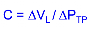
Static Compliance of the Lung
Represents the lung compliance, or the elastic properties of the lungs, when no air is flowing through
dynamic compliance
Represents the lung compliance during periods of gas flow (inspiration/expiration)
Lung compliance in Pulmonary fibrosis
low lung compliance
stiff lungs due to overproduction of collagen
Lung compliance in Emphysema
high lung compliance
“floppy” lungs due to lost alveolar tissue
Hysteresis
defines the different behaviour of the inflation and the deflation curves in pathological conditions as well as in physiological conditions
Lung compliance is determined by…
elastic components
surface tension (interface of alveoli)
Elastin properties
low tensile strength
extensible
Collagen properties
high tensile strength
inextensible
Emphysema and elastin
floppy lungs result from elastin destruction
Pulmonary fibrosis and collagen
stiff lungs due to collagen deposition in alveolar walls
How does surface tension affect alveoli?
creates inward recoil by decreases vol of gas inside of alveoli and increasing pressure— leading to alveolar collapse
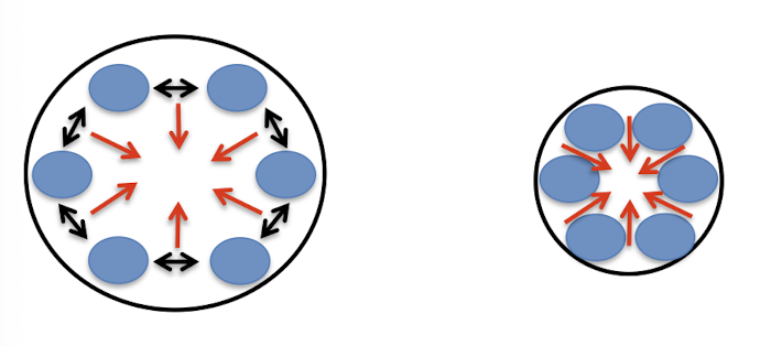
What is the function of surfactant?
lowers surface tension so we can breath w/o too much effort
makes alveoli stable against collapse
increase compliance
How does surfactant affect the pressures between alveoli of different sizes?
thickness of surfactant varies inversely w/ SA, equalizing pressures between different sized alveoli and preventing collapse
how do regional differences affect ventilation
weight of lungs increases pressures + makes intrapleural pressures less negative.
Thus alveoli at the bottom have a less negative PIP leading to a larger increase in lung vol
Dalton’s law
states that in a mixture of gas, such as air, each gas has its specific pressure and the total pressure of this mixture of gas is given by the sum of the individual pressures

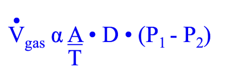
Fick’s law
the rate of transfer of a gas (V; L/min) through a sheet of tissue per unit of time is proportional to the surface area of the membrane (A) and depends on the difference in partial pressures between the two environments and inversely proportional to the thickness (T) of the membrane
Henry’s Law
the amount of gas dissolved in a liquid is directly proportional to the partial pressure of gas in which the liquid is in equilibrium
what affects the amount of gas dissolved in a liquid?
partial pressure, solubility
only a gas that is dissolved contributes to pp
The partial pressure of oxygen — in alveolar air while carbon dioxide —
decreases; increases
How will Increasing alveolar ventilation affect PO2 and PCO2?
increase alveolar PO2 and decrease PCO2
How will increasing metabolic rate affect PO2 and PCO2?
consuming more oxygen and producing more carbon dioxide thus, this will decrease alveolar PO2 and increase alveolar PCO2
characteristics of pulmonary circulatory system
low pressure system
low resistance system
high compliance vessels
— air must be delivered to regions of the lung where the — is going and vice versa
Inspired; blood
The greater the ventilation, the more the PO2 and PCO2 of the alveoli will be — to the atmospheric pressures
similar