Anatomy 6511 Histology Pictures Exam II 낱말 카드
 Call Kai
 Learn
Learn Practice Test
 Spaced Repetition
 Match
Match Flashcards
 Knowt Play
1/100

There's no tags or description
Looks like no tags are added yet.
Name | Mastery | Learn | Test | Matching | Spaced | Call with Kai | Chat |
|---|
No analytics yet
Send a link to your students to track their progress
101 Terms
Perimysium
What is labeled 1?
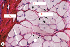
Endomysium
What is labeled 2?

Epimysium
What is labeled 3?

Perimysium
What part of skeletal muscle is labeled 1?
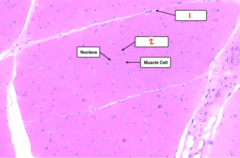
Endomysium
What part of skeletal muscle is labeled 2?

I band
What part of the sarcomere is labeled 1?
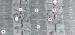
A band
What part of the sarcomere is labeled 2?

Z line or disc
What part of the sarcomere is labeled 3?
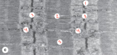
M line
What part of the sarcomere is labeled 4?
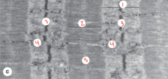
H band
What part of the sarcomere is labeled 5?

Sarcomere
What is labeled 1?
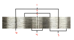
A band
What part of the sarcomere is labeled 2?
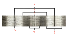
M line
What part of the sarcomere is labeled 3?
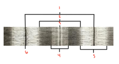
H band
What part of the sarcomere is labeled 4?

I band
What part of the sarcomere is labeled 5?
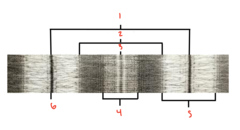
Z disc
What part of the sarcomere is labeled 6?

Myofilaments
What is labeled 1?
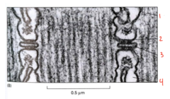
Terminal cisternae (SR)
What is labeled 2?
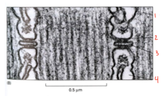
T tubule
What is labeled 3?

Sarcoplasmic reticulum
What is labeled 4?

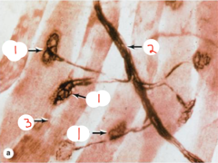
Motor end plates
What is the arrow pointing to on the skeletal muscle?

Motor end plates
What is labeled 1?

Neural body
What is labeled 2?

Sarcomere
What is labeled 3?
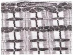
Type I
Is this type I or II muscle fibers?
Type II
Is this type I or II muscle fibers?

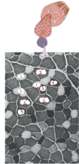
Type IIb
What muscle fiber type is lightest in color and labeled 1?

Type IIa
What muscle fiber type is in between in color and labeled 2?
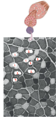
Type I
What muscle fiber type is darkest in color and labeled 3?
Intercalated discs
What part of cardiac muscle is being pointed to by the arrows?

Smooth
What muscle type is shown?

Smooth
What muscle type is shown?

Skeletal
What muscle type is shown?

Cardiac
What muscle type is shown?

Cardiac
What muscle type is shown?

Skeletal
What type of muscle tissue is this?

Smooth
This is a longitudinal orientation of what muscle type?
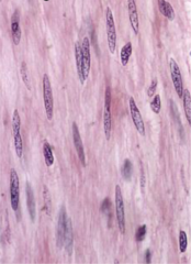
Smooth
This is a transverse orientation of what muscle type?
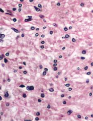
Smooth
What muscle type is shown?

Skeletal
What type of muscle tissue is this?
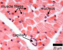
Skeletal
What type of muscle tissue is this?

Smooth
What muscle type is shown?

Skeletal
What muscle type is this?

Cardiac
What muscle type is this?

Osteoclast
What cell is labeled A?

Osteoblast
What cell is labeled B?

Osteoclast
What type of cell is this?

Chondrocyte
What cell is shown in this EM image?
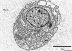
Osteoblast
What cell is labeled A?

Osteocytes
What cell is labeled B?

Osteoclast
What cell type is shown in the lower corner?

Hyaline
What cartilage type is shown?

Elastic
What cartilage type is shown?

Fibrocartilage
What cartilage type is shown?

Hyaline
What cartilage type is shown?
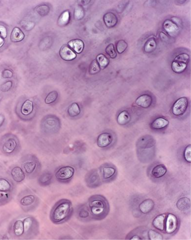
Endochondral
What type of bone formation is taking place?

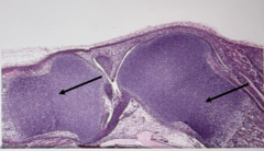
Endochondral bone development
What type process is occurring where the black arrows are pointing?
Territorial matrix
What are the yellow arrows pointing to?
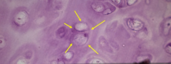
Interterritorial matrix
What is being identified by the yellow arrows?

Territorial matrix
Which part of the hyaline cartilage ECM is the arrow pointing to (in between light and dark staining)?
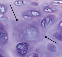
Capsular matrix
Which part of the hyaline cartilage ECM is the arrow pointing to (darkest staining)?
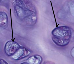
Interterritorial matrix
Which part of the hyaline cartilage ECM is the arrow pointing to (lightest staining)?

Perichondrium
What layer of hyaline cartilage is the arrow pointing to?

Superficial
What zone of articular cartilage is labeled 1?
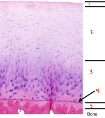
Intermediate
What zone of articular cartilage is labeled 2?
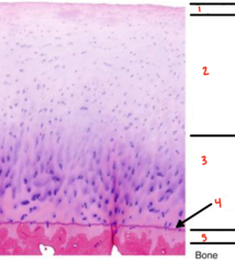
Deep
What zone of articular cartilage is labeled 3?

Tide mark
What line of articular cartilage is labeled 4?
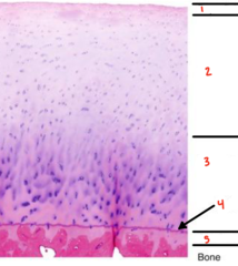
Calcified
What zone of articular cartilage is labeled 5?

Hyaline
What type of cartilage is shown?

Fibrocartilage
What type of cartilage is shown?

Elastic
What type of cartilage is shown?
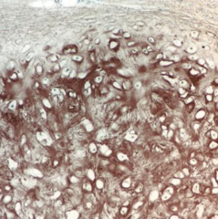
Elastic
What type of cartilage is shown?
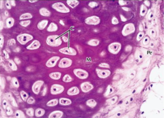
Hyaline
What type of cartilage is shown?

Fibrocartilage
What type of cartilage is shown?

Hyaline
What type of cartilage is shown?

Fibrocartilage
What type of cartilage is shown?

Hyaline
What type of cartilage is shown?
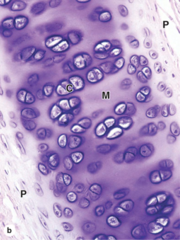
Elastic
What type of cartilage is shown?

Fibrocartilage
What type of cartilage is shown?

Red
What type of bone marrow is pictured?

Yellow
What type of bone marrow is pictured?

Reserve zone
Which zone in endochondral bone formation is shown?

Proliferation zone ("stacked pennies")
Which zone in endochondral bone formation is shown?

Hypertrophy zone (large cells/clear cytoplasm)
Which zone in endochondral bone formation is shown?

Calcification/Mineralization zone
Which zone in endochondral bone formation is shown?

Woven/immature
Which type of bone is pictured?

Compact (lamellar)
Which type of bone is pictured?

Spongy (trabecular)
Which type of bone is pictured?
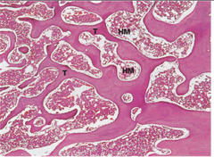
Osteons
What are the arrows pointing to?

Mature
Is this mature or immature bone?

Compact
What type of bone is this?
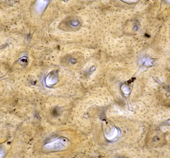
Spongy
What type of bone is this?

Spongy
What bone type is labeled 1?
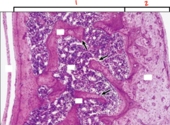
Compact
What bone type is labeled 2?

Bone marrow
What is outlined with stars in it?

Periosteum
What are the arrows pointing to?
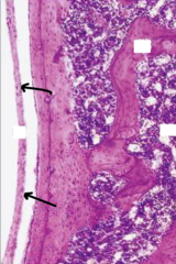
Spongy
What bone type is shown?

Volkmanns canals
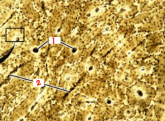
What component of compact bone is labeled 1?

Haversian canals
What component of compact bone is labeled 2?

Haversian canals
What component of compact bone is labeled 1?