Adv EP Lesson 8: Mapping Basics
 Call Kai
 Learn
Learn Practice Test
 Spaced Repetition
 Match
Match Flashcards
 Knowt Play
1/28

There's no tags or description
Looks like no tags are added yet.
Name | Mastery | Learn | Test | Matching | Spaced | Call with Kai | Chat |
|---|
No analytics yet
Send a link to your students to track their progress
29 Terms
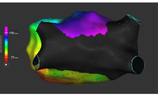
what is this and purpose
activation map
helps visualize a rhythm with additional tools like propagation animation and can show areas of slow conduction or block

what is this and purpose
voltage map
displays tissue voltage as a color and is determined by physician preference
helps visualize tissue health based on relative voltage values
can show areas of scar or isolcation/block; useful for checking PVI
trigger signal
tells the system when a new beat occurs
IVC anatomy
inferior portion of the venous return that connects to the RA
where the IVC meets the floor of the RA is the
Eustachian ridge
AV node anatomy
sits superior to the CS ostium on the near the septal leaflet of the TV
SVC anatomy
superior portion to the venous return that connects the RA
what makes the group of cells that makes the SA node
lateral portion of the SVC/RA junction contains this group of cells
what sits close the the RSPV
septal side of the SVC
CS anatomy right regions and left regions
right: located low on the septum of the RA near the TV
left: epicardial to the LA running from the inferior side, up the lateral wall by the appendage, and anterior
the right phrenic nerve is close to what
RSPV
coumadin ridge anatomy
sits bw the left PVs and the LAA
what is the most anterior sturcture of the heart
RVOT
blood flow with RVOT
blood flows from the RV to the lungs through the pulmonary valve and Pulmonic artery
what are more commonly found in RVOT vs LVOT
outflow tract PVCs
LVOT place
posterior to the RVOT leading tot he aorta and the rest of the arterial system
focal rhtyhms
Atrial tachycardia
PVC/non ischemic VT
how do we map atrial tachycardia
goal: find the earliest atrial activation
ideal ablation target: completely negative unipolar signal
treating AT
once earliest site is located, it is surrounded by points with a later activation time
post-ablation response: ectopy matching the tachycardia or acceleration, termination of tachycardia, inability to induce tachycardia
PVC/Ischemic VT mapping
comparing the 12lead ECG tot he clinical PVC can confirm early activation is a good target for ablation
PVC/Ischemic VT treatment
focal RF ablation
reentry arrythmias
typical Atrial flutter, atypical atrial flutter, AVNRT, AVRT, ischemic ventricular tachycardia
treatment for typical atrial flutter
CTI ablation
treatment for atypical atrial flutter
linear ablation anchored to sites of previous ablation, scar, etc
in a safe anatomical location
as short as possible
how do we map AVNRT
once confirmed it’s AVNRT, typically treated as an anatomical ablation and rarely mapped
two primary methods of mapping AVNRT
wavefront collision during sinus rhtyhm
low voltage bridge mapping during sinus rhythm
both to visualize near the slow pathway
treatment of AVNRT
ablation of slow pathway
treatment for AVRT
ablate the pathway closest tot he AV timing
treatment for AF
PVI via RF/PF or cryo