HLTH1004 Human Bioscience
1/88
Earn XP
Description and Tags
if really skinny, don't view in full screen mode.
Name | Mastery | Learn | Test | Matching | Spaced |
|---|
No study sessions yet.
89 Terms
WEEK 3: SKELETAL SYSTEM
add all the expectation thingy’s/learning objectives
state functions of skeletal system
SYSTEM: Skeletal system
SUBSECTION: General
NOTE: None
CUE: None
support
posture maintenance (facilitates upright position)
movement
articulation and leverage
protection
brain, lungs, pelvis, spinal chord, other vital organs
storage
minerals (phosphate, ca²+)
lipids and triglycerides (fat)
haematopoiesis
formation of blood cells
hormone production
explain structure of typical long bone (gross anatomy)
SYSTEM: Skeletal system
SUBSECTION: Gross anatomy
NOTE: include subpoints.
CUE: None
diaphysis (shaft)
long cylindrical; main portion of bone.
possesses hollow centre known as the medullary cavity (which stores yellow bone marrow in adults, and red bone marrow in new born infants).
epiphysis (bone ends)
proximal epiphysis (close to joint)
distal epiphysis (far from origin)
membranes
periosteum
outer layer connective tissue surrounding the bone (particularly the epiphysis).
functions:
associated with blood supply.
protects and assists bone in fracture repair.
helps nourish bone & tissue.
serves as attachment point for ligaments and tendons.
endosteum
surrounds/lines the medullary cavity.
very thin membrane.
WEEK 6: NERVOUS SYSTEM
LEARNING OBJECTIVES
Know the location and names of major brain and spinal cord structures
Have an overview of central nervous system functions
Understand the how the brain communicates with the body
Know the main afferent and efferent pathways and the modalities of information that they convey.
To understand the role of the peripheral nervous system (PNS) in communication within the body
To understand how the PNS connects and functions with the CNS
To know the basic structure of a neurone
To know the basic structure of a nerve
To understand how neurones communicate with other neurons and with other tissues (neurotransmission)
SUMMARIES
CNS is comprised of the brain and the spinal cord
The CNS is functionally divided into the autonomic and somatic nervous system
The main function of the brain is to synthesise and respond to information from the body in order to maintain homeostasis
Some functions are lateralised to one side of the brain, including some parts of language
Neurones are the communicating elements of the CNS
The PNS has two main functional divisions:
Sensory (afferent) – sensation and perception
Motor (efferent) – movement (autonomic and voluntary) and secretion
Neurones have a basic floorplan but have modifications to
structure depending on their function
Neurones receive information via dendrites and transmit messages that reach a threshold depolarisation (electrical transmission)
Signal is communicated to other cells via neurotransmitters across the synaptic cleft (chemical transmission)
Nerves are bundles of neurone axons plus connective tissue and blood vessels
what is the central nervous system ?
SYSTEM: Nervous System
SUBSECTION: Central Nervous System
NOTE: include structures.
CUE: None
what is it
the electrical connectivity system of the body.
allows body to process external and internal information.
where the brain and spinal chord are connected to the rest of body through the peripheral nervous system.
interacts with body via sensory receptors, providing external information (somatic sensory system) (afferent), that usually stimulates an autonomic response from the brain via efferent system.
structures
brain and spinal chord
the neural tissue (neurons and glial cells)
glial cells: supporting cells.
blood vessels
various connective tissues that protect and support the CNS.
what are the functions of the central nervous system (CNS) ?
SYSTEM: Central Nervous System
SUBSECTION: Central Nervous System
NOTE: None
CUE: None
maintain homeostasis by integrating, processing, and coordinating sensory data and motor commands.
coordinating sensory data: provide information about the conditions inside and/or outside the body.
motor commands: control or adjust the peripheral organs (e.g. skeletal muscles).
the control of intelligence, memory, learning, and emotion for each individual (the brain).
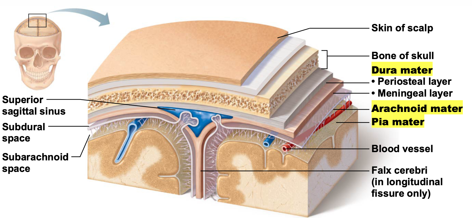
state and describe the structures that protect the brain
SYSTEM: Central Nervous System
SUBSECTION: Brain
NOTE: None
CUE: None
skull
bony, hard outer case, that offers brain physical protection from damage. the skull securely houses the brain, preventing movement of the brain inside.
limitation of skull (bony case): no room for expansion.
if damage occurs that results in swelling, there is no space for brain to go, causing pressure to accumulate, possible leading to further brain damage.
for spinal chord: it is the vertebrae.
meninges
series of three connective tissue layers.
dura mater
first of the meninges
thick, leathery, outer protective layer.
subdural space
a space between dura mater and arachnoid mater.
arachnoid mater
second layer
subarachnoid space
spider-like processes
a space between arachnoid mater and pia mater.
contains major blood vessels
takes ~20% of body’s blood glucose and blood supply to maintain the brain.
pia mater
final layer
fine layer of connective tissues that follows all the contours of the gyri and sulci of the cerebral cortex
blood brain barrier
semi-permeable layer of epithelial cells
allows the passage of:
certain nutrients via facilitated diffusion
fat-soluble substances, including alcohol, nicotine, and anaesthetics
denies passage of:
metabolic wastes, proteins, toxins, most drugs, small-non-essential amino acids, K+
BBB is absent in some areas, such as the vomiting centre and hypothalamus.
necessary to monitor the chemical composition and temperature of blood (circulating the brain).
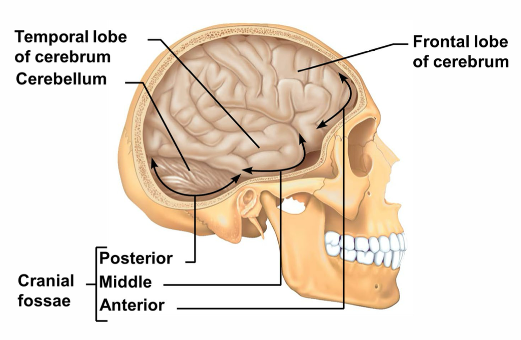
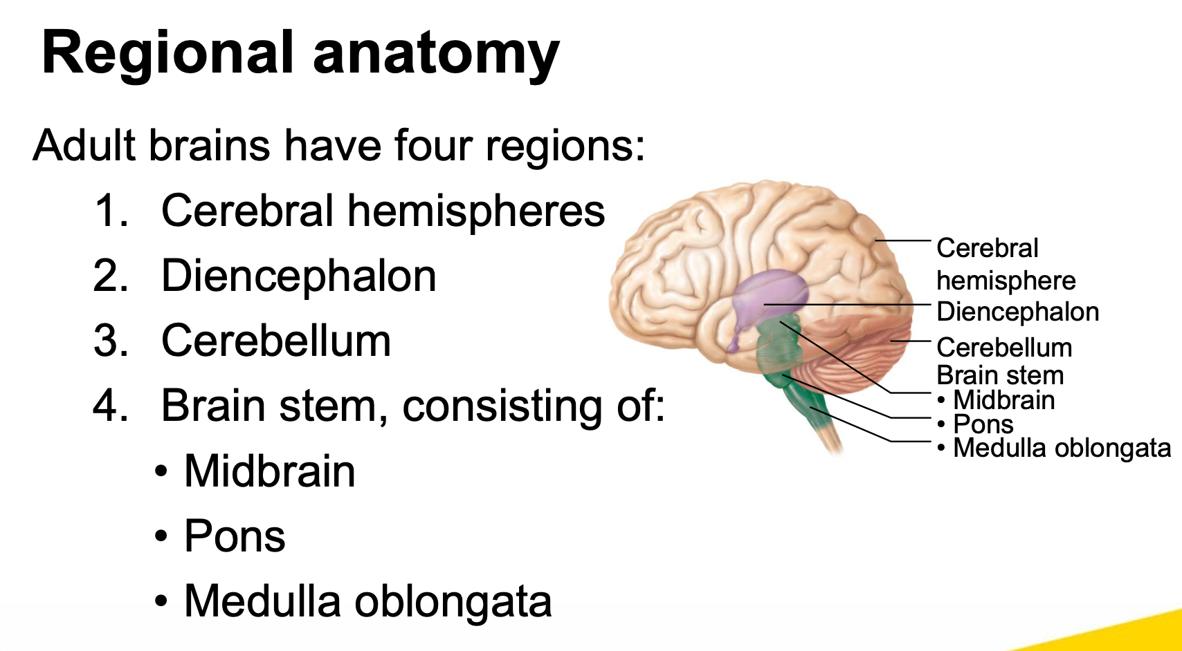
state the major regions of the adult brain
SYSTEM: Central Nervous System
SUBSECTION: Regional anatomy
NOTE: None
CUE: Not the lobes
Adult brains have four regions:
cerebral hemispheres
form superior part of brain
accounts for ~83% of brain mass
diencephalon, consisting of:
thalamus
provides relay station for incoming information.
hypothalamus
epithalamus
cerebellum
brain stem, consisting of:
midbrain
pons
medulla oblongata
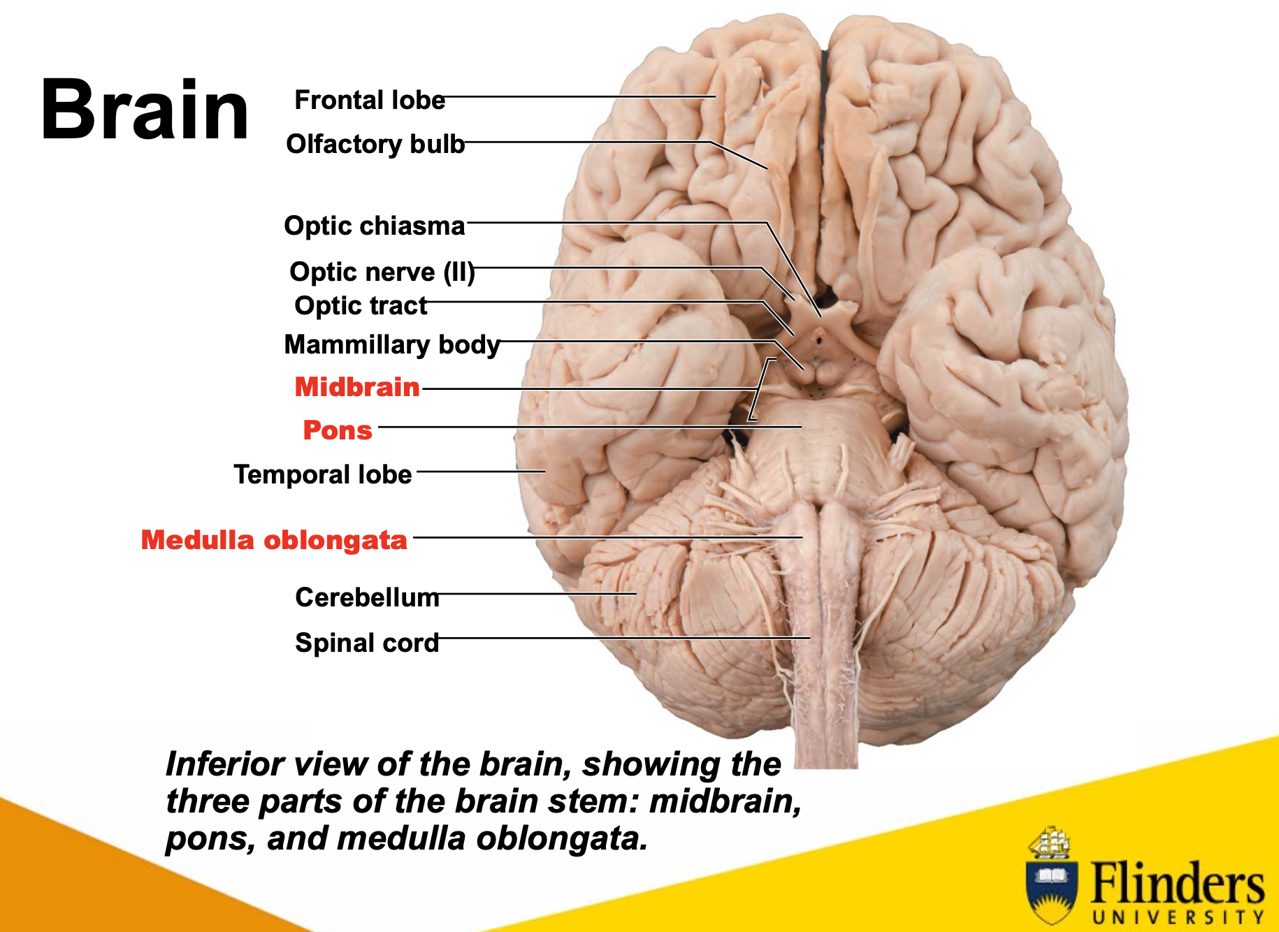
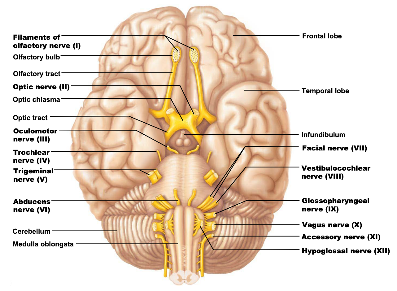
also about this diagram (that she mentions in lecture):
olfactory bulb: long, slender part
optic nerve (II): look for characteristic cross (this is the optic chiasm, where optic tract crosses)
mammillary body: small, round structures
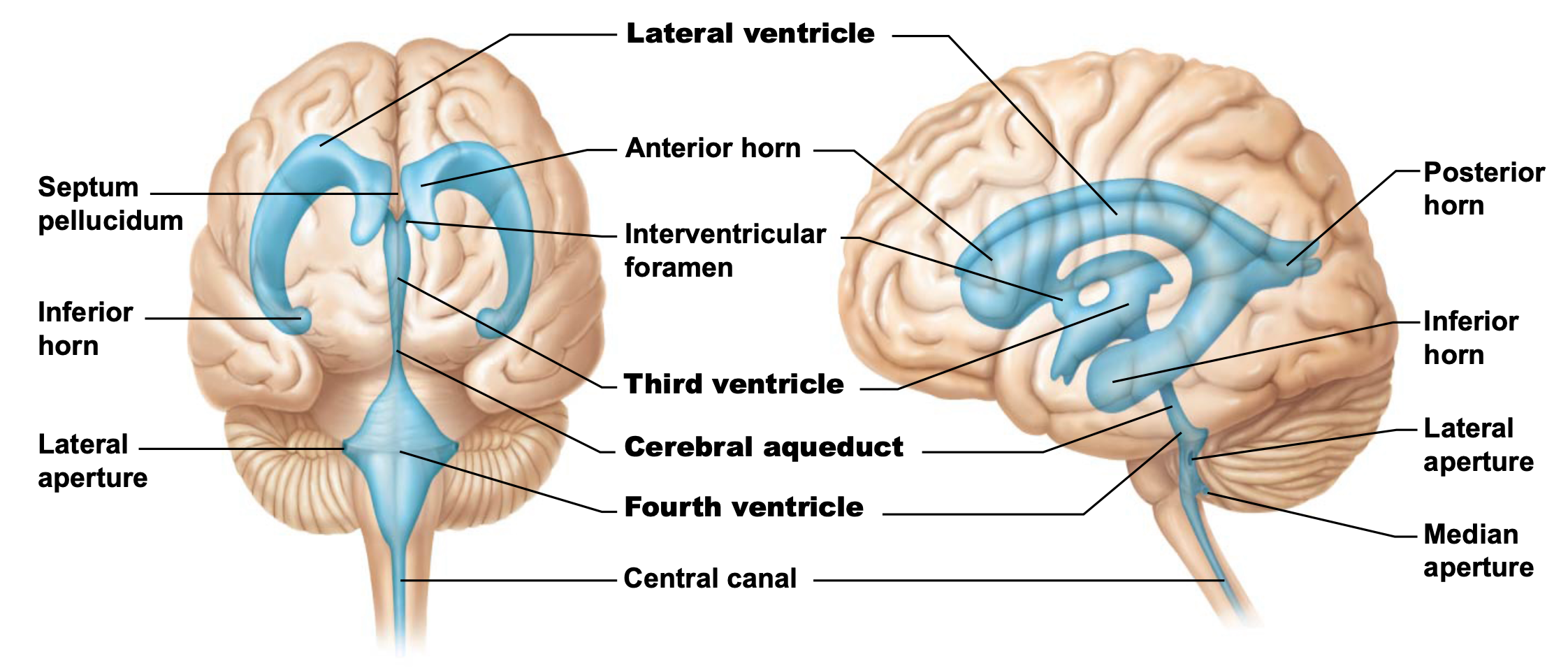
what are the ventricles of the brain
SYSTEM: Central Nervous System
SUBSECTION: Brain
NOTE: None
CUE: None
holy yap: the brain has a hollow space, in embryological development, brain starts off as being a flat disc, which folds up to be a tube. the spinal chord and midbrain sort of have remnants of this tube. the cerebral cortex grows and rapidly expands, and folds forwards on itself, forming a c-shaped space inside. this increases surface area, providing more space for neurons. The ventricles follow this space.
ventricles:
fluid-filled chambers continuous to one another and to the central canal of the spinal chord (allowing circulation of CSF)
filled with cerebrospinal fluid (CSF)
lined by ependymal cells (neuroglial cells)
there are four ventricles.
lateral ventricles: (2)
paired, large C-shaped chambers located deep in each hemisphere
separated from one another by a membranous septum
each lateral ventricle is connected to the third ventricle via interventricular foramen
third ventricle:
lies in the diencephalon and is connected to the fourth ventricle through the cerebral aqueduct
fourth ventricle:
lies in the hindbrain, and is continuous with the central canal of spinal chord.
three openings connect the forth ventricle to the subarachnoid space that surrounds the brain.
paired lateral apertures in side walls
median aperture in roof
state the cerebral hemispheres (lobes)
SYSTEM: Central Nervous System
SUBSECTION: Brian, Regional Anatomy
NOTE: None
CUE: None
Several sulci divide each hemisphere into five lobes
frontal lobe
parietal lobe
temporal lobe
occipital lobe
insula lobe
frontal, parietal, temporal, and occipital lobes are all visible from the surface.
insula lobe is a not visible; buried under the other lobes. only visible if move temporal lobe out of the way.
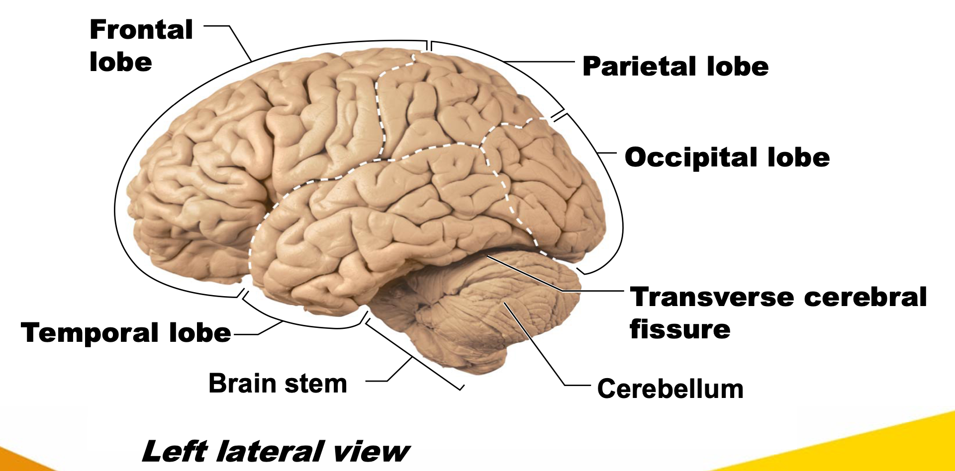
lobes are separated from the cerebellum by the transverse cerebral fissure.
describe the surface markings of the brain
SYSTEM: Central Nervous System
SUBSECTION: Brain, Regional Anatomy
NOTE: None
CUE: None
gyri: ridges
sulci: shallow grooves
central sulcus separates parietal and frontal lobes.
fissures: deep grooves
longitudinal fissure
separates two hemispheres
transverse cerebral fissure
separates cerebrum and cerebellum
describe grey and white matter in CNS
SYSTEM: Central Nervous System
SUBSECTION: Brain and Spinal Cord, Neuronal Pathways
NOTE: heard this stuff will definitely be in exam
CUE: None
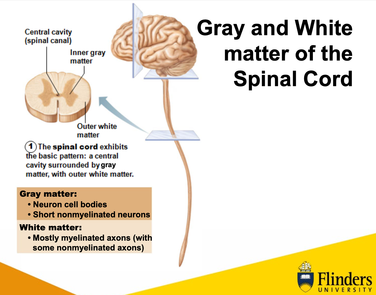
holy yap (brain): brain has bark-like structure (cortex=bark in latin). thin, couple of millimetres thick area, where cell bodies of CNS are limited to. when brains are stained, cell bodies stain differently to the axons, (due to myelinated sheaths of the axons). refer to the area where cell bodies are found as “grey matter”, and areas where axons are found, “white matter.”
holy yap (spinal cord): unlike brain where cell bodies were in the outer cortex, in the spinal cord, grey matter is internal (located within the butterfly), and white matter (myelinated axons) are surrounding.
grey matter:
neuron cell bodies
short, non-myelinated neurons
organised in areas
white matter (1):
mostly myelinated axons (with some non-myelinated axons).
organised in tracts
white matter in CNS (2):
(theres some more cards based on these pathways below too)
myelinated and non-myelinated nerve fibres allow communication between parts of the spinal cord, and spinal cord and brain.
white matter is organised into tracts that run in three directions:
Ascending: up to high centres (the brain) (sensory inputs)
Descending: from brain to spinal cord, or lower cord levels (to exit muscles) (motor inputs).
Transverse: from one side to the other (commissural fibres).
each spinal tract is composed of axons with similar destination’s and functions.
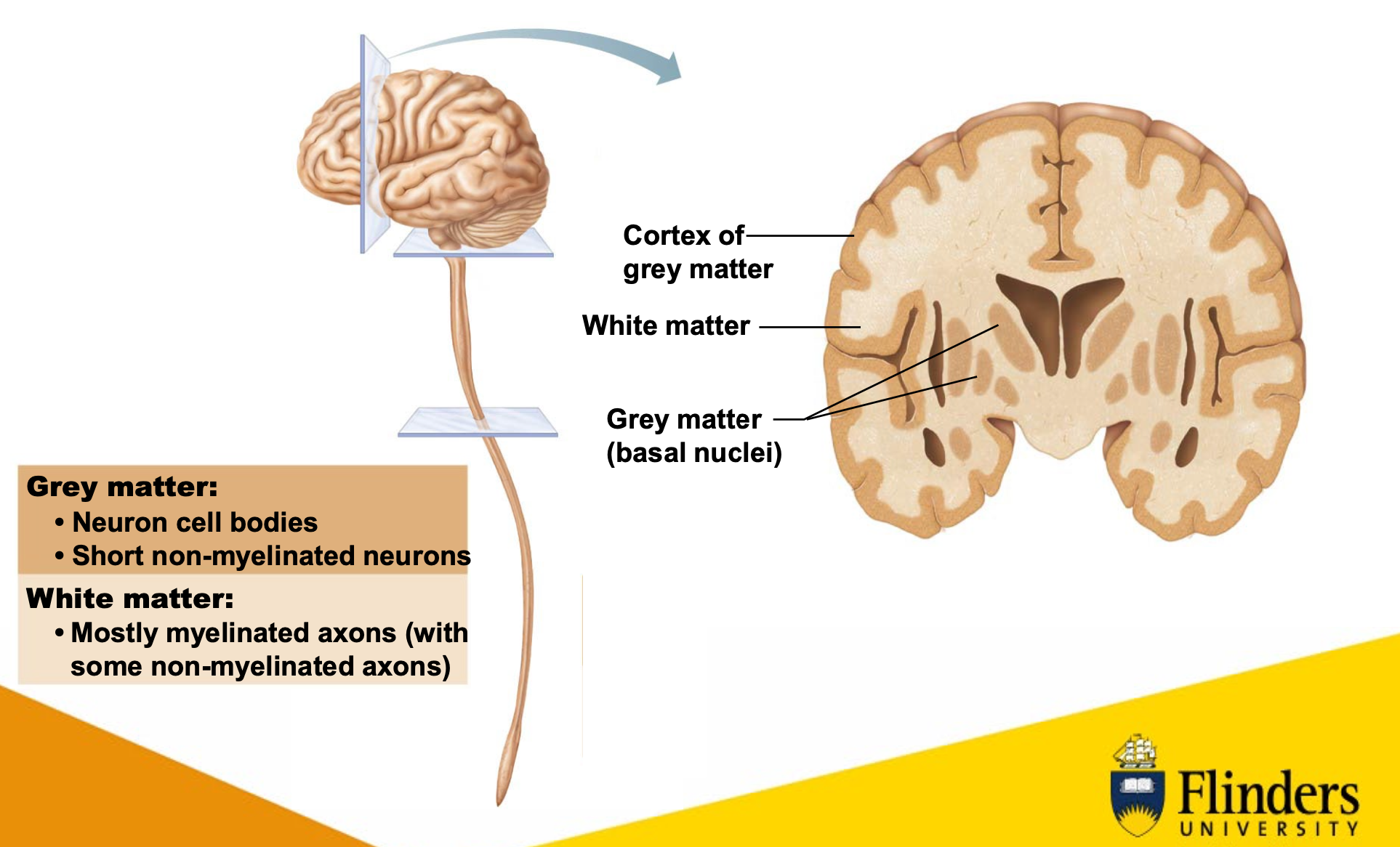
in diagram: can see collections of cell bodies deeper, away from the cortex, called ganglia. the ones indicated are basal ganglia (basal nuclei). can also see part of longitudinal fissure separating the hemispheres (crevice looking thing going down middle), can also see part of ventricular system (hollow looking thing) where CSF flows through.
idk if this is really important but yeah
non-myelinated axons are usually associated with pain.
note:
spinal tracts, are basically just white matter.
cerebral cortex is the grey matter of brain.
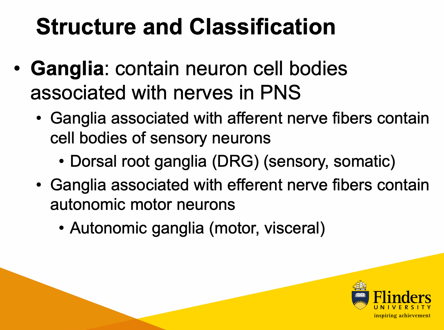
idk if we need this either since lecturer skipped the slide in the lecture, but here is more on ganglia:
ganglia associated with afferent nerve fibres contain grey matter (cell bodies) of sensory neurons
dorsal root ganglia (DRG; sensory, somatic)
ganglia associated with efferent nerve fibres contain grey matter of autonomic motor neurons
autonomic ganglia (motor, visceral)
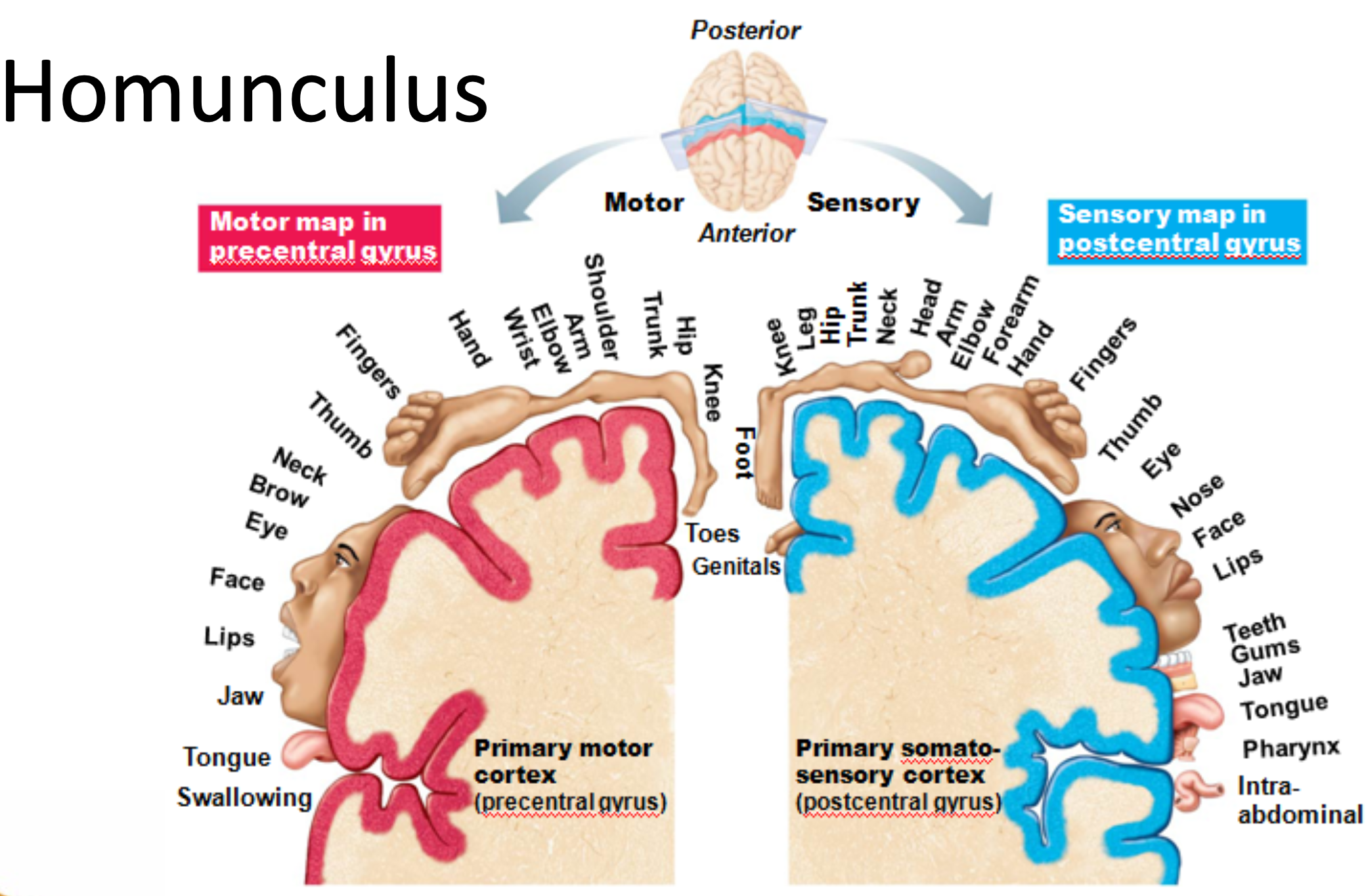
how is body represented in the brain ?
SYSTEM: Central Nervous System
SUBSECTION: Brain
NOTE: None
CUE: None
holy yap: not all regions of body are represented equally in the brain. some are given more brain neurons than others.
- idrk what they want us to know about this.
homunculus
representation of how brain visualises the
body.
hands, tongue, lips
high amount of sensory receptors/chemoreceptors/mechanoreceptors, lots of sensory information from the hands, tongue, and lips.
more neurons devoted to these sensors as opposed to other body components, such as the legs and trunk.
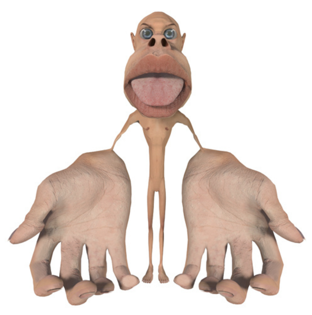
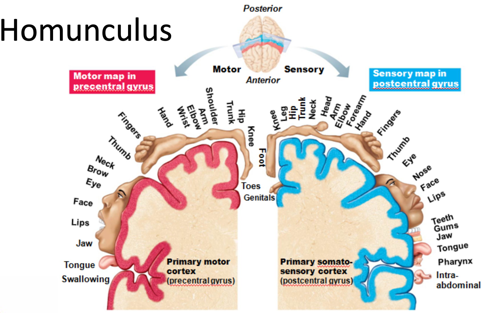
homunculus applies to sensory and motor neurons.
can see homunculus around the central sulcus.
what is the cerebral cortex and its function ?
SYSTEM: Central Nervous System
SUBSECTION: Brain
NOTE: None
CUE: None
holy yap: when think about cerebral cortex, there are three types of functionals area: motor areas (controlling voluntary movement), sensory areas (respond to conscious awareness of sensation), association areas (where integration of diverse information occurs). each hemisphere is concerned with the contralateral (opposite) side of the body. Incoming information from right arm, is represented on the left postcentral gyrus; and motor control of right arm is represented on left primary motor cortex (left side of brain) (with reference to the homunculus diagram thing). we do get some specialisation of cortical function, meaning some functions are only found in one hemisphere (e.g. language and speech are functions found on left side only).
cerebral cortex:
thin (2-4mm) superficial layer of grey matter.
composed of neuron cell bodies, dendrites, glial cells, and blood vessels - but NO axons (or short, non-myelinated axons).
constitutes 40% of brain mass
it is the site of the conscious mind (higher cognitive function) involving:
awareness
sensory perception
voluntary motor initiation
communication
memory storage
understanding.
lateralisation and cortical functioning:
hemispheres are not identical.
cerebral dominance:
90% of humans have left-sided dominance, resulting in right-handedness.
in other 10%, roles of hemispheres are reversed.
left hemisphere controls:
languages, math, logic
right hemisphere controls:
visual, spatial skills, emotion, music, art
the hemispheres communicate almost instantaneously via fibre tracts and functional integration (through white matter tracts).
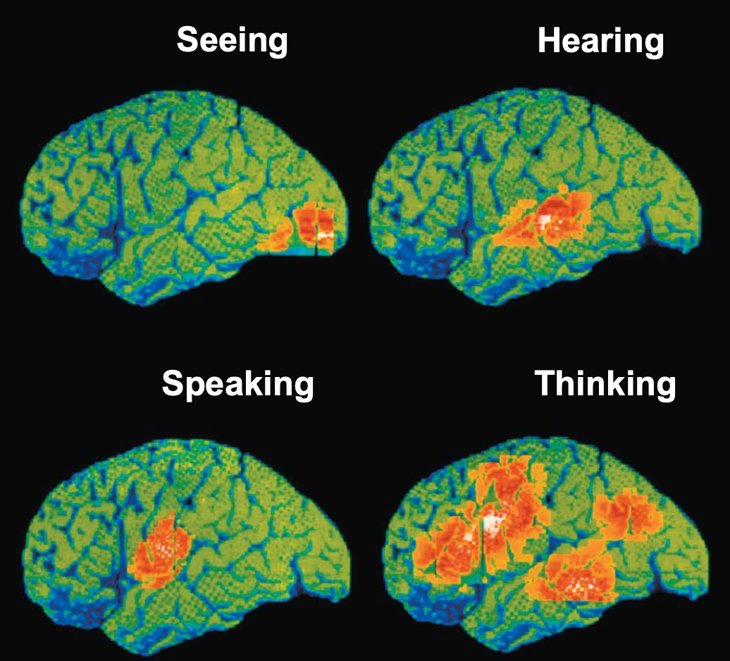
below shows: functional neuroimaging (fMRI) of the cerebral cortex (using radioactive glucose; shows which parts of the brain are active when performing particular tasks).
explain traumatic brain injury, and the types.
SYSTEM: Central Nervous System
SUBSECTION: Brain
NOTE: None
CUE: None
traumatic brain injury (TBI)
form of brain injury caused by sudden damage to the brain.
two types of injuries that can lead to brain trauma.
open head injuries (penetrating injuries):
occur when an object (e.g. a bullet) enters the body, and causes damage to specific brain parts.
closed head injuries:
result from a blow to the head (e.g. head strikes windshield in a car accident).
what are the types of cerebrovascular accidents (CVAs)
SYSTEM: Central Nervous System
SUBSECTION: Brain
NOTE: None
CUE: None
aka. strokes
strokes may arise from ischemia or hemiplegia.
ischemia occurs when:
tissue is deprived of blood supply, leading to death of brain tissue.
this can be caused by blockage of a cerebral artery caused by blood clot
could also be caused by excitation damage, where raising of glutamate overexcites neurons, worsening the condition (excitotoxin).
hemiplegia is the paralysis on one side, causing deficits in sensory input and speech/communication, depending on where the damage is.
paralysis of body occurs on the contralateral side of the brain.
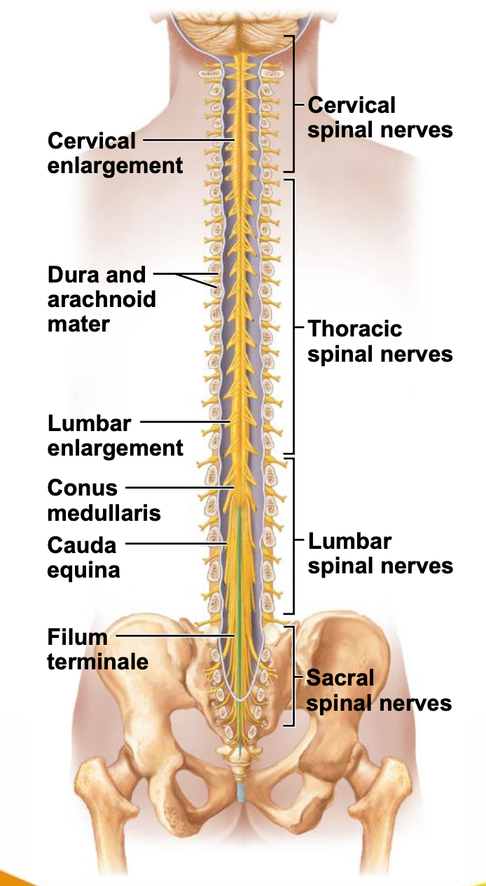
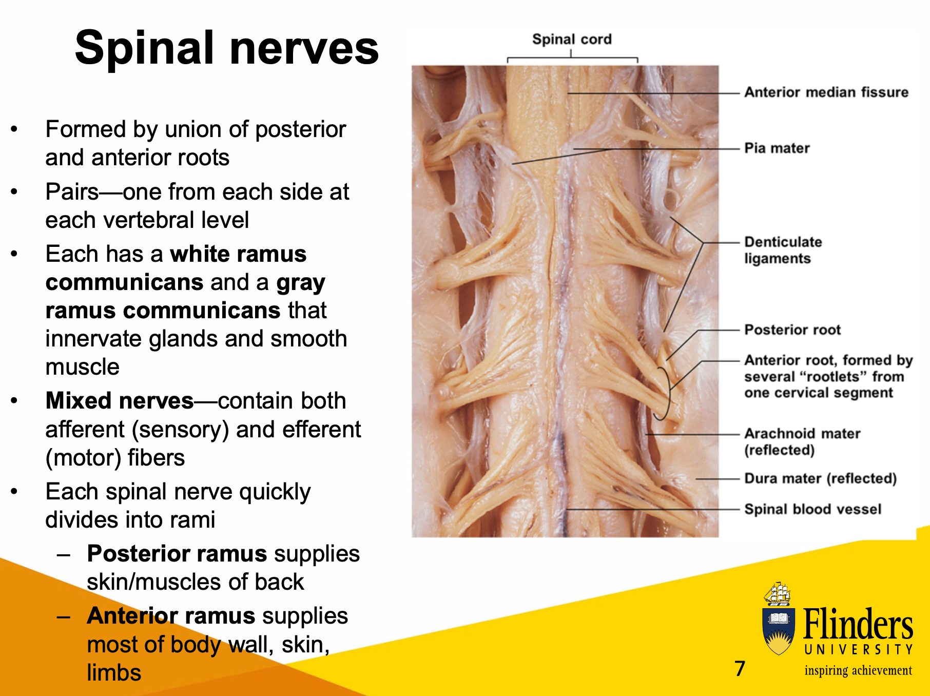
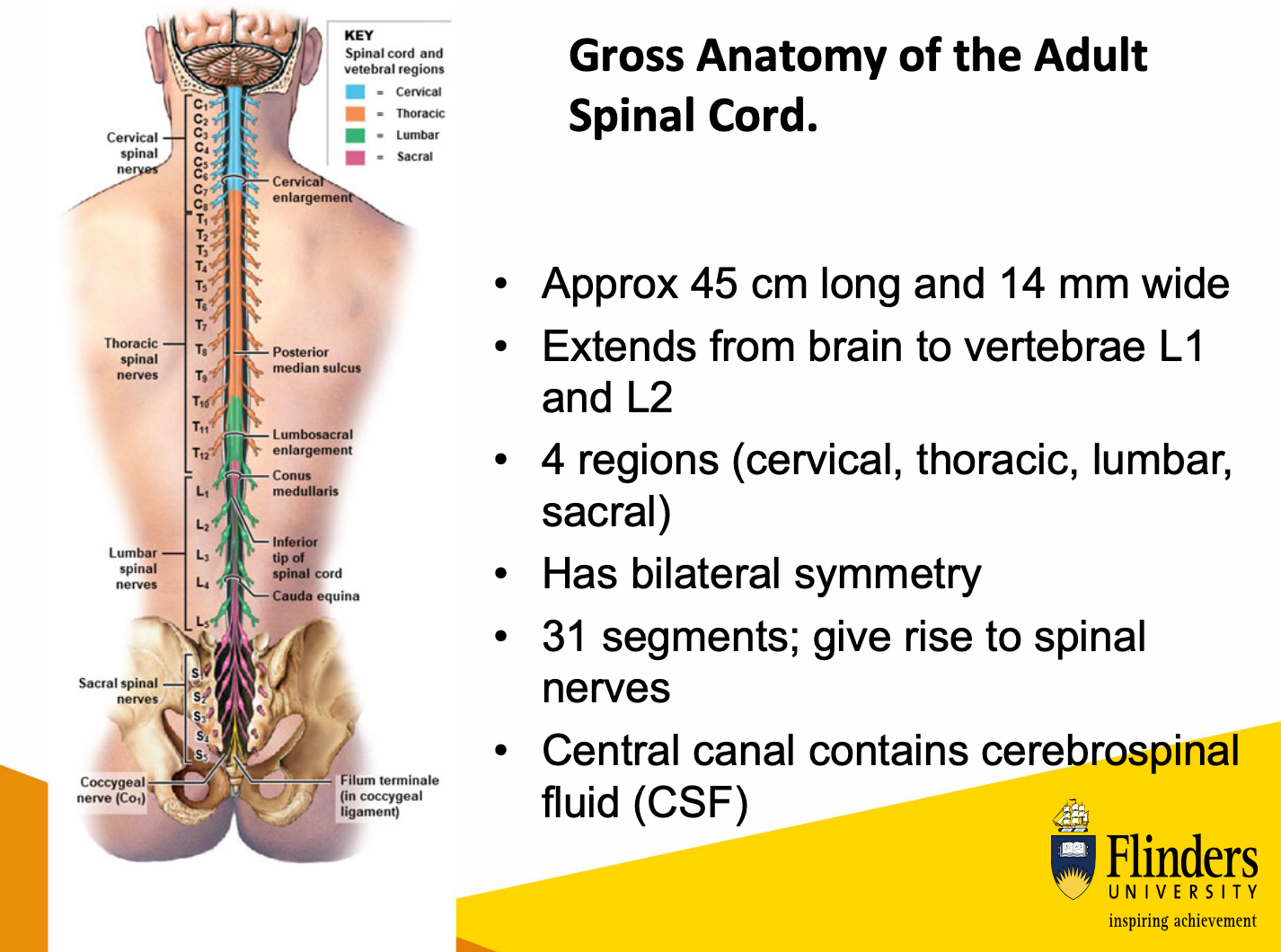
describe gross structure of spinal cord
SYSTEM: Central Nervous System
SUBSECTION: Spinal Cord
NOTE: I VERY VERY STRONGLY DISLIKE THIS LECTURER. WTF. Sorry future me, this card is in shambles~ 🤗
CUE: girl…. just flip the card… don’t even try…
the spinal chord consists of the cervical, thoracic, lumbar, and sacral segments.
however, the entire spinal chord can be divided into 31 segments based on where the spinal nerves originate.
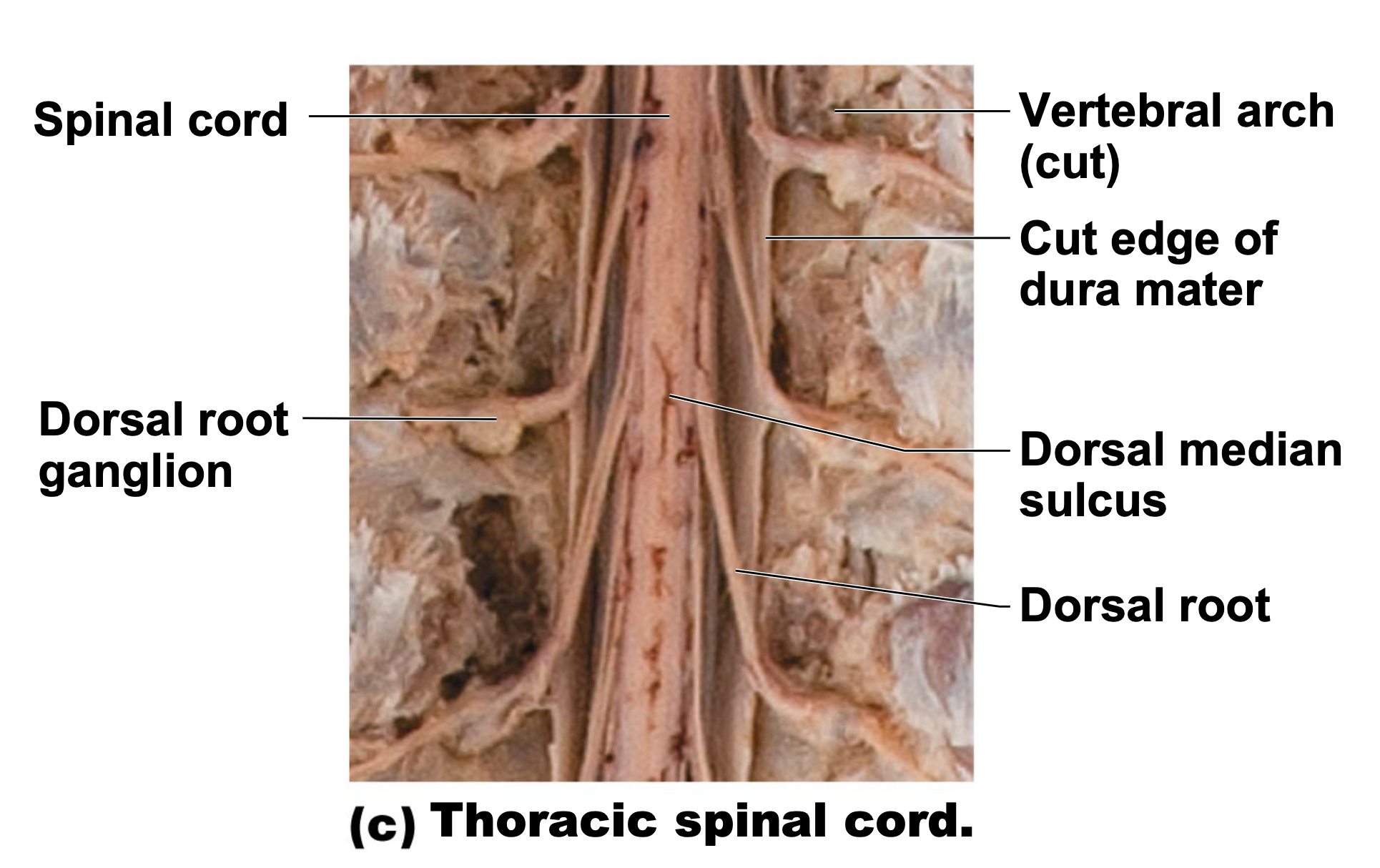
each spinal spinal segment is associated with a pair of dorsal root ganglia (located near spinal chord).
ganglia contains cell bodies of sensory neuron, where the axons of these neurons form the dorsal roots that bring sensory information to the spinal chord.
another one: dorsal root ganglia (aka. dorsal root ganglia) contain cell bodies of sensory neurons that form the posterior (dorsal) root.
located between pedicles of adjacent vertebrae.
spinal nerves contain sensory and motor neurons which
connect to the periphery
extend in the gaps between vertebra
serves the tissue in that particular area
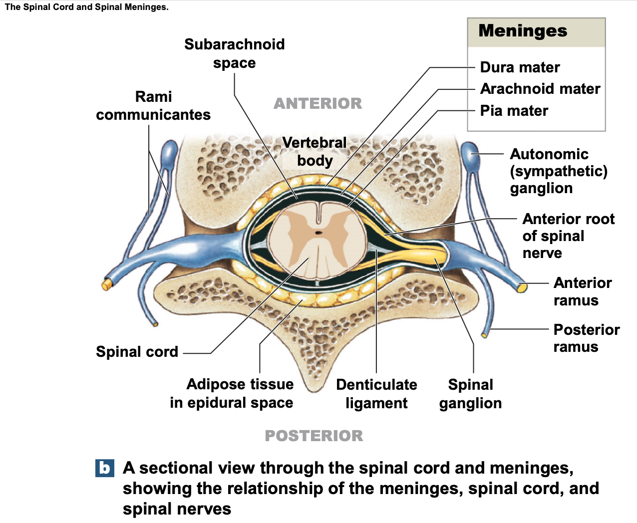
about diagrams:
like brain, spinal cord has meninges
dura mater
arachnoid mater
pia mater
dorsal root ganglion: bringing sensory information to spinal chord, the dorsal root ganglion is one of the relay stations, where sensory information detected by receptors in the periphery/around viscera is sent to the dorsal root ganglion, and then to the CNS by entering the spinal chord.
cell bodies sitting here, axons travel up as tracts in particular areas/columns in spinal chord.
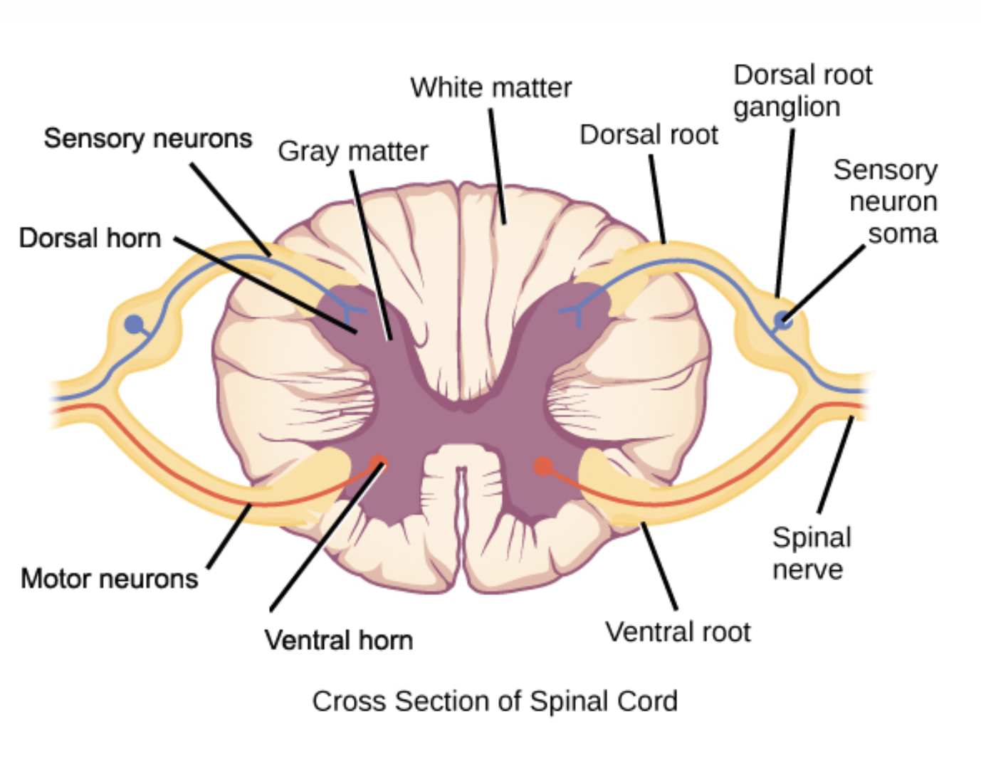
about diagram:
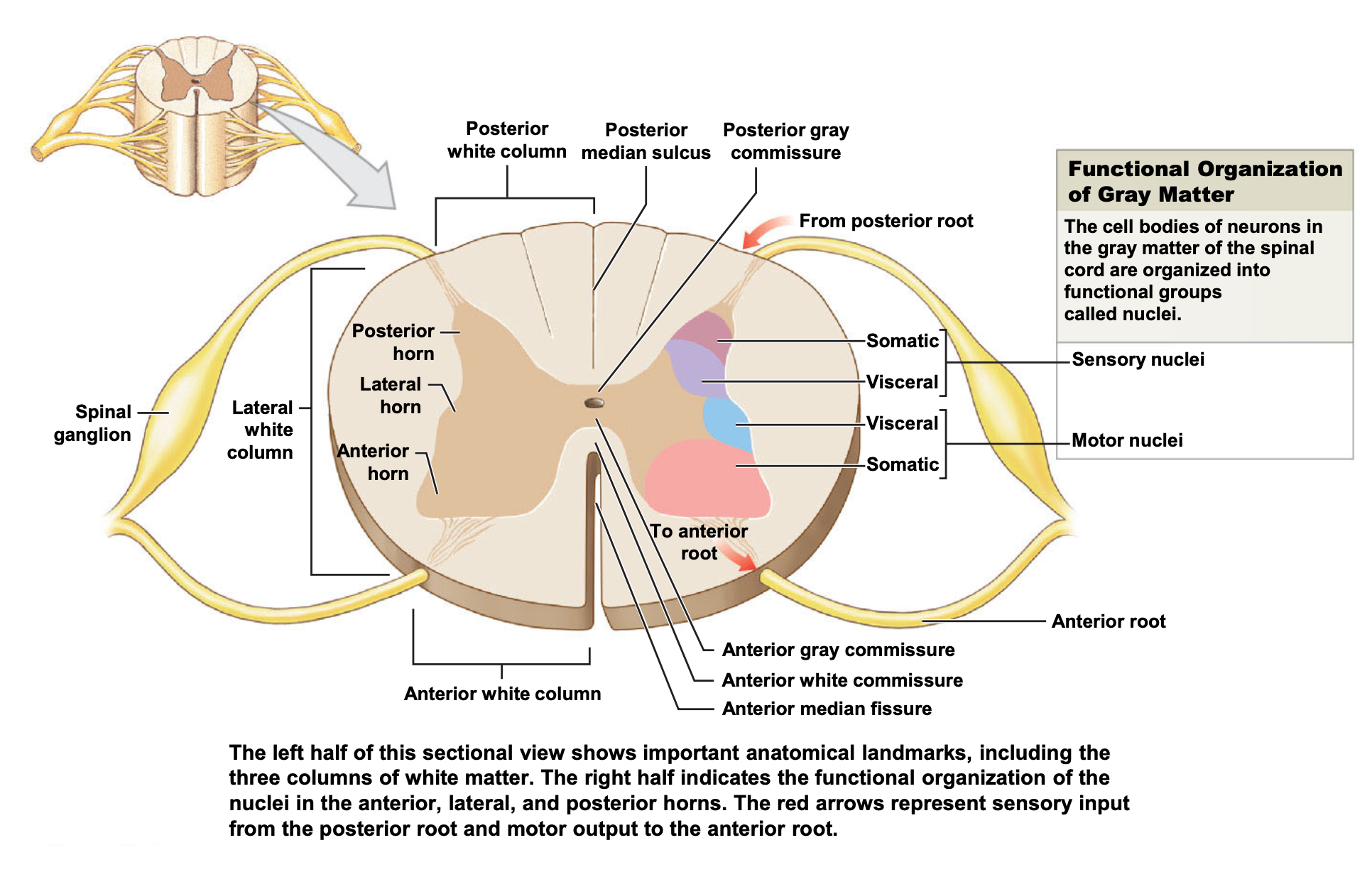
can see typical H/butterfly shape arrangement.
unlike brain where cell bodies were in the outer cortex, in the spinal cord, grey matter is internal (located within the butterfly), and white matter (myelinated axons) are surrounding.
they’re mirror images
two sides
dorsal side (associated with incoming sensory/afferent information).
dorsal horn, dorsal root, dorsal root ganglion
ventral side (associated with motor function).
ventral horn, ventral root (extending into ventral part of spinal chord)
in the thoracic and superior lumbar regions, there are sympathetic neurons using this pathway as well (wtf is she YAPPING about without any CONTEXT ????)
also got commissural neurons; bridge of grey matter connecting the ventral and dorsal on either side.
spinal nerves are where sensory neuron axons and motor neuron axons travel together as nerves.
which are covered in myelin sheaths, to maintain the speed, and integrity of electrical communication.
spinal nerves exit through vertebrae, and serve that particular region of the body.
spinal nerves collects sensory information, and conveys motor information.
hence, if spinal nerve is damaged, losing sensation and motor control of that particular area.
describe the organisation of the spinal cord and how signals get sent around to spinal cord
SYSTEM: Central Nervous System
SUBSECTION: Spinal Cord
NOTE: idk this is a chaotic card, but yeah
CUE: None
discussed in previous card:
four segments (looking at entire spinal cord)
cervical, thoracic, lumbar, and sacral segments.
two sides (looking at the cross-section)
dorsal side (associated with incoming sensory/afferent information).
dorsal horn, dorsal root, dorsal root ganglion
ventral side (associated with motor function).
ventral horn, ventral root (extending into ventral part of spinal chord)
grey matter (cell bodies) is organised in areas; white matter is organised in columns/tracts.
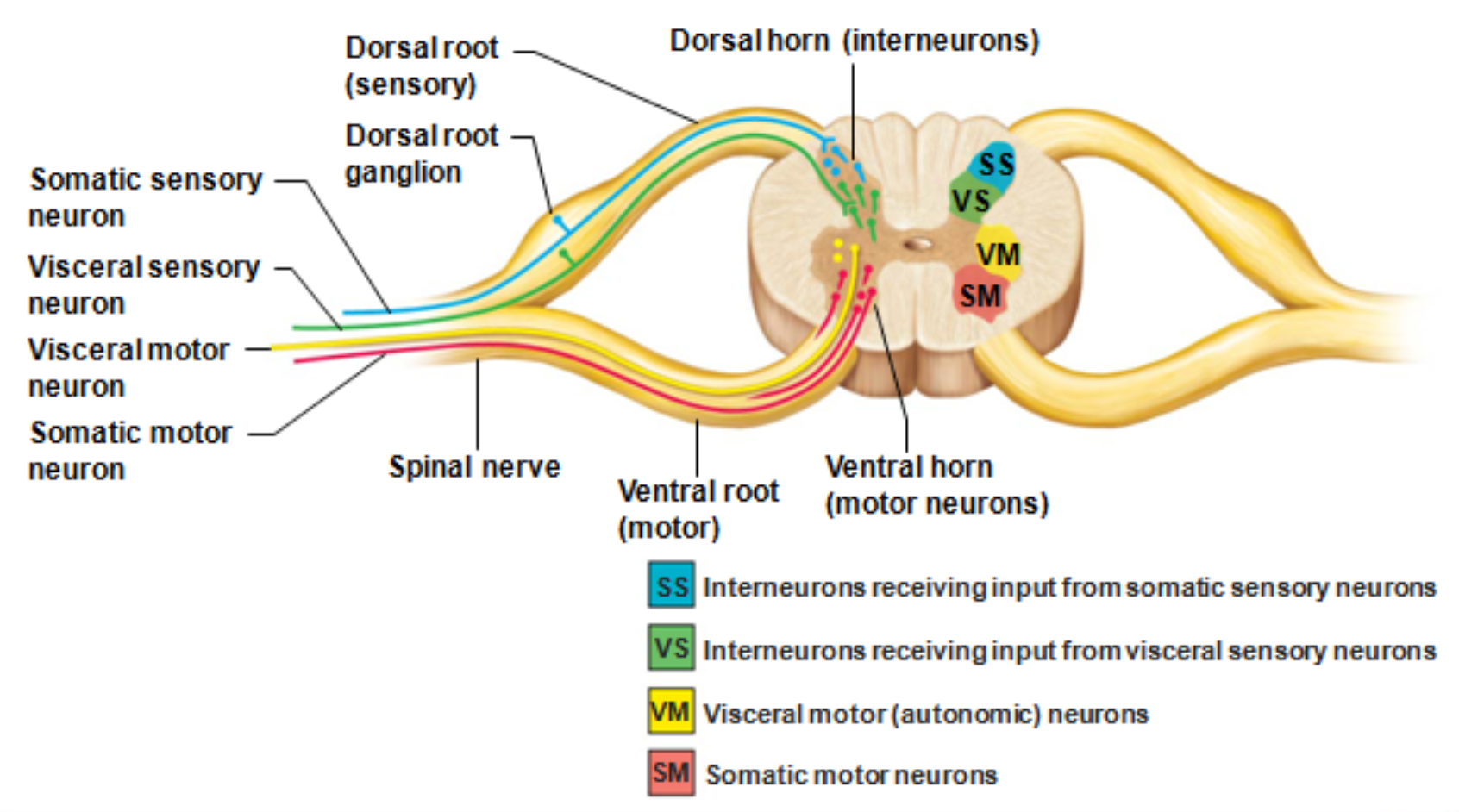
about diagram:
think about spinal chord as a highway, making local connections with cell bodies, and the acons either moving up to brain for further processing, or moving out to body to convey motor commons
somatic and visceral sensory neurons in dorsal horn.
somatic and visceral motor neurons in ventral horn.
visceral refers to internal environment.
somatic refers to voluntary control/decisions.
these neurons are carried together in spinal nerves (carry information to and from CNS).
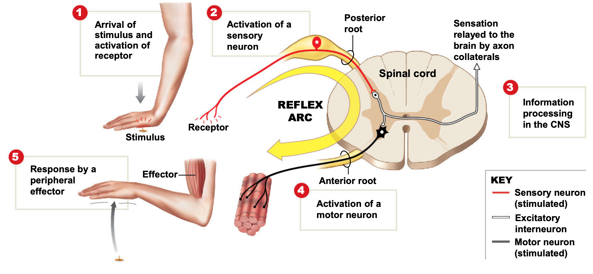
how signals get sent around spinal cord:
basically its the reflex arc, but she explains it TERRIBLY. ill write her holy yap; might be best to refer to year 12 notes though.
holy yap
interneurons that connect the dorsal and ventral horns means can bypass going all the way up to brain for thought and processing at that level. can have certain amount of local processing at spinal cord, speeding process up for a bit.
e.g. hand coming into contact with a flame.
high temp causes tissue damage, which is detected by sensory receptors (temperature receptors, pain receptors), and conveyed into the dorsal horn of the spinal cord at that level. communication/connections with interneurons to the ventral side, and then motor reflex comes back, pulling hand away from fire very quickly, since information is under a reflex speed (very quick).
spinal reflex arc (also explained in another card).
stimulus activates a receptor.
with enough stimulation, action potential is generated in sensory neuron. Axon (from sensory neuron) enters spinal cord through the posterior (dorsal) root.
information processing in spinal cord usually occurs at one or more interneurons.
i.e. sensory information is processed in the spinal cord at one or more interneurons.
interneurons stimulate action potentials in motor neuron; its axon leaves via anterior (ventral) root
motor neuron stimulates effector (muscle/gland).
what are the characteristics of neuronal pathways
SYSTEM: Central Nervous System
SUBSECTION: Neuronal pathways
NOTE: None
CUE: None
major spinal tracts are part of multi-neuron pathways (meaning they are relay stations).
in nervous system, relay stations facilitate information integration (important for analysing and processing information).
four key points about spinal tracts (white matter) and pathways:
decussation (crossing place): most pathways cross from one side of CNS to other at some point.
allows for some integration
fits with contralateral characteristic of brain; needs to be crossing over at some point.
relay: consist of chain of two or three neurons.
somatotopy: precise spacial relationship in CNS corresponds to spatial relationship in body.
mapping of body parts on the motor and somatosensory cortexes.
body parts close to each other on body, are likely mapped near each other on the cortexes as well.
symmetry: pathways are paired symmetrically (left and right)
what is the ascending pathway (describe the neuron chain), and what are main sensory pathways?
SYSTEM: Central Nervous System
SUBSECTION: Neuronal pathways
NOTE: None
CUE: three pathways
ascending pathways conduct sensory information upwards (from the body; visceral or somatic sensory neurons) through a chain of three neurons.
first-order neuron:
sensory information from receptors
refers to the sensory receptor.
skin receptors, pain receptors, proprioreceptors.
branch diffusely as they enter spinal cord and medulla, and where they synapse with second-order neuron.
second-order neuron
interneuron
cell body is located in the dorsal horn of the spinal cord, or in the medulla.
axon extends to the thalamus or cerebellum where they synapse with the third-order neuron.
third-order neuron
interneuron
with the cell body in the thalamus.
axon extends to the somatosensory complex
there are three main sensory pathways on each side of the spinal cord:
two pathways transmit somatosensory information to the sensory cortex via thalamus
dorsal column-medial lemniscal pathways (touch, vibration)
spinothalamic pathways (pain, temperature, coarse touch, pressure).
third pathway, spinocerebellar tracts, terminate in the cerebellum in the primary sensory cortex (muscle tendon or stretch).
both ventral and dorsal tracts
useful in coordinating muscle activity.
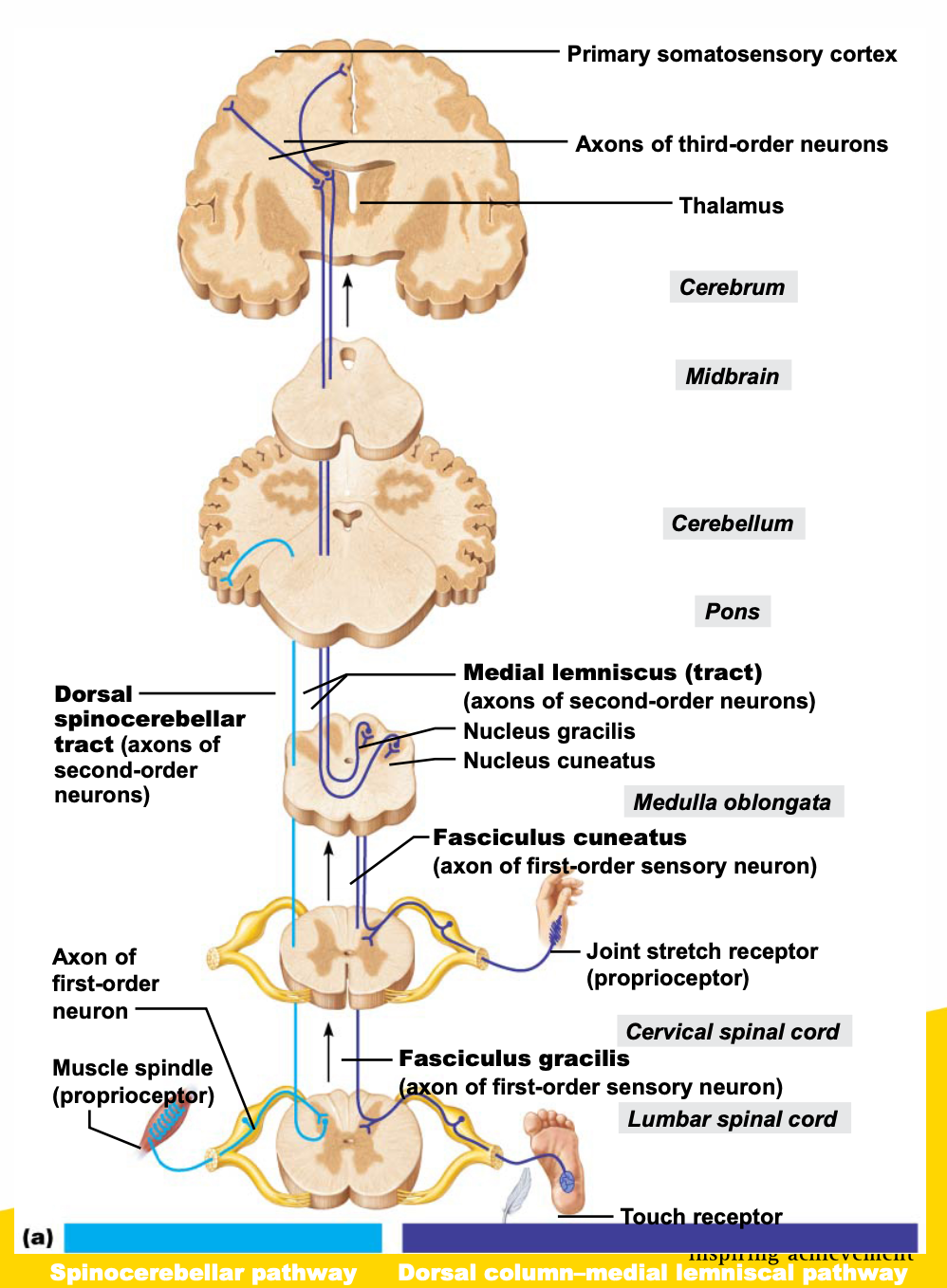
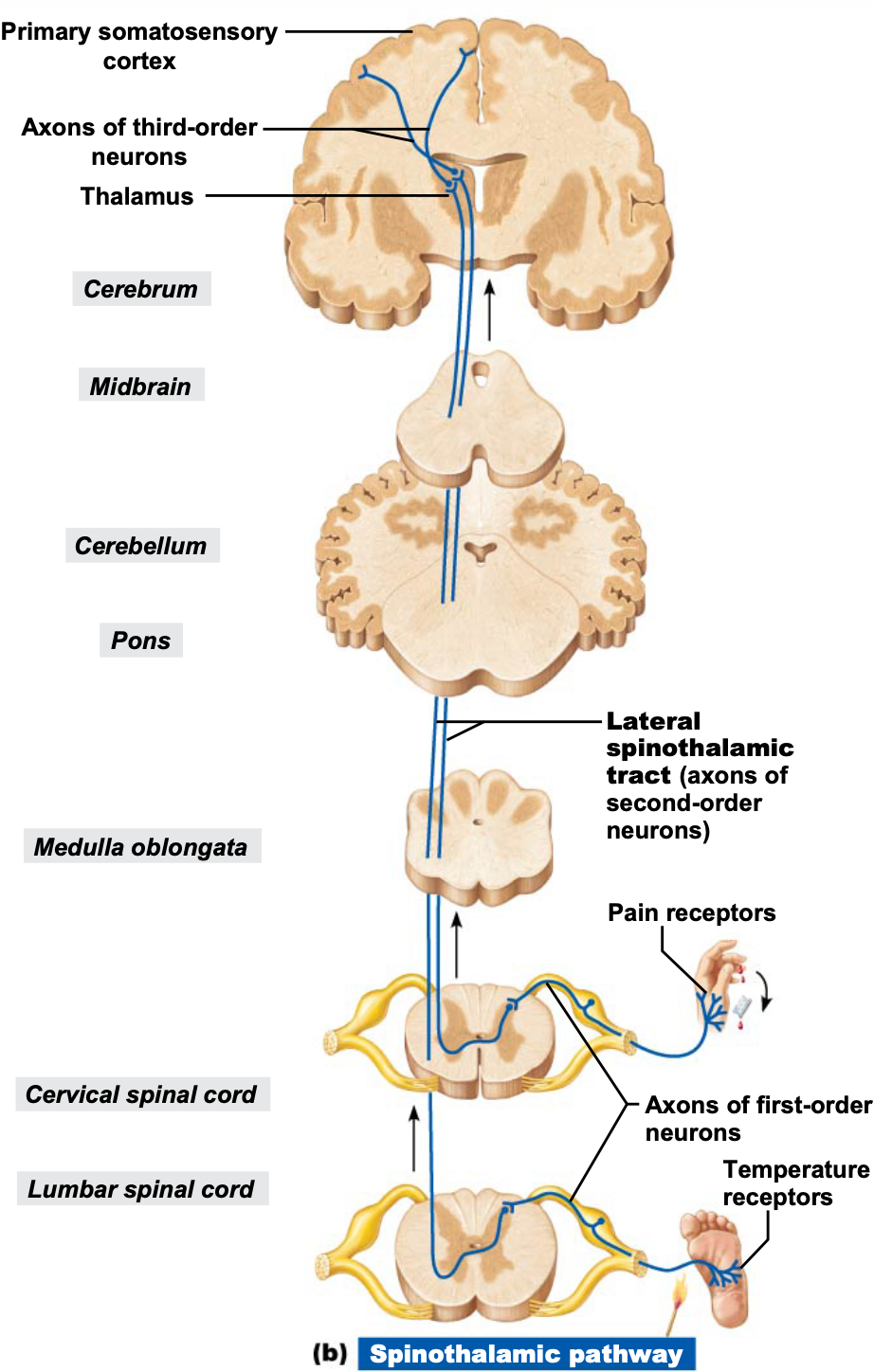
about diagrams: GIRLLLLLLLLLLLLLLLLLLLLLLLLLLLLLLL YOU WRONG. FOR NOT UPLOADING THE TRANSCRIPT…. YOU WRONG FOR THATTTTTTTTTTT.
can see where the pathways cross.
dorsal column-medial lemniscal pathway transmits input to the somatosensory cortex for touch and vibration.
composed of paired fasciculus cuneatus, fasciculus gracillis, and medial lemniscus (from medulla to thalamus) in the spinal cord
spinothalamic pathway is comprised of the lateral and ventral spinothalamic tract, transmitting pain, temperture, touch, and pressure information.
spinocerebellar tract travels in ventral and dorsal tract and carries information about muscle tendons and stretch to cerebellum.
remember:
white matter is organised into tracts that run in three directions:
Ascending: up to high centres (the brain) (sensory inputs)
Descending: from brain to spinal cord, or lower cord levels (to exit muscles) (motor inputs).
Transverse: from one side to the other (commissural fibres).
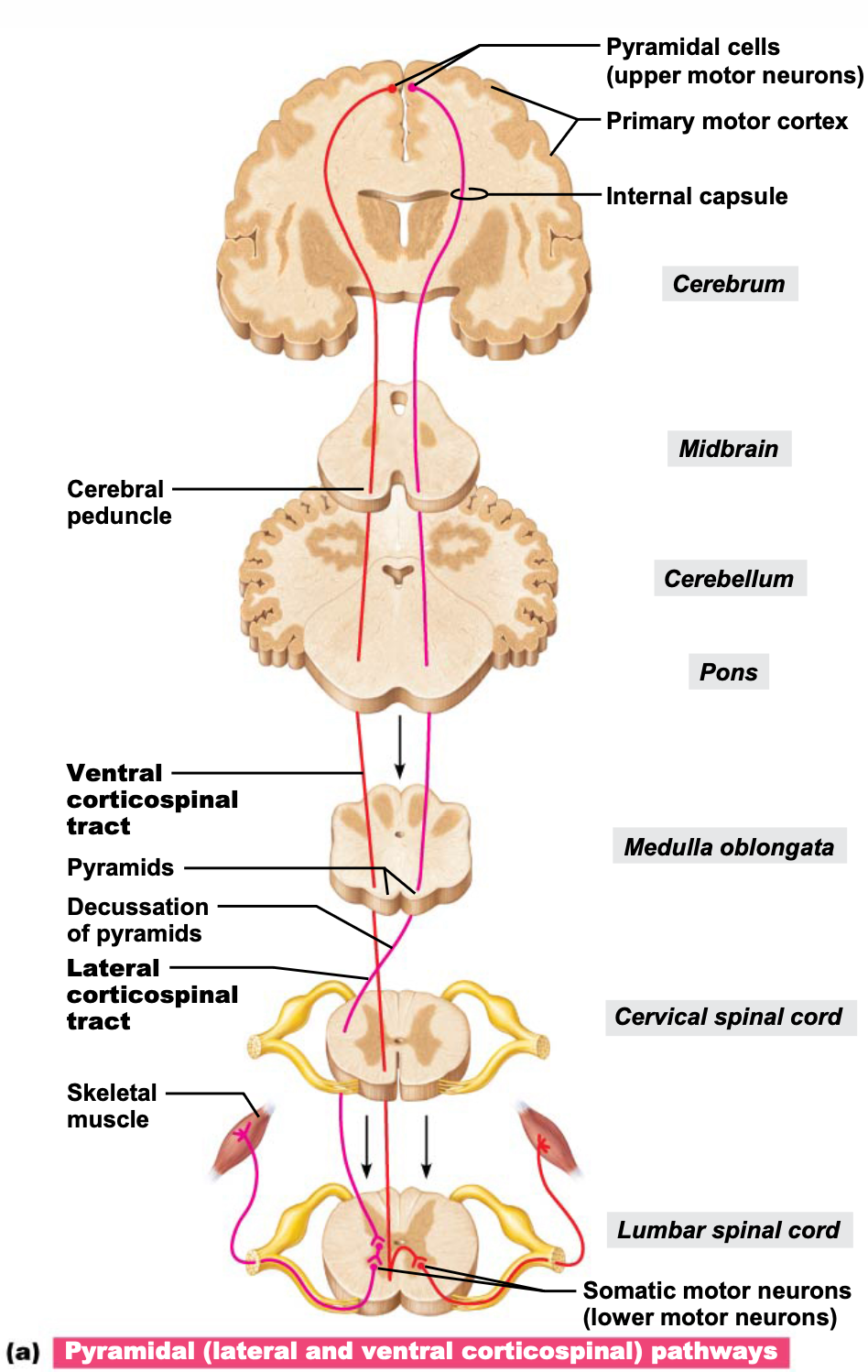
what are direct and indirect descending pathways, and what is the motor chain/pathway ?
SYSTEM: Central Nervous System
SUBSECTION: Neuronal pathways
NOTE: None
CUE 1: two types
CUE 2: describe direct (pyramidal) pathways
descending pathways deliver efferent impulses from brain to spinal cord.
i.e. carry motor information from brain to spinal cord.
there are two groups/types of descending pathways:
direct (pyramidal) pathways: through pyramidal tracts.
impulses from pyramidal neurons in precentral gyri pass through pyramidal (lateral and ventral corticospinal) tracts.
impulses descend directly without synapsing until axon reaches end of tract in spinal cord.
in spinal cord, axons synapse with interneurons (in lateral tract) or ventral horn motor neurons (in ventral tract)
direct pathways regulate fast and fine (skilled) movements.
every time a synapse is introduced; transmission is slowed down slightly.
indirect pathways: through all others
aka. multineuronal pathways.
complex and multisynaptic (take longer)
includes brain stem motor nuclei, and all motor pathways except pyramidal pathways.
these pathways regulate:
axial muscles, maintaining balance and posture
i.e. provide motor information to axial muscles to maintain posture and balance.
muscles controlling coarse limb movements
head, neck, and eye movements that follow objects in visual field.
there are four major indirect pathways:
reticulospinal and vestibulospinal tracts:
maintain balance by varying tone of postural muscles.
rubrospinal tracts:
control flexor muscles.
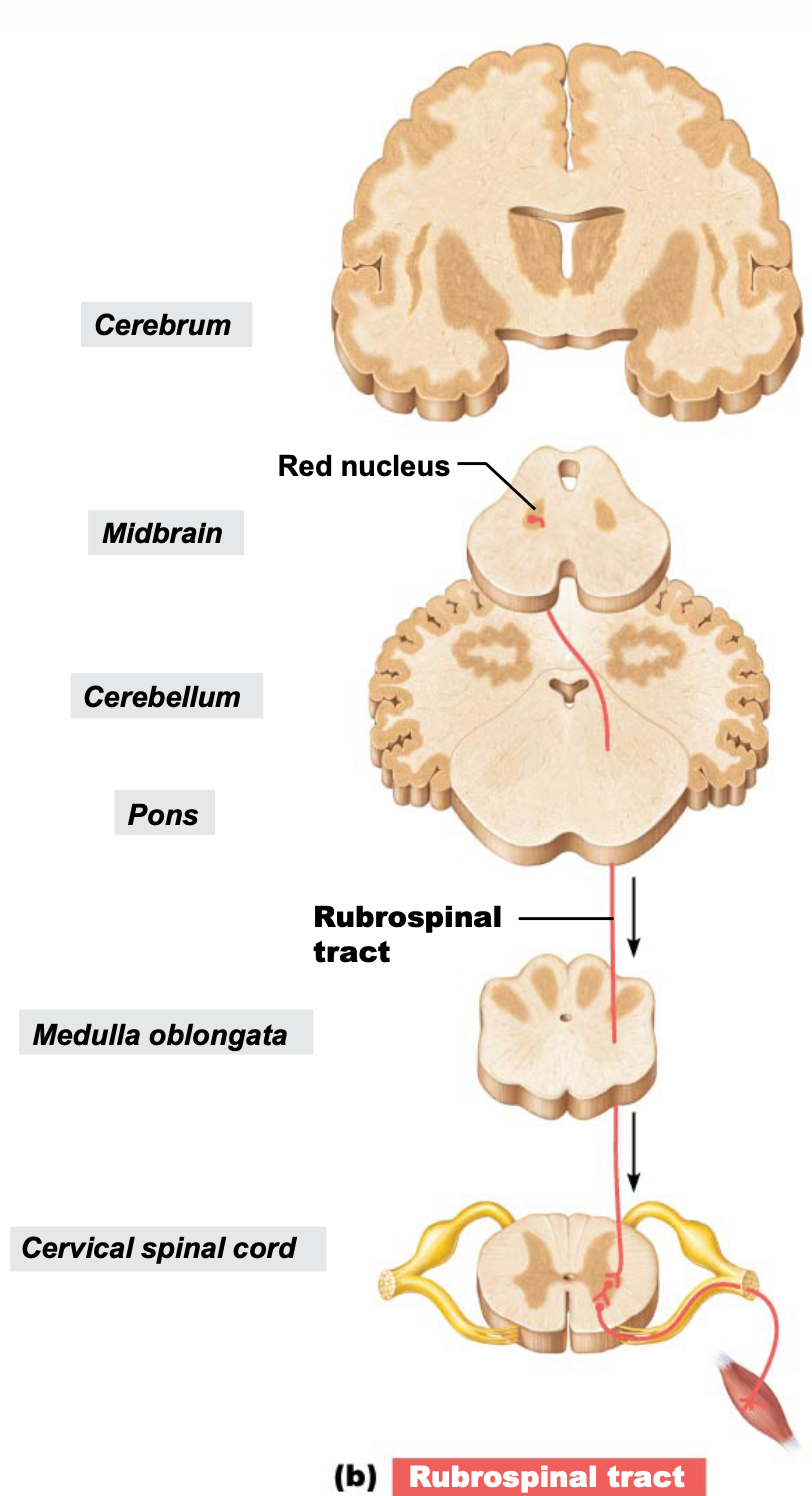
tectospinal tracts:
neurons in this tract originate from the superior colliculi, and mediate head movements in response to visual stimuli.
motor pathways involve two neurons:
upper motor neurons
pyramidal cells in primary motor cortex.
lower motor neurons
ventral horn motor neurons
which, for example, innervate skeletal muscles.
remember:
white matter is organised into tracts that run in three directions:
Ascending: up to high centres (the brain) (sensory inputs)
Descending: from brain to spinal cord, or lower cord levels (to exit muscles) (motor inputs).
Transverse: from one side to the other (commissural fibres).
state the divisions of the peripheral nervous system, and describe its function.
SYSTEM: Peripheral Nervous System
SUBSECTION: Peripheral Nervous System
NOTE: None
CUE: None
divisions of peripheral nervous system:
sensory (afferent) division
brings information from sensory receptors (e.g. from viscera/external environment) into the body (through PNS) for processing at CNS.
motor (efferent) division
motor decisions to move muscle (e.g. voluntary skeletal muscle), or something that is autonomic.
initiated by CNS, conveyed through PNS, then different divisions depending on whether movement is voluntary or involuntary.
somatic nervous system
autonomic nervous system (ANS)
sympathetic nervous system
fight or flight
parasympathetic nervous system
rest and digest
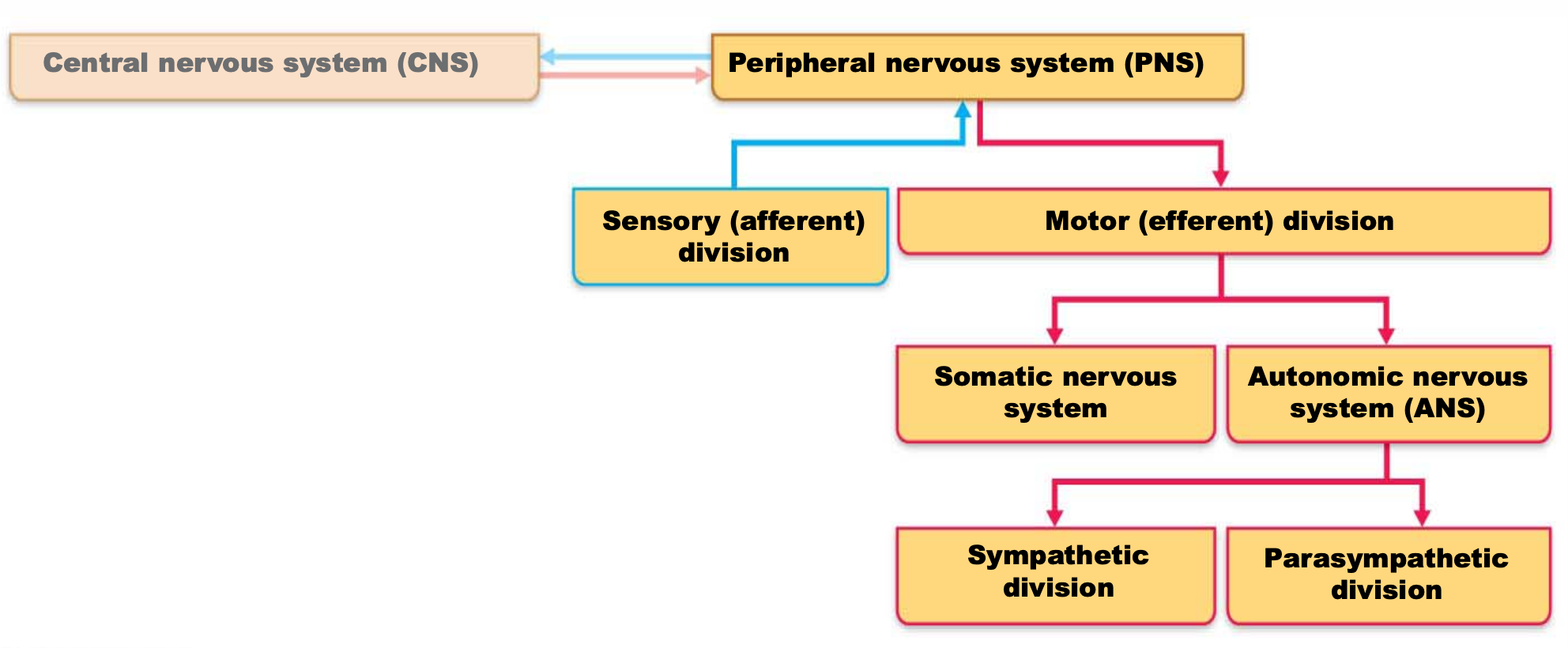
function:
PNS provides crucial links to and from the world outside the body, and consists of all neural structures outside of the brain and spinal cord. The neural structures can be divided into the following:
detecting incoming information (afferent, sensory)
from viscera or surrounding environment (internal or external to body)
transmission lines (neurones/nerves)
neurons bundled together form nerves.
motor endings and motor activity.
or something like this:
PNS interacts with body via sensory receptors, providing internal and external sensory information (afferent), that usually stimulates an autonomic or somatic response from the brain via efferent system.
state:
meaning of sensory receptors and sensation
why it is important
the ways that receptors may be classified as
SYSTEM: Peripheral Nervous System
SUBSECTION: Sensory Receptors and Sensation
NOTE: None
CUE: None
what it is
sensory receptors are the detectors which respond to changes in the environment (stimuli)
sensation refers to the awareness of stimuli, and the brains interpretation of the stimulus’ meaning (perception).
processing that occurs in brain.
importance
survival depends on sensation and perception.
sensation: the awareness of changes in internal and external environment
importance for homeostasis maintenance
perception: the conscious interpretation of stimuli
three ways to classify receptors:
stimulus type
body location
structural complexity
there’s also
non-encapsulated
encapsulated
state the sensory receptors based on each classification.
stimulus type
body location
structural complexity
discuss about non-encapsulated and capsulated receptors (?)
SYSTEM: Peripheral Nervous System
SUBSECTION: Sensory Receptors and Sensation
NOTE: None
CUE: None
stimulus type
mechanoreceptors
respond to touch, pressure, vibration, stretch
thermoreceptors
respond to changes in temperature
photoreceptors
respond to light energy
(found in retina)
chemoreceptors
respond to chemicals
(e.g. smell, taste)
(e.g. O2, and CO2 detection in main arteries)
nociceptors
respond to pain-causing stimuli
(e.g. extreme heat/cold, excessive pressure)
body location
exteroceptors
respond to stimuli arising outside body
(i.e. on external surface of body)
(e.g. receptors in skin responding to touch, pressure, pain, and temperature)
interoceptors (visceroceptors)
respond to stimuli arising in internal viscera and blood vessels
sensitive to chemical changes, temperature changes, tissue stretch
sometimes cause discomfort, but usually person is unaware of their workings
body locations
proprioceptors
respond to stretch in skeletal muscles, tendons, joints, ligaments, and connective tissue coverings of bones and muscles
where the brain looks to see where body is in three dimensional space.
informs brain of ones’ movements
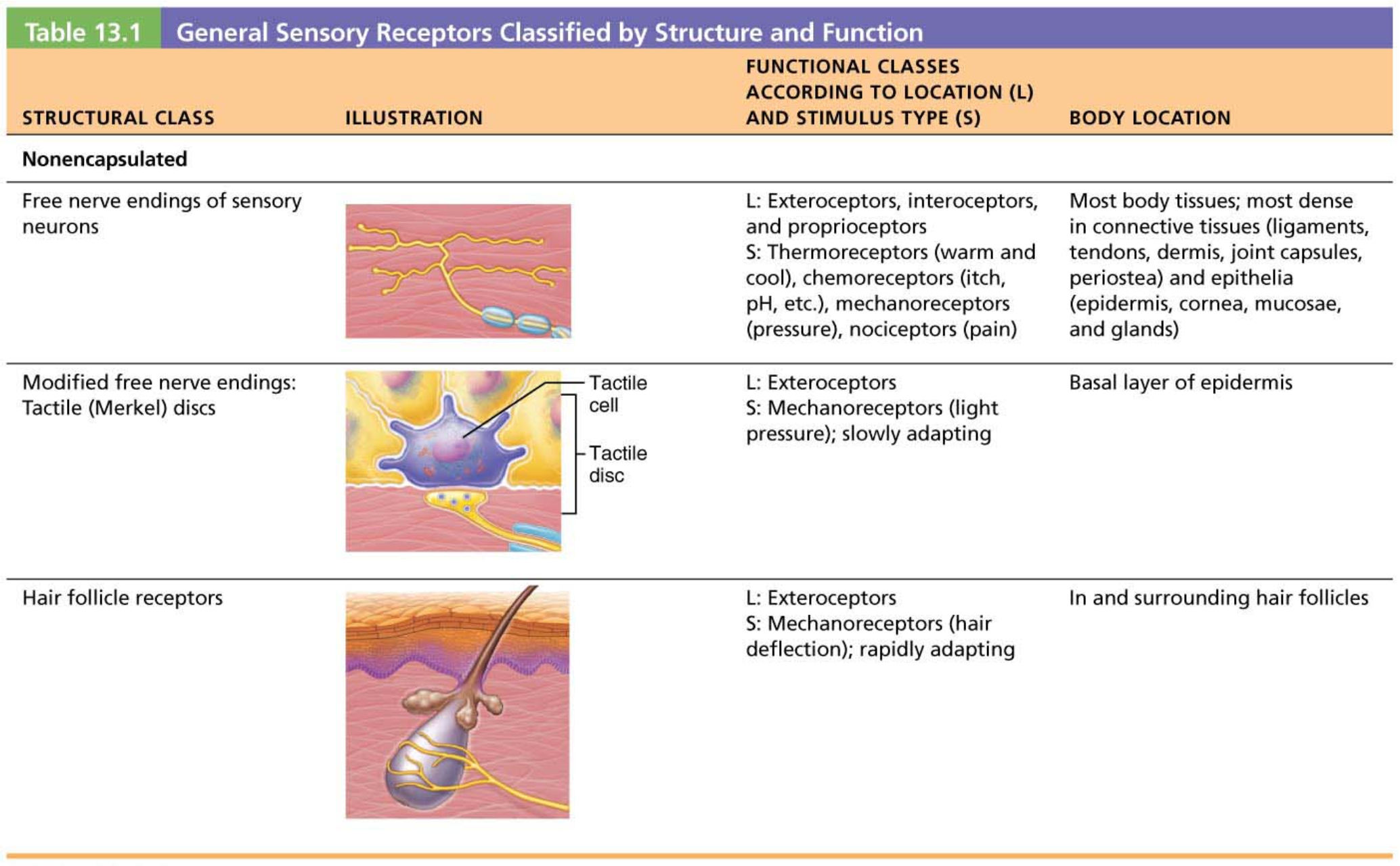
modified free nerve endings:
adaption e.g.: wearing of clothes, we feel it at first, but then we adapt to the pressure, get used to it, and stop responding to its stimuli after a certain amount of time etc.
lecturer does not necessarily delve into too much detail for this
just kind of reads the table.
state what inputs the somatosensory system receives, and describe the three main levels that the inputs are processed.
SYSTEM: Peripheral Nervous System
SUBSECTION: Sensory Receptors and Sensation
NOTE: tw: crashout
CUE: None
the somatosensory system is part of the sensory system serving body wall and limbs
receives input from
exteroceptors
interoceptors
proprioceptors
input is relayed to the brain (sensory areas), but is processed along the way.
it is processed at three main levels.
There are three levels of neural integration in sensory systems.
receptor level:
sensory receptors
circuit level:
processing in ascending pathways
perceptual level:
processing in cortical sensory areas
holy yap: IDEFK WHAT SHES TALKING ABOUT. SHES SO FUCKING WRONG FOR NOT PROVIDING A TRANSCRIPT OR ANYTHING CONTAINING THE WORDING OF THIS INFORMATION. YOU ACTUALLY NEED HELP IF YOU THINK PEOPLE CAN LEARN LIKE THIS. IDFK WHAT YOURE SAYING. 👎 . LIKE HOW DID YOU SAY ALL THIS ON ONE SLIDE WITH A FUCKING IMAGE ON IT. 👎.
for a sensation to occur, the stimulus must excite the receptor to a particular threshold that leads to an action potential being generated.
stimulus energy must match receptor specificity (e.g. touch receptor won’t respond to light), and stimulus must occur within receptive field of that receptor (i.e. occur in region where receptor is).
transduction must occur: energy of stimulus is converted into a graded action potential; threshold needs to be met to generate action potential.
information therefore ascends, where it is processed further (ascending pathways)
processing at circuit level: pathways integrate information on their way upwards on their way to the appropriate cortical regions.
first-order sensory neurons conduct impulses from the receptors to the spinal cord (reflexes or to second-order neurons).
second-order neurons send impulses to third-order neurons.
third-order neurons conduct impulses from thalamus to somatosensory cortex. when action potential reaches the somatosensory cortex, this is where sensation occurs/become aware of sensation.
perceptual level: where processing around meaning and interpretation occurs.
sensory perception detection requires a summation of impulses.
one action potential from one neuron may not be enough to bring the awareness to the particular stimulus; may require activation of several neurons sending train of action potentials.
intensity is encoded in frequency of impulses.
action potential is “all or nothing”
depolarisation either meets threshold, or it doesn’t.
so how we decode if something is hotter or not, it is detected by the frequency of action potentials.
spatial discrimination is also important; this identifies the site or the pattern of the stimulus.
two point discrimination test: tests how far two stimuli need to be in order to be detected as two stimuli.
hence spacial awareness/discrimination is also coded.
she said all this. on THIS SLIDE??? ARE YOUUUUUU JOOOOKINNGGGGGGGG BOUTTA START SAYING SLURSSSSSSSSSSSSS 👎

pain perception
what is its purpose
what are possible stimuli
how is pain transmitted
state what pain tolerance is, and what determines it.
describe the two types of pain.
SYSTEM: Peripheral Nervous System
SUBSECTION: Sensory Receptors and Sensation
NOTE: i need this lecturer to be fired, genuinely.
CUE: None
warns of actual or impending tissue damage so that protective action can be taken.
stimuli include:
extreme pressure and temperature
histamine
K+
ATP
acids
pain is transmitted via impulses which travel on fibres that release glutamate and substance P neurotransmitters.
some pain impulses are blocked by endogenous opioids (e.g. endorphins)
(acetylcholine is the neurotransmitter associated with motor neurones, glutamate and substance P are the transmitters associated with pain-related sensory neurons)
pain tolerance refers to how one perceives pain when exposed to the same stimulus intensity, and is a very individual experience.
pain tolerance varies per individual.
“sensitive to pain” means low pain tolerance, not low pain threshold.
i.e. refers to how much one can tolerate pain, and not the threshold that neurons are activated at.
genes help determine pain tolerance, as well as response to pain medications, and previous experiences to pain.
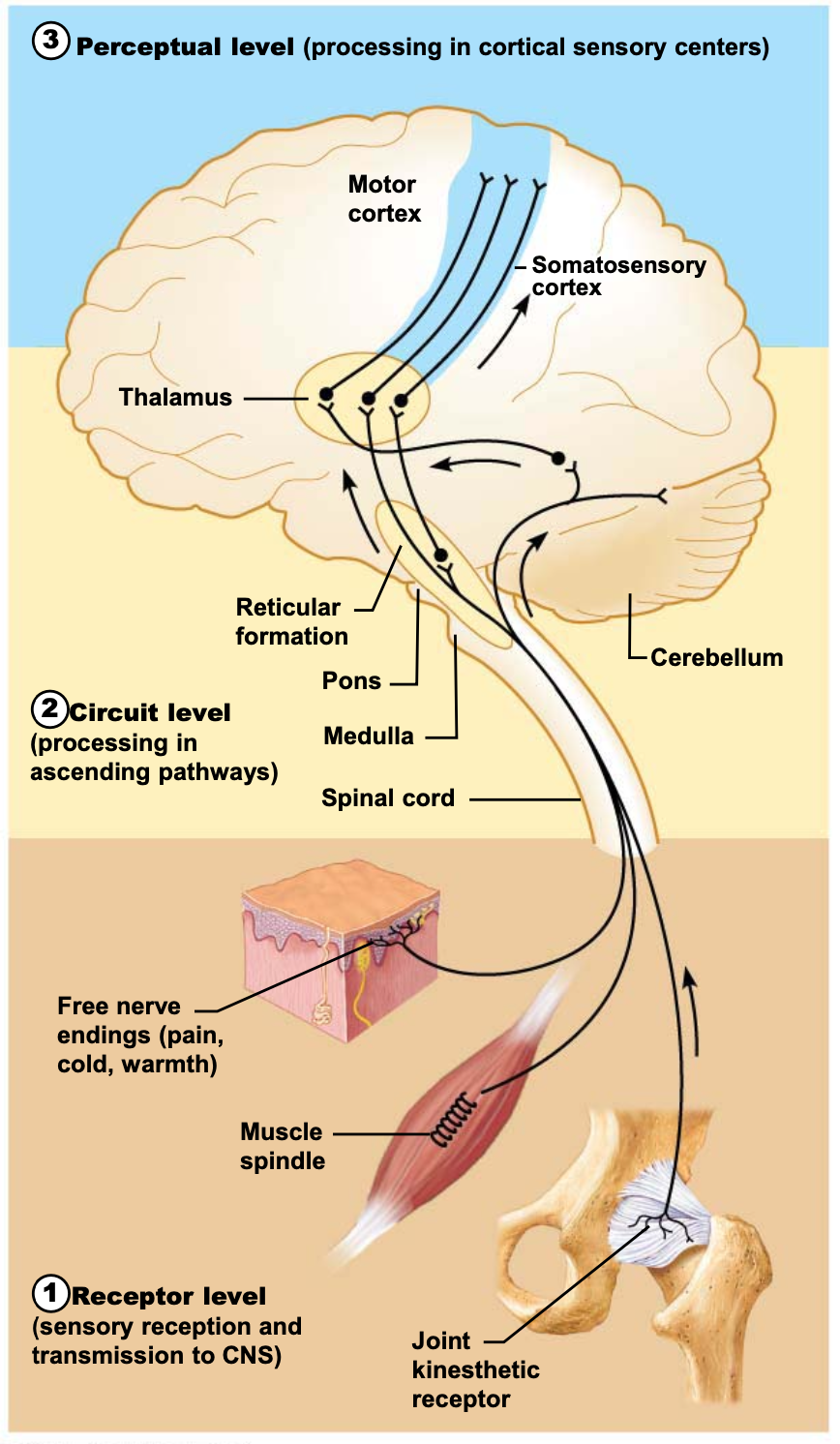
visceral pain and referred pain.
visceral pain:
results from stimulation of visceral organ receptors.
felt as vague aching, gnawing, burning.
attributed to the free endings look like around the viscera. DOES SHE KNOW WHAT PROVIDING FUCKING CONTEXT IS.
activated by tissues stretching, ischemia (reduced blood supply), chemicals, muscle spasms
referred pain:
pain from one body region perceived as coming from a different region to where it is being detected. (pain stemming from sharing of spinal nerves).
this is because visceral and somatic pain fibres travel along the same nerves, so brain assumes come from common (somatic) region.
e.g. feeling pain in left arm during heart attack.
state the difference between a neurone and a nerve
state the two nerve types.
SYSTEM: Nervous System
SUBSECTION: Transmission Lines: Neurones, Nerves, and their Structure and Repair
NOTE: None
CUE: None
Neurone: individual communicating cell
Nerve: cordlike organ of PNS; a bundle of myelinated and nonmyelinated peripheral axons enclosed by connective tissue.Cranial nerves; Spinal nerves
draw the general structure of a neurone; label it.
SYSTEM: Nervous System
SUBSECTION: Transmission Lines: Neurones, Nerves, and their Structure and Repair
NOTE: None
CUE: None
cell body has a nucleus, where proteins are made, DNA is found, and where neurotransmitters are produced and transmitted along the axon to the axon terminal where they are released when the neurone has an action potential.
dendrites (finger-like processes): connection point to other neurones, sensory neurones have much more developed dendrite pattern (lots more connection sites for incoming information). conversely motor neurons have more structural changes near the axon terminal.
myelin sheath: fatty layer of connective wrapped around a Schwann cell; insulates the action potential/electrical depolarisation from becoming weaker or bleeding out.
action potential: jumps fom node to node (conduction of action potential is much faster in myelinated neurones; speeds it up).
the small area where the cell body joins the axon is called the axon hillock; where the depolarisation reaches the threshold to produce an action potential, or it doesn’t and dissipates (site where action potential is initiated).
state three types of neurones, and their specialisations relative to their function.
SYSTEM: Nervous System
SUBSECTION: Transmission Lines: Neurones, Nerves, and their Structure and Repair
NOTE: None
CUE: None
motor neurone
small dendritic tree
generally myelinated axon
modified motor end plate (particularly for those innervating smooth muscle).
sensory neurone (most diverse neuron type)
receptor cell (this is adaptive depending on type; for detecting particular stimuli)
receptor cell detects signal; and transmits along axon to the cell body, and then to the axon terminals
cell body modified, sort of within the axon
generally myelinated axon
in diagram, it is a bipolar cell, so axon in both ways (one major, and one minor)
interneurone
multipolar; cell body has a highly arrange dendritic tree for bringing together incoming information
generally non-myelinated axon
axon terminal
describe how the action potential is produced, and what it does.
SYSTEM: Nervous System
SUBSECTION: Transmission Lines: Neurones, Nerves, and their Structure and Repair
NOTE: (not NMJ process, that’s on another card).
CUE: None
Electrical signal (depolarisation) passes along the neurone from the dendrites (the information in).
depolarisation (holy yap): opening of sodium channels to allow relative positive ions into cell, leading to positive charge on inside of membrane relative to the outside.
If depolarisation reaches a threshold value at the axon hillock, it produces an action potential, conducting it along the axon to the axon terminal.
this is an ‘all or nothing ‘ event, either threshold is reached, or signal dissipates.
At axon terminal, depolarisation leads to neurotransmitter release, where neurotransmitter chemicals carry the signal across the synaptic cleft to the postsynaptic cell (e.g. another neuron, smooth muscle cell, skeletal muscle cell, gland).
neurotransmitter released depends on the function and type of neurone.
Action can either excite or inhibit postsynaptic cell.
what are the connective tissue coverings of nerves
SYSTEM: Nervous System
SUBSECTION: Transmission Lines: Neurones, Nerves, and their Structure and Repair
NOTE: general structure of a nerve
CUE: None
connective tissue coverings include:
Endoneurium:
loose connective tissue that encloses axons and their myelin sheaths (Schwann cells)
Perineurium:
coarse connective tissue that bundles fibres (note: fibres are basically just a bunch of axons) into fascicles.
Epineurium:
tough fibrous sheath around all fascicles to form the nerve.
basically: nerves are a bunch of axons wrapped in three layers of connective tissue.
axon can either be sensory or motor; nerves can have bundles of sensory or motor, or a mix of both.
spinal nerves: are a mixture of both
cranial nerves: can be sensory, motor, or mixed
most nerves are mixtures of afferent and efferent fibres, and somatic and autonomic (visceral) fibres
pure sensory (afferent) or pure motor (efferent) nerves are rare; most nerves are mixed.
types of fibres in mixed nerves:
somatic afferent (sensory from muscle to brain)
somatic efferent (motor from brain to muscle)
visceral afferent (sensory from organs to brain)
visceral efferent (motor from brain to organs)
state how nerves are classified
SYSTEM: Nervous System
SUBSECTION: Transmission Lines: Neurones, Nerves, and their Structure and Repair
NOTE: None
CUE: None
most nerves are mixtures of afferent and efferent fibres, and somatic and autonomic (visceral) fibres
nerves are classified according to the direction they transmit impulses
mixed nerves: impulses travel to and from CNS
sensory (afferent) nerves: impulses only toward CNS
motor (efferent) nerves: impulses only away from CNS
question 2: what are the different types of fibres in mixed nerves?
nerves can have bundles of sensory or motor, or a mix of both.
spinal nerves: are a mixture of both
cranial nerves: can be sensory, motor, or mixed
pure sensory (afferent) or pure motor (efferent) nerves are rare; most nerves are mixed.
types of fibres in mixed nerves:
somatic afferent (sensory from muscle to brain)
somatic efferent (motor from brain to muscle)
visceral afferent (sensory from organs to brain)
visceral efferent (motor from brain to organs)
when do damaged nerve fibres regenerate, and how ?
include for:
PNS axons
CNS axons
SYSTEM: Nervous System
SUBSECTION: Transmission Lines: Neurones, Nerves, and their Structure and Repair
NOTE: None
CUE: None
If damaged, mature neurons do not regenerate (are amitotic), but if the soma (cell body) of the damaged nerve is intact, the peripheral axon may regeneration in PNS.
basically: central nervous system neurons do not recover (damage to CNS neurons/nerves, damage generally does not repair, and the neurons die). In PNS, some regeneration may occur if the cell body is intact. This is because the neurons are mature, and no longer undergo mitosis, so they are not able to regenerate, however, if cell body is intact, the axon of peripheral nerve might regenerate.
PNS axons
PNS axons can regenerate if damage is not severe, and cell body is intact.
axon fragments and myelin sheaths distal to the injury degenerate (called Wallerian degeneration); degeneration spreads down axon.
macrophages clean dead axon debris; Schwann cells are stimulated to divide.
(stimulated to divide to make more myelin)
axon filaments grow through regeneration tube (where Schwann cells stimulated to divide)
axon regenerations, and new myelin sheath forms.
CNS axons
most CNS fibres never regenerate.
CNS oligodendrocytes bear growth-inhibiting proteins that prevent CNS fibre regeneration
astrocytes (a type of glial cell) at injury site form scar tissue
treatments: neutralising growth inhibitors, blocking receptors for inhibitory proteins, destroying scar tissue components
i.e. both of the above have been targets for research
remember: fibre is basically just a term for axons
state the cranial nerves and their numbers ?
SYSTEM: Nervous System
SUBSECTION: Transmission Lines: Neurones, Nerves, and their Structure and Repair
NOTE: need to be able to name and number the nerves
CUE: None
int:
12 pairs of cranial nerves associated with brain
two attach to forebrain, rest with brain stem
first two pairs are purely sensory
each numbered (I-XII), and named from rostral to caudal (areass)
olfactory nerve (I)
optic nerve (II)
occulomotor nerve (III)
trochlear nerve (IV)
trigeminal nerve (V)
abducens nerve (VI)
facial nerve (VII)
vestibulocochlear nerve (VIII)
glassopharyngeal nerve (IX)
vagus nerve (X)
accessory nerve (XI)
hypoglossal nerve (XII)
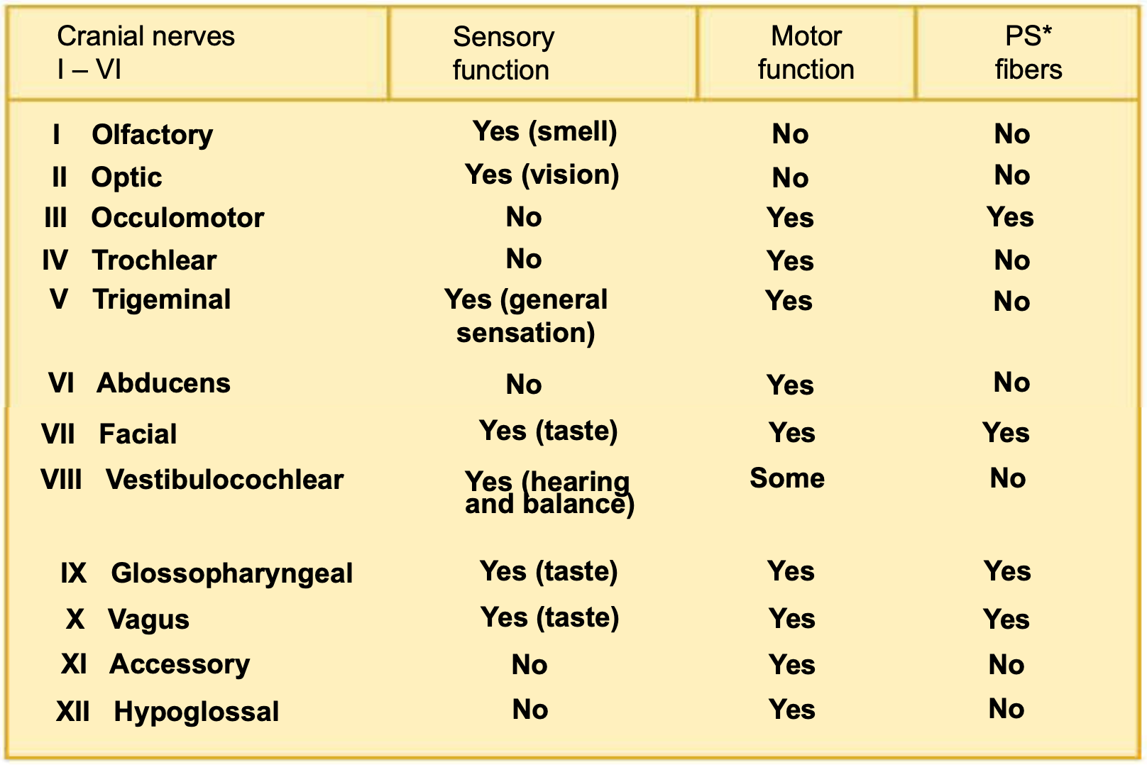
mnemonics to remember:
On occasion, our trusty truck acts funny—very good vehicle anyhow
or
Oh once one takes the anatomy final, very good vacations are heavenly
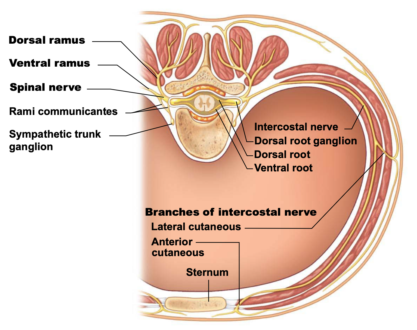
state the spinal nerve divisions, and the two roots that spinal nerves are connected to the spinal cord by.
describe characteristics of spinal nerves including:
where they emerge from
length (?)
branches
SYSTEM: Nervous System
SUBSECTION: Transmission Lines: Neurones, Nerves, and their Structure and Repair
NOTE: None
CUE: anterior and posterior roots and rami
there are 31 pairs of spinal nerves
all are mixed nerves named for point of issue from spinal cord (i.e where they exit spinal cord)
supply all body parts except for head and part of neck
divisions:
8 pairs of cervical nerves (C1-C8)
12 pairs of thoracic nerves (T1-T12)
5 pairs of lumber nerves (L1-L5)
5 pairs of sacral nerves (S1-S5)
1 pair of tiny coccygeal nerves (C0)
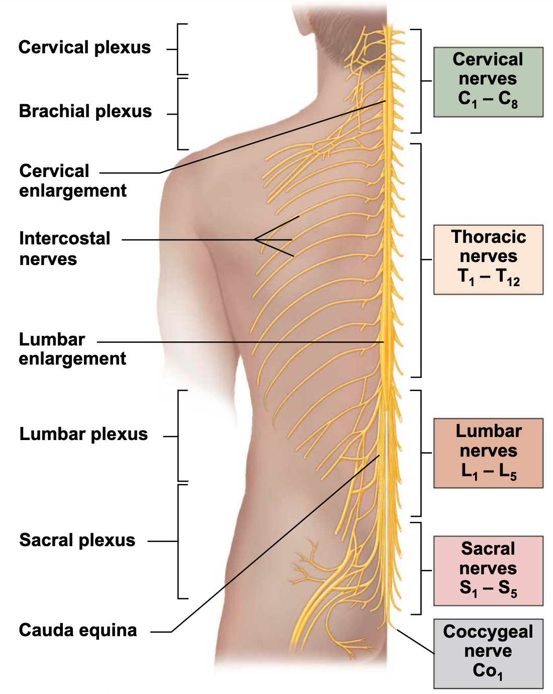
about diagram:
plexus is an area where several nerves come together: benefit of having an overlapping function.
each spinal nerve is connected to the spinal cord via two roots.
ventral roots (anterior root)
contain motor (efferent) fibres from motor neurons in the ventral horn
innervates skeletal muscles
dorsal roots (posterior root)
contain sensory (afferent) fibres from sensory neurons in dorsal root ganglia
conduct impulses from peripheral receptors
both ventral and dorsal roots are branched medially as rootlets which then join laterally to form the spinal nerve
spinal nerve roots divide into rootlets between entering or leaving the spinal cord.
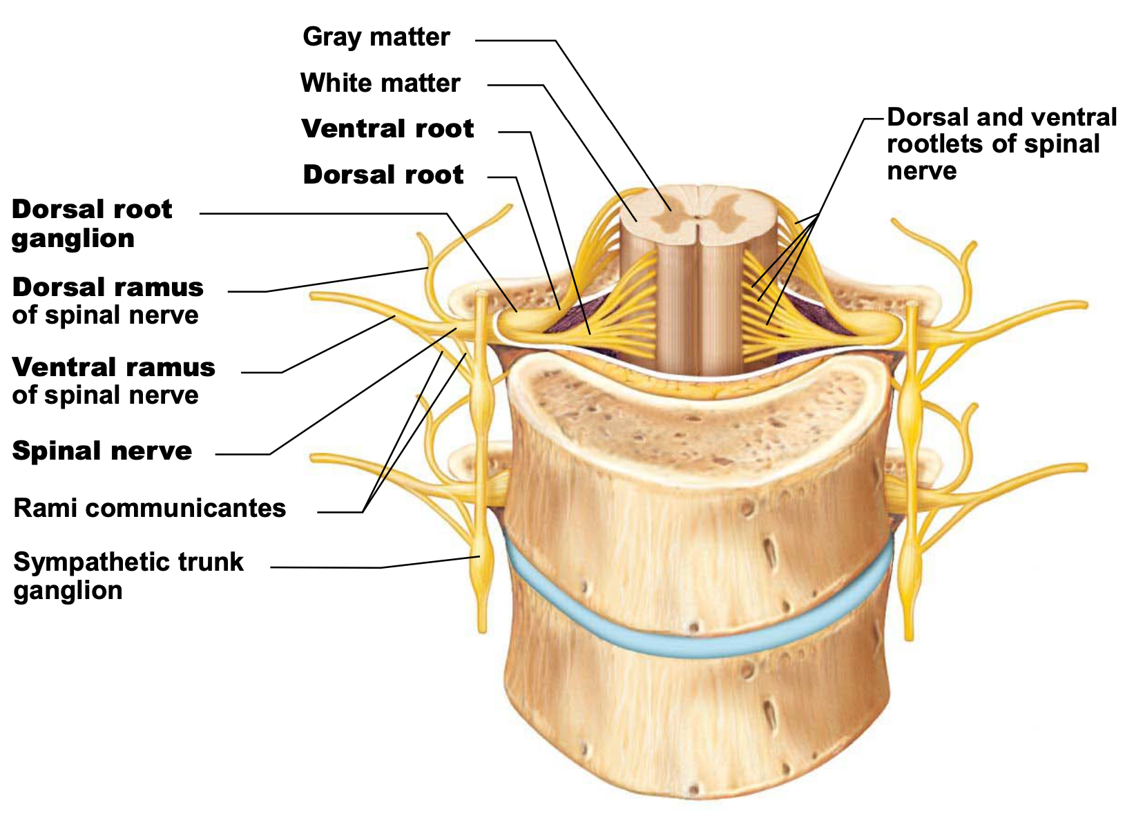
spinal nerves emerge from the vertebral column via respective intervertebral foramina (the holes between vertical discs).
spinal roots become progressively longer superiorly to inferiorly down the spinal cord (since have longer spaces to innervate)
lumber and sacral roots are very long, and extend through lower vertebral canal (and lower limbs) as the cauda equine (horses tail in Latin).
spinal nerves are relatively short (~1-2 cm)
almost immediately after exiting foramen, spinal nerves divide into three branches.
dorsal ramus (posterior ramus)
smaller branch
ventral ramus (anterior ramus)
larger branch
meningeal branch
tiny branch that reenters vertebral canal to innervate meninges and blood vessels
rami communicantes contain autonomic nerve fibres that join ventral rami in the thoracic region.
summary card ig?????
state what peripheral motor endings are
describe the innervation of
skeletal muscle
visceral muscle
glands
SYSTEM: Nervous System
SUBSECTION: Motor Endings and Motor Activity
NOTE: None
CUE: None
peripheral motor endings
motor endings are PNS elements that activate effectors by releasing neurotransmitters.
these elements innervate skeletal muscle, visceral muscle, and glands.
innervation of skeletal muscle
takes place as neuromuscular junction
where there is the motor unit (motor neuron and all of the muscle fibres that it innervates).
when action potential is transmitted along the motor neuron, depolarisation at the axon terminal stimulates the release of acetylcholine (ACh)
ACh binds to receptors in muscle resulting in:
movement of NA+ and K+ across membrane
leading to depolarisation of muscle cell
an end plate potential spreads to adjacent areas of the sarcolemma (membrane surrounding muscle cell), which triggers the opening of Na+ voltage-gated channels
results in the depolarisation of the sarcolemma (action potential), leading to muscle contraction.
innervation of visceral muscle and glands (smooth muscle)
autonomic motor endings and visceral effectors are simpler than neuromuscular (somatic) junctions
branches (are not as defined as the neuromuscular junction) form synapses en passant (“synapses in passing”) with effector (smooth muscle) cells via structures called varicosities (containing synaptic vesicles storing neurotransmitters).
varicosities appear as little pearls, containing neurotransmitters
acetylcholine and noradrenaline (aka. norepinephrine) act indirectly via second messengers
visceral motor responses are slower than somatic responses (due to this secondary messenger system and overall arrangement).
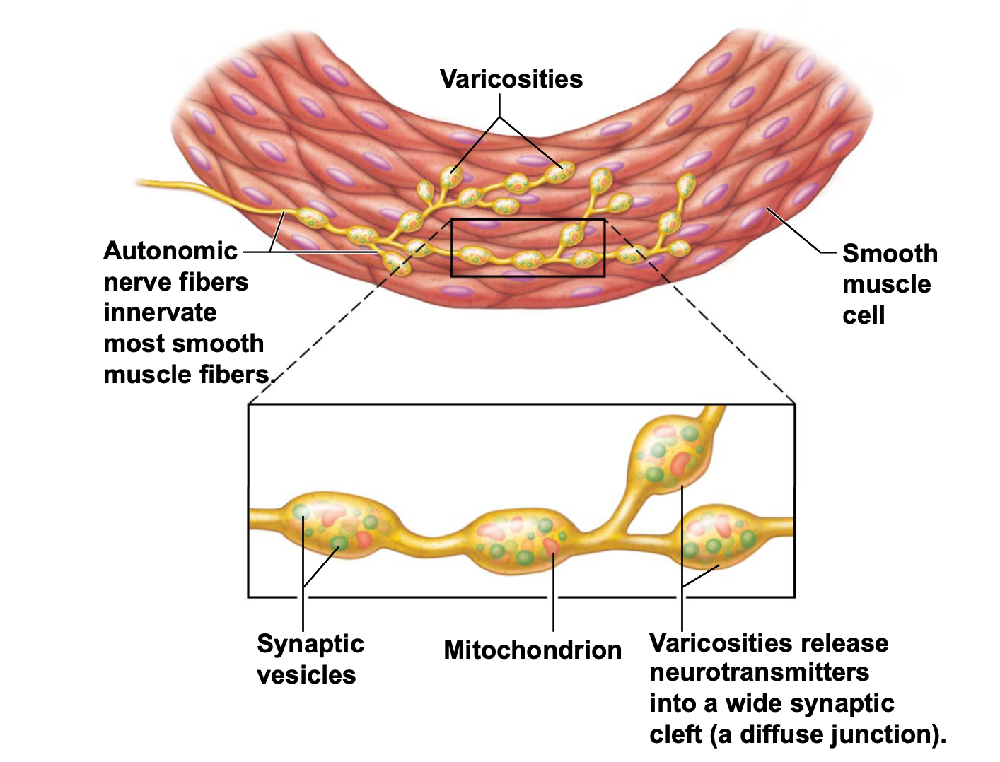
about diagram:
got smooth muscle, with autonomic nerve fibres coming in with varicosities that contain neurotransmitter-containing synaptic vesicles. when action potential comes along autonomic nerve, neurotransmitters are released into smooth muscle, leading to depolarisation and contraction of smooth muscle.
much more widespread and less discrete than NMJ.
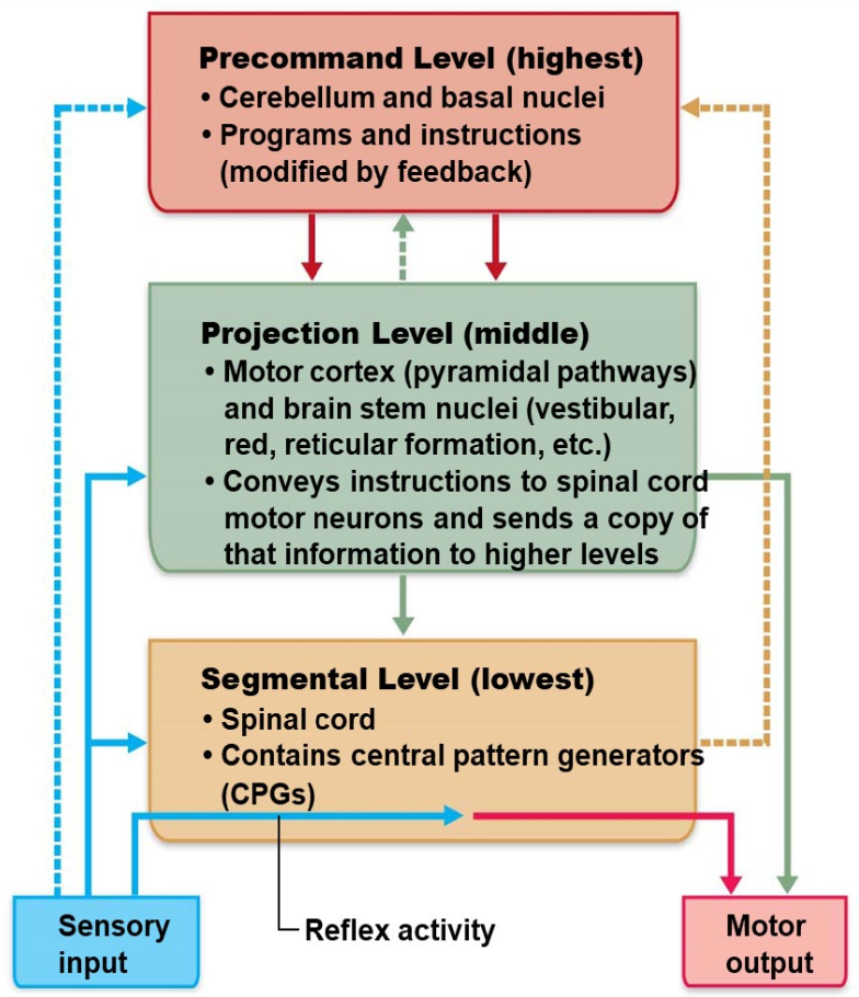
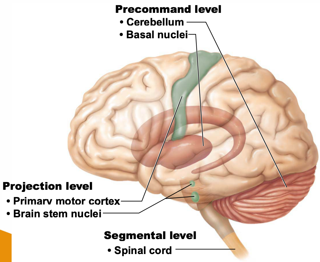
discuss the three levels of motor control
SYSTEM: Nervous System
SUBSECTION: Motor Endings and Motor Activity
NOTE: None
CUE: None
similarly to sensory information being processed at three different levels, see the same processing at motor control.
cerebellum and basal nuclei are the ultimate planners and coordinators of complex motor activities.
complex motor behaviour depends on complex patterns of control
e.g. patterns of control of sequences of muscles (sequential relaxation and contraction of muscle; relaxing antagonist while contracting agonist)
lots of patterns of control in order to do things voluntarily and for things to happen under ANS control.
segmental level
lowest level of the motor hierarchy
segmental circuits activate networks of the ventral horn neurons to stimulate specific groups of muscle.
includes:
reflexes and autonomic movement
central pattern generators (CPGs)
circuits that control locomotion, and specific (often repeated) motor activity
networks of oscillating inhibitory and excitatory neurons, allowing for crude rhythms and patterns of movement to occur.
projection level
consists of:
upper motor neurons that initiate direct pyramidal system to produce voluntary skeletal muscle movements.
brain stem motor areas that oversee and direct extrapyramidal system to control reflex and CPG controlled motor actions
projection motor pathways send information to lower motor neurons, keeping higher command levels informed of what is happening.
projection level communicates with precommand level and segmental level.
precommand level
neurons in the cerebellum and basal ganglia.
regulate motor activity patterns
precisely start and stop motor movements
coordinate movements with posture
block unwanted movements
override system
for example the reflex; e.g. picking up something hot, but instead of reflexively letting go of it, keep holding onto it despite knowing it will burn etc.
monitor muscle tone
perform unconscious planning and discharge of events in advanced of willed movements
WEEK 7: REFLEXES
LEARNING OBJECTIVES
Describe the steps in a neural reflex and classify the types of reflexes.
Distinguish among the types of motor responses produced by various reflexes and explain how reflexes interact to produce complex behaviors.
Explain how higher brain centers control and modify reflex responses.
SUMMARIES
define neural reflexes
SYSTEM: Nervous System
SUBSECTION: Reflexes
NOTE: None
CUE: None
neural reflexes are
rapid, automatic responses to specific stimuli
they are the basic building blocks of neural function
a particular reflexes will produce the same motor response each time
here is a recap on the gross anatomy of the adult spinal cord:
about diagram:
rami communicantes is a communicative nerve that allows two nerves to communicate with each other.
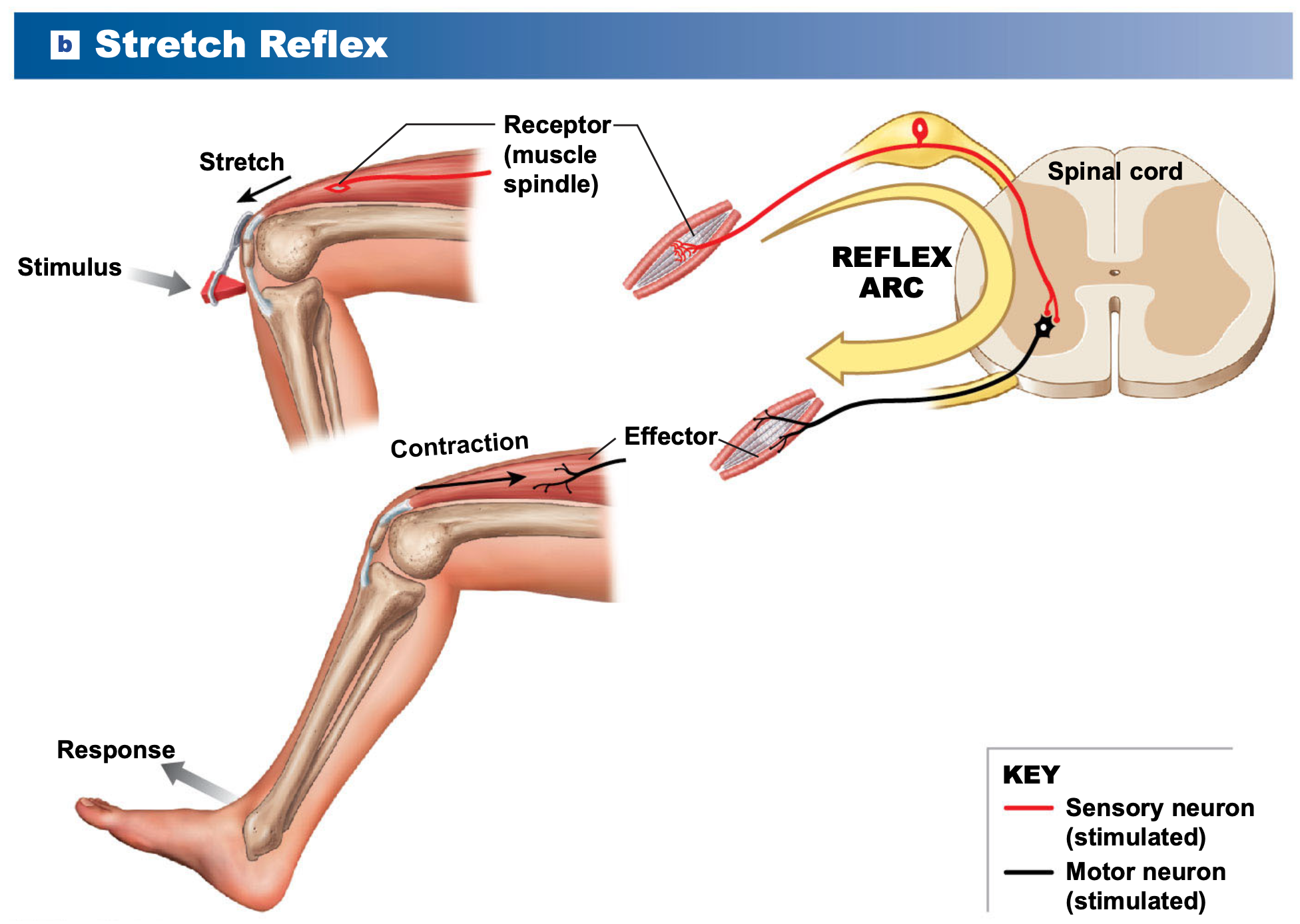
what are the five components of a reflex arc?
what is the reflex arc process?
SYSTEM: Nervous System
SUBSECTION: Reflexes
NOTE: None
CUE: None
five components of a reflex arc:
sensory receptor
sensory neuron
information processing in CNS
motor neuron
effector
spinal reflex arc:
stimulus activates a receptor.
with enough stimulation, action potential is generated in sensory neuron. Axon (from sensory neuron) enters spinal cord through the posterior (dorsal) root.
information processing in spinal cord usually occurs at one or more interneurons.
i.e. sensory information is processed in the spinal cord at one or more interneurons.
interneurons stimulate action potentials in motor neuron; its axon leaves via anterior (ventral) root
motor neuron stimulates effector (muscle/gland).
state the four classifications of reflexes
explain which they are
SYSTEM: Nervous System
SUBSECTION: Reflexes
NOTE: Classification of reflexes, not the different types of reflexes.
CUE: None
four classifications:
development
motor response
complexity of neural circuit (on the basis of complexity)
site of information processing
development of reflexes
innate reflexes
basic neural reflexes formed before birth
genetically programmed (inborn)
e.g. withdrawal, chewing, visual tracking
acquired reflexes
rapid, automatic learned motor patterns
repetition enhances them
e.g. braking car in emergency
motor response
somatic reflexes
control skeletal muscle (voluntary) contractions
superficial reflexes - stimuli in skin/mucous membranes
stretch/deep tendon reflexes
e.g. patellar/”knee-jerk” reflex
immediate—important in emergencies
slipping, cutting finger
visceral reflexes
control other effectors
smooth muscle, cardiac muscle, glands
complexity of neural circuit
monosynaptic reflex
single synapses—simplest reflex arc
sensory neuron synapses directly with motor neuron
fast response
polysynaptic reflex
at least one interneuron between sensory neuron and motor neuron; most common
slower response
delay increases with number of synapses involved (longer path=longer delay)
intersegmental reflex arcs—many spinal cord segments interaction, producing a variable response (different parts of the body can be involved)
sites of information processing
spinal reflexes
processing occurs in spinal cord
cranial reflexes
processing occurs in brain
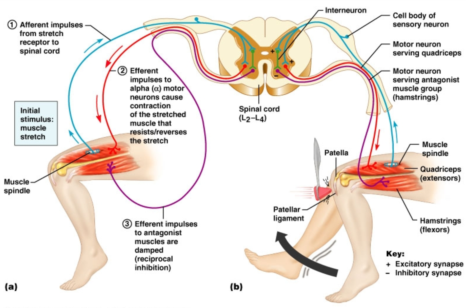
Describe the steps in a stretch reflex
SYSTEM: Nervous System
SUBSECTION: Somatic Reflexes
NOTE: None
CUE: None
stimulus = muscle stretching
distortion of receptor sends an action potential through sensory neuron
(stretching muscle distorts the sensory receptors).
sensory neuron synapses with motor neurons in the spinal cord
motor neurons send signals to motor units; which triggers reflexive contraction of stretched muscle.
the stretch reflex is monosynaptic.
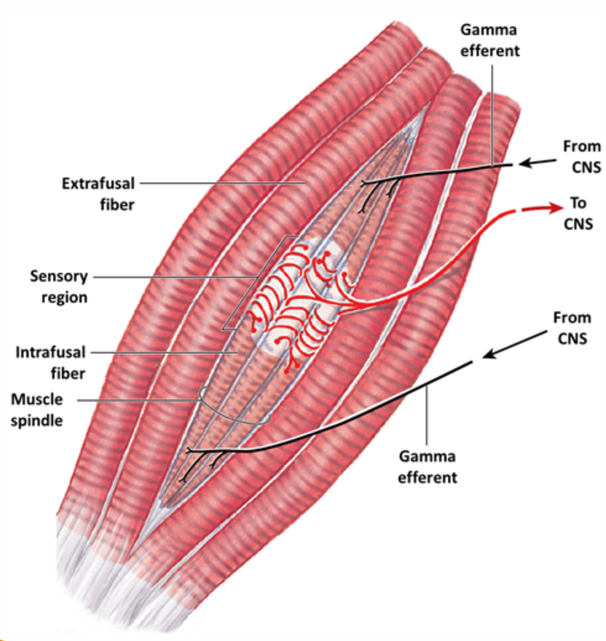
What are muscle spindles
SYSTEM: Nervous System
SUBSECTION: Somatic Reflexes
NOTE: None
CUE: None
Muscle spindles are the:
receptors in stretch reflexes
bundles of specialised intrafusal muscle fibres
dendrites of sensory neurons wind around central region of intrafusal fibres
sensory neuron: synapses in the spinal cord directly with (gamma) motor neurons
(which make gamma efferent fibres)
important for maintaining muscle tone of body and muscle contractions
gamma efferents complete reflex arc by synapsing back at the intrafusal fibres.
muscle contracts back to its resting length.
what are postural reflexes
SYSTEM: Nervous System
SUBSECTION: Somatic Reflexes
NOTE: None
CUE: None
postural reflexes:
include both stretch reflexes (monosynaptic) and also complex polysynaptic reflexes
postural reflexes maintain normal upright posture, and
often involve multiple muscle groups
e.g. back and abdominal muscles
they maintain firm muscle tone, and are
extremely sensitive receptors, allowing constant fine adjustments to be made as needed.
what are polysynaptic somatic reflexes ?
explain three examples
include an explanation for reciprocal inhibition
and the two types of reflex arcs
SYSTEM: Nervous System
SUBSECTION: Somatic Reflexes
NOTE: None
CUE: None
polysynaptic reflexes are
more complicated than monosynaptic reflexes
interneurons can control multiple muscle groups
facilitating flexibility of the response
they can stimulate some muscles (to contract), while others are inhibited
good when have groups of opposing muscles.
examples include
tendon reflex
withdrawal reflexes
crossed-extensor reflexes
tendon reflex
prevents skeletal muscles from:
developing too much tension
which puts them at risk for tearing or breaking (of tendons)
in the tendon, the sensory receptors are Golgi tendon organs.
these are stimulated (producing action potential) when collagen fibres (of the tendon) are overstretched, which
stimulates inhibitory interneurons in the spinal cord, where
increased muscle tension leads to increased muscle inhibition.
protective mechanism.
withdrawal reflexes
move body part away from stimulus (pain or pressure)
e.g. flexor flex in limbs; pulls hand away from hot pan.
strength and extent of response depends on intensity and location of stimulus.
might have several synapses involved if different muscle groups are involved.
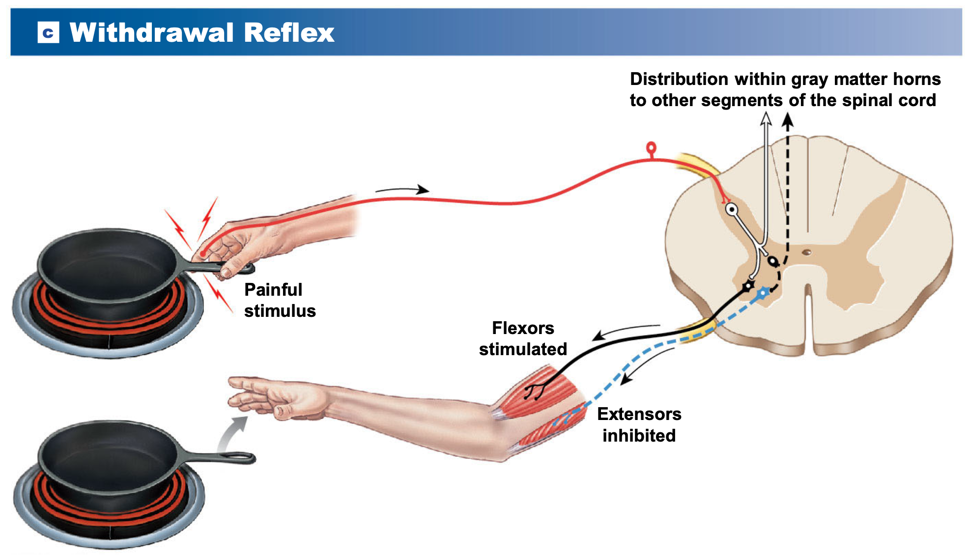
about diagram:
got heat registration coming in through dorsal horn
interneuron, motor neuron
flex activation of both flexors and extensors
flexors are stimulated to contract, moving hang up and away from hot pan, to facilitate this action, the extensors are inhibited. (this is called reciprocal inhibition)
in the case that the pan was very precious, and we do not want to drop the pan
there is also ascending information, allowing a cognitive override decision to be made if necessary during withdrawal reflex.
(can override pain of stimulus to move the fragile/object, and put it down somewhere and put it away)
reciprocal inhibition
for flexor reflex to work, stretch reflex of antagonist (extensor) muscles must be inhibited (reciprocal inhibition) by interneurons in spinal cord.
when flexors contract, extensors relax.
when extensors contract, flexors relax.
crossed extensor reflexes
coordinated with flexor reflex
e.g. step on something sharp: before flexor reflex can lift food, crossed extensor reflex straightens opposite limb to receive body weight, then flexor reflex can occur.
maintained by reverberating circuits.
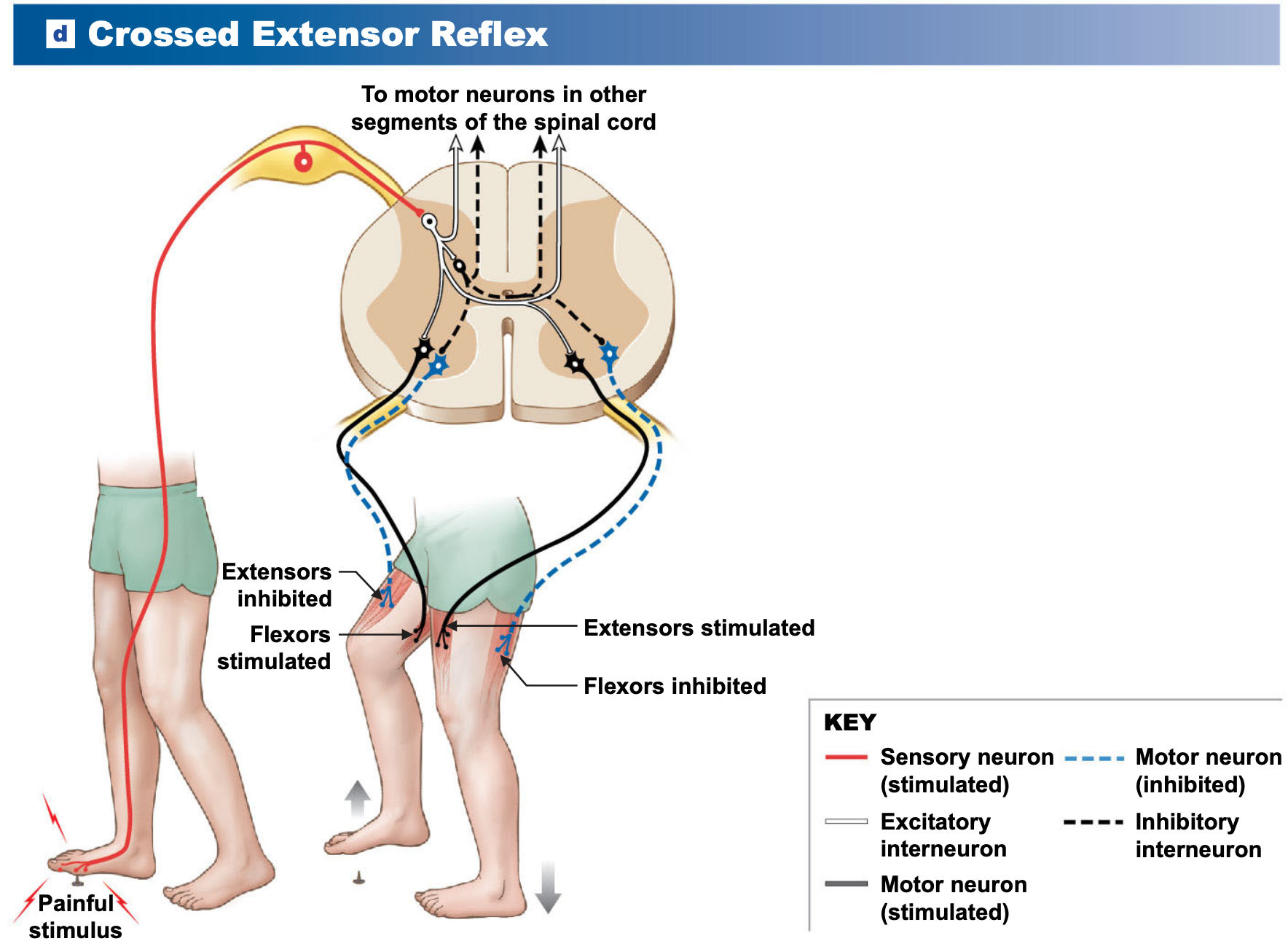
reflex arcs
ipsilateral reflex arcs
occur on the same side of body as stimulus
e.g. stretch, tendon, and withdrawal reflexes
contralateral reflex arcs
occur on the opposite side of the body as stimuluss
e.g. crossed extensor reflexes
e.g. walking and step on sharp item; need to lift foot; but to do that, need to transfer weight to opposite site (other foot); crossing over in spinal chord allows this action without falling over.
reminder of five general characteristics of polysynaptic reflexes
involve pools of interneurons
may cause excitation or inhibition
allows flexibility of response; having a pool increases size (could be large response or small response); pool means response can be modulated to be excitatory or inhibitory depending on the need.
involve more than one spinal segment
can activate muscles in multiple areas of the body
involve reciprocal inhibition
coordinates contractions and reduces resistance
for flexor reflex to work, stretch reflex of antagonist (extensor) muscles must be inhibited (reciprocal inhibition) by interneurons in spinal cord.
when flexors contract, extensors relax.
when extensors contract, flexors relax.
have reverberating circuits
prolongs reflexive motor response
allows reflexive response to be prolonged
several reflexes may cooperate
to produce coordinated, controlled response.
in case of stepping on sharp object, and needing to transfer body weight to the opposite side of body; taking in lots of balance information may also be required when transferring weight onto the contralateral side.
what are the five general characteristics of polysynaptic reflexes?
SYSTEM: Nervous System
SUBSECTION: Reflexes
NOTE: None
CUE: None
involve pools of interneurons
may cause excitation or inhibition
allows flexibility of response; having a pool increases size (could be large response or small response); pool means response can be modulated to be excitatory or inhibitory depending on the need.
involve more than one spinal segment
can activate muscles in multiple areas of the body
involve reciprocal inhibition
coordinates contractions and reduces resistance
for flexor reflex to work, stretch reflex of antagonist (extensor) muscles must be inhibited (reciprocal inhibition) by interneurons in spinal cord.
when flexors contract, extensors relax.
when extensors contract, flexors relax.
have reverberating circuits
prolongs reflexive motor response
allows reflexive response to be prolonged
several reflexes may cooperate
to produce coordinated, controlled response.
in case of stepping on sharp object, and needing to transfer body weight to the opposite side of body; taking in lots of balance information may also be required when transferring weight onto the contralateral side.
integration and control of spinal reflexes
reflex behaviours are autonomic, but processing centres in the brain can facilitate or inhibit spinal reflex motor patterns.
ascending contralateral interneurons that go up to brain, allow brain to be notified of reflexes, and also allow brain to override reflexes where necessary.
explain how the brain can alter spinal reflexes
SYSTEM: Nervous System
SUBSECTION: Somatic Reflexes
NOTE: None
CUE: None
voluntary movements and reflex motor patterns
spinal reflexes produce a characteristic response for a given stimulus.
the brain cal also activate these same motor patterns through descending pathways.
can facilitate, inhibit. or “fine-tune” the established motor response.
e.g. walking, running, jumping
requires coordination of where body is put in space, and where deciding to put body during these activities; requires coordination of relaxation and inhibition of leg muscles in order to move.
reinforcement of spinal reflexes
higher centres can adjust the sensitivity of reflexes by stimulating excitatory or inhibitory interneurons in the brainstem or spinal cord.
when excitatory synapses are chronically stimulated, postsynaptic neurons can be in general facilitation.
which reinforces spinal reflexes.
This reinforcement enhances spinal reflexes.
inhibition of spinal reflexes
higher centres inhibit spinal reflexes by
stimulating inhibitory neurons, which
creates IPSPs at reflex motor neurons, thereby
IPSPs = inhibitory post synaptic potentials
opposite of excitatory postsynaptic potentials
suppressing postsynaptic neurons, thus inhibiting the reflex.
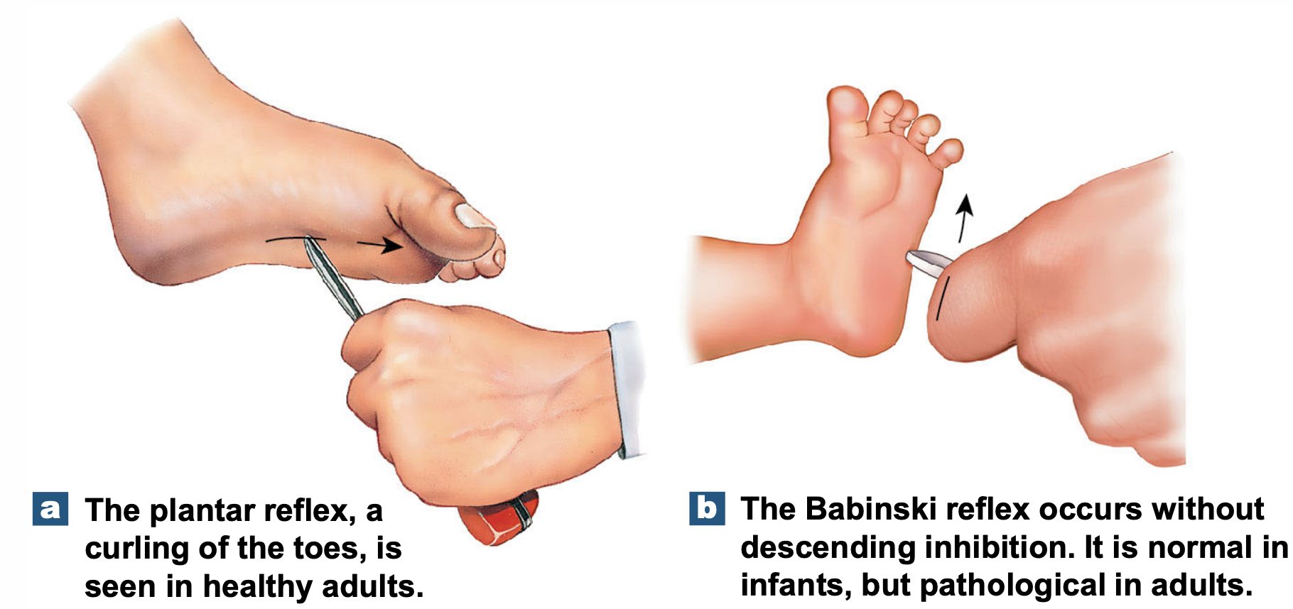
e.g. plantar reflex
normal in adults
where if stroke the lateral sole of foot, it causes reflexive toe curling
e.g. babinski reflex
normal in infants (lost when infant starts walking; not present in adults) (lost as infant grows since develop descending inhibition that dampens down the response).
if seen in adults, may indicate CNS damage
if descending inhibition is removed (e.g. due to neurodegeneration of descending motor pathways), babinski reflex may be observed again.
where if stroke lateral sole of foot, the foot broadens out (big toe moves upwards/towards top surface of foot).
talk about visceral reflexes…?
provide examples of sympathetic and parasympathetic visceral reflexes.
SYSTEM: Nervous System
SUBSECTION: Visceral Reflexes
NOTE: None
CUE: None
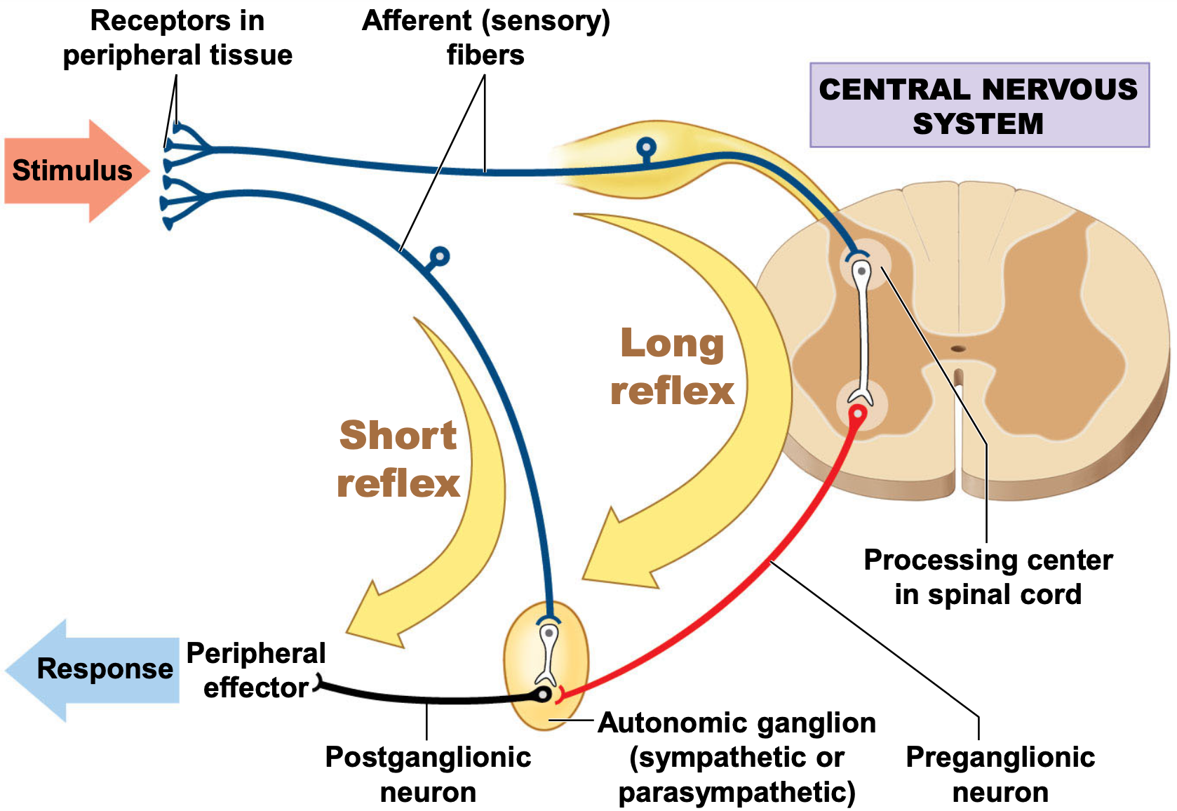
about diagram
visceral reflexes rely on spinal cord
have stimulus, which could be stretch receptors in smooth muscle
detected in through dorsal root to dorsal horn
involves interneuron and motor response
can see a short and long reflex
long reflex goes through spinal cord
short reflex goes straight from the receptor being stimulated, directly to autonomic ganglia (could be sympathetic or parasympathetic; can initiate the peripheral response; and be even quicker).
sympathetic visceral reflexes (fight or flight)
parasympathetic reflexes (rest and digest)
define what is meant by ‘special senses’
state the ‘special senses’
SYSTEM: Nervous System
SUBSECTION: Special Senses
NOTE: None
CUE: None
special senses are where sensory cells have specialised structures adapted for a unique function (usually at the dendrites)
e.g. cells of the retina have photoreceptors which capture photons of light, that only respond to light photons (not touch, taste, etc).
and have a dedicated neuronal pathway
e.g. cranial nerves that are responsible for carrying just that set of information.
special senses include:
olfaction (smell)
gustation (taste)
vision
equilibrium (balance)
hearing
state the sense that humans rely on more than any other special sense in the body
SYSTEM: Nervous System
SUBSECTION: Special Senses
NOTE: None
CUE: None
vision.
about vision and the structures of the eye (introduction)
it is unique, such as it provides visual information about the world.
the accessory structures of the eye provide:
protection
lubrication
support
and include:
eyelids
superficial epithelium of eye
lacrimal apparatus.
label the gross and superficial anatomy of the accessory structures of the eye
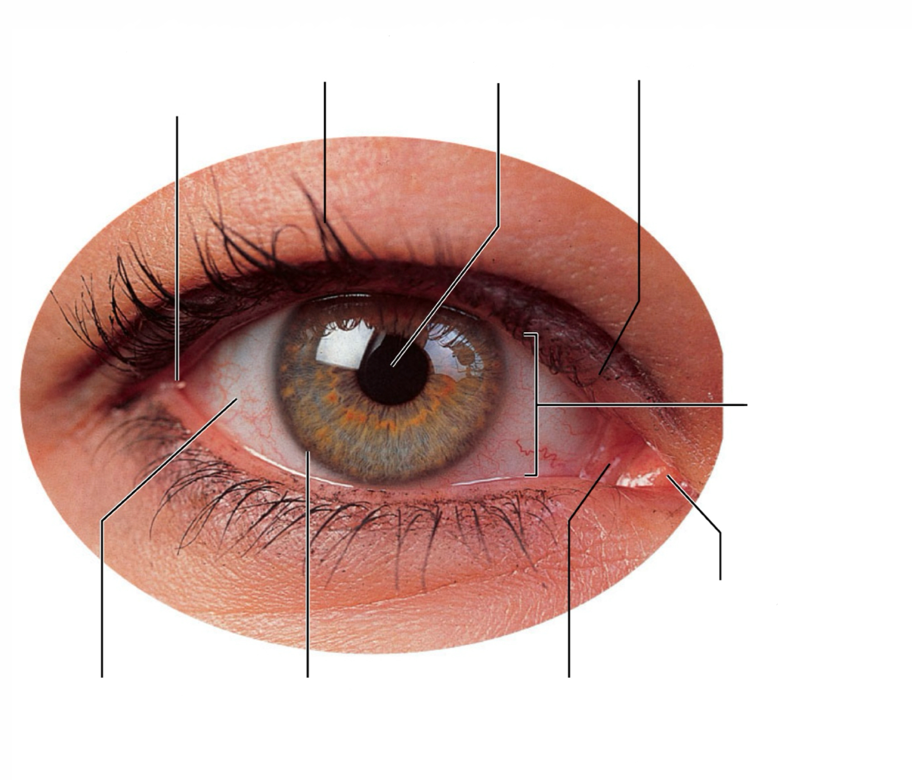
label the organisation of the lacrimal apparatus
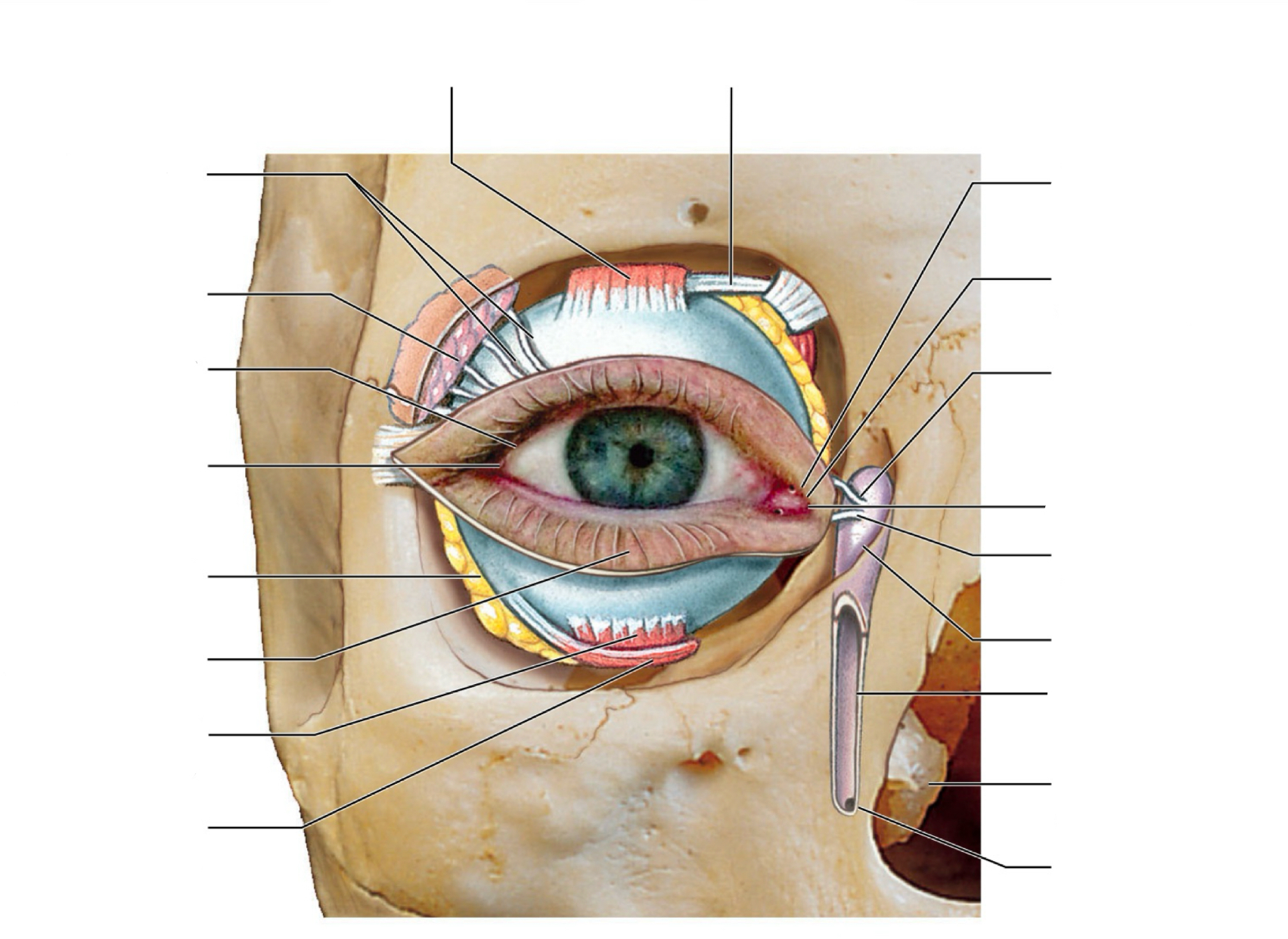
label sectional anatomy of eye
SYSTEM: Nervous System
SUBSECTION: Special Senses - Vision
NOTE: (idk if we actually need to label these, but good since its optometry related) (better labelling one below)
CUE: None
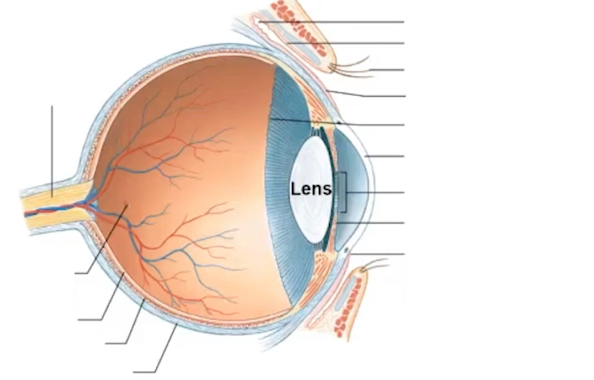
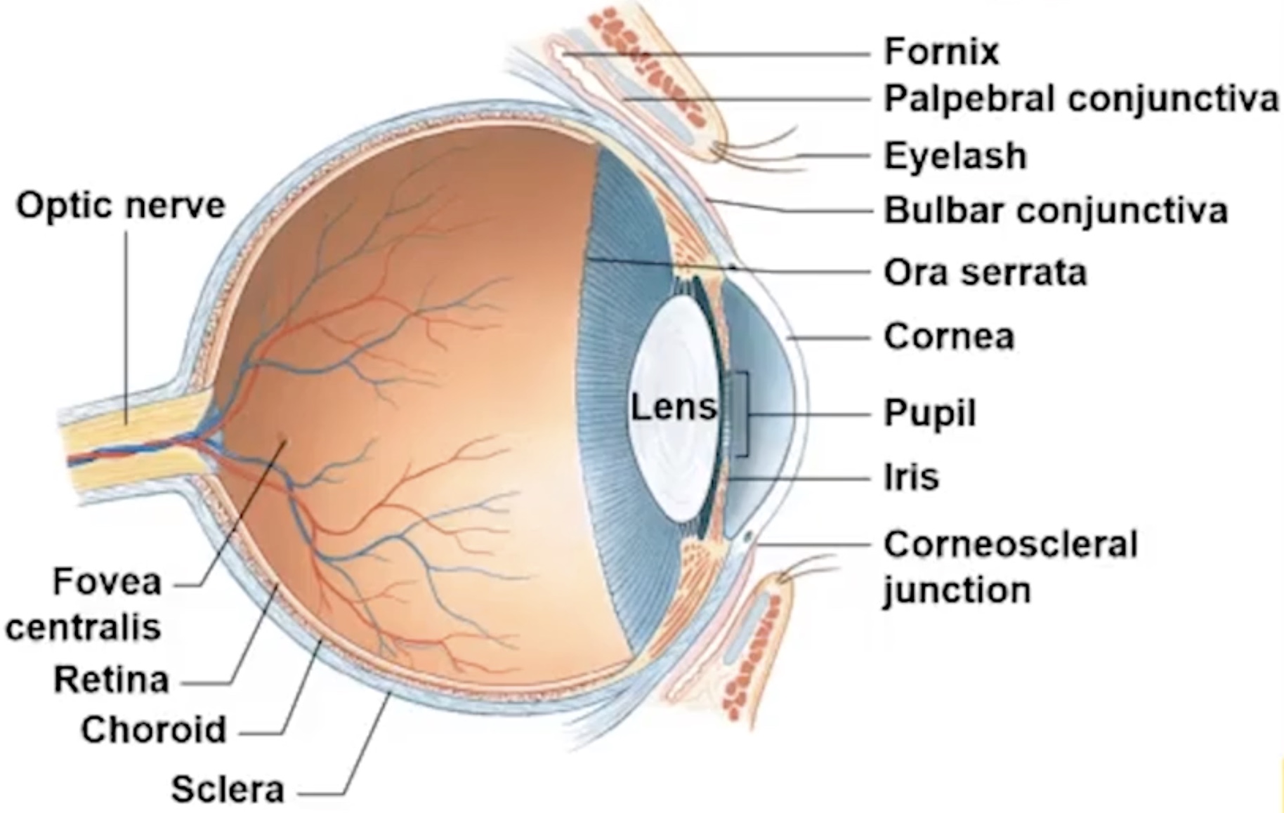
lateral angle
medial angle (nose is next to this)
lacrimal caruncle
associated with producing tears
eyelashes
prevent debris from entering eyes
sclera
outer coating of eye
corneoscleral junction
where sclera meets the cornea
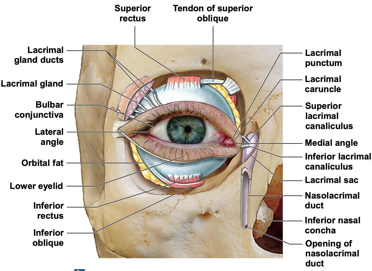
extraocular muscles that insert into the sclera
superior and inferior rectus; inferior and superior oblique.
when contract, they pull eye to certain directions
eye also has fat pads that help protect the eye (i think lecturer is referring to the thing in blue).
lacrimal gland; lacrimal duct produces tears and secretions that wash across the eye
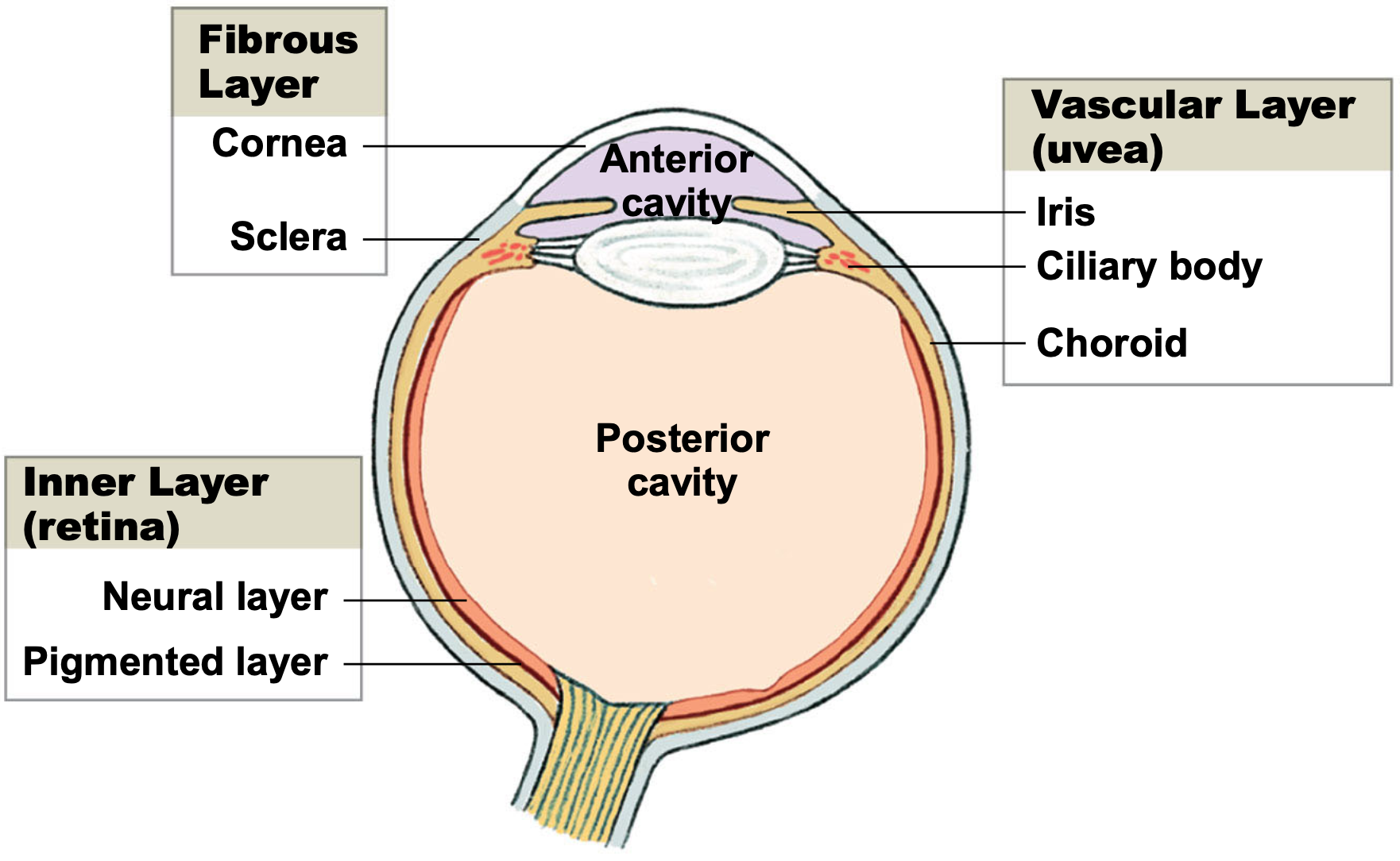
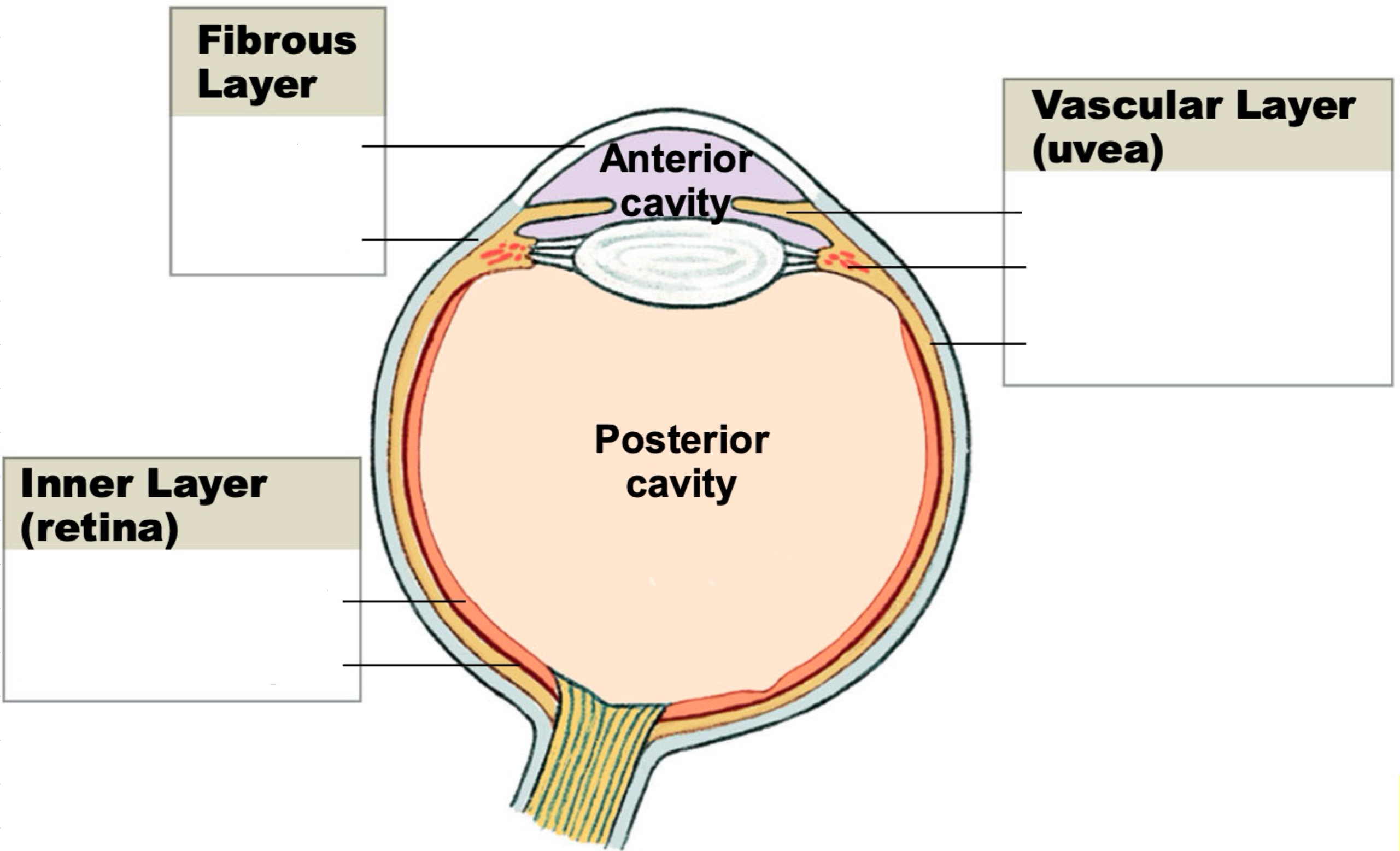
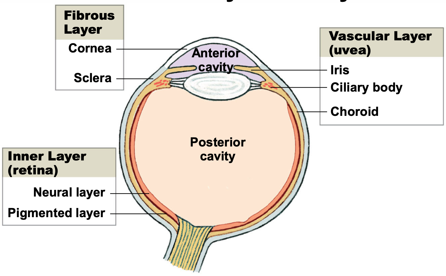
eye has three main layers
layers of the wall of the eyeball
help to maintain shape of eye
eye is basically hollow, filled with fluid
has two chambers (anterior and posterior cavities)
anterior cavity contains aqueous humour; transparent, more fluid-like than jelly
light comes in through anterior cavity, and hits the lens
lens can change shape; has suspensory ligaments around the outside that accommodates and changes the shape of the lens so that can focus on near/far objects
posterior cavity contains vitreous humour; thick and dark, more jelly-like than fluid
optic nerve
emerging from inner layer
fibres, outgoing fibres carrying visual information from the eye to the brain
label sectional anatomy of eye
SYSTEM: Nervous System
SUBSECTION: Special Senses - Vision
NOTE: None
CUE: None
external to the lens is the pupil
pupil has a circular ring of smooth muscle, innervated by the autonomic nervous system
if pupil is too dilated, resulting in too much incoming light; can bleach the pigments, and damage the photoreceptors of the retina.
hence, in presence of bright light, pupils constrict (an autonomic reflex)
presence of reflex reflects health optical system/healthy optic nerve
ora serrata
serrated junction
marks division between non-neural and neural
neural retina and non-neural tissue
bulba conjunctiva
thin membrane that covers the eye
palpebral conjunctiva
part of conjunctiva that lines the eyelids
fornix
loose soft tissue
allows movement of eyelids (so that they can close)
direction of light
comes through cornea, through pupil, through lens, through vitreous humour, to activate photoreceptors in the retina
other stuff about the eye:
lateral angle
medial angle (nose is next to this)
lacrimal caruncle
associated with producing tears
eyelashes
prevent debris from entering eyes
sclera
outer coating of eye
corneoscleral junction
where sclera meets the cornea
extraocular muscles that insert into the sclera
superior and inferior rectus; inferior and superior oblique.
when contract, they pull eye to certain directions
eye also has fat pads that help protect the eye (i think lecturer is referring to the thing in blue).
lacrimal gland; lacrimal duct produces tears and secretions that wash across the eye
eye has three main layers
layers of the wall of the eyeball
help to maintain shape of eye
eye is basically hollow, filled with fluid
has two chambers (anterior and posterior cavities)
anterior cavity contains aqueous humour; transparent, more fluid-like than jelly
light comes in through anterior cavity, and hits the lens
lens can change shape; has suspensory ligaments around the outside that accommodates and changes the shape of the lens so that can focus on near/far objects
posterior cavity contains vitreous humour; thick and dark, more jelly-like than fluid
optic nerve
emerging from inner layer
fibres, outgoing fibres carrying visual information from the eye to the brain
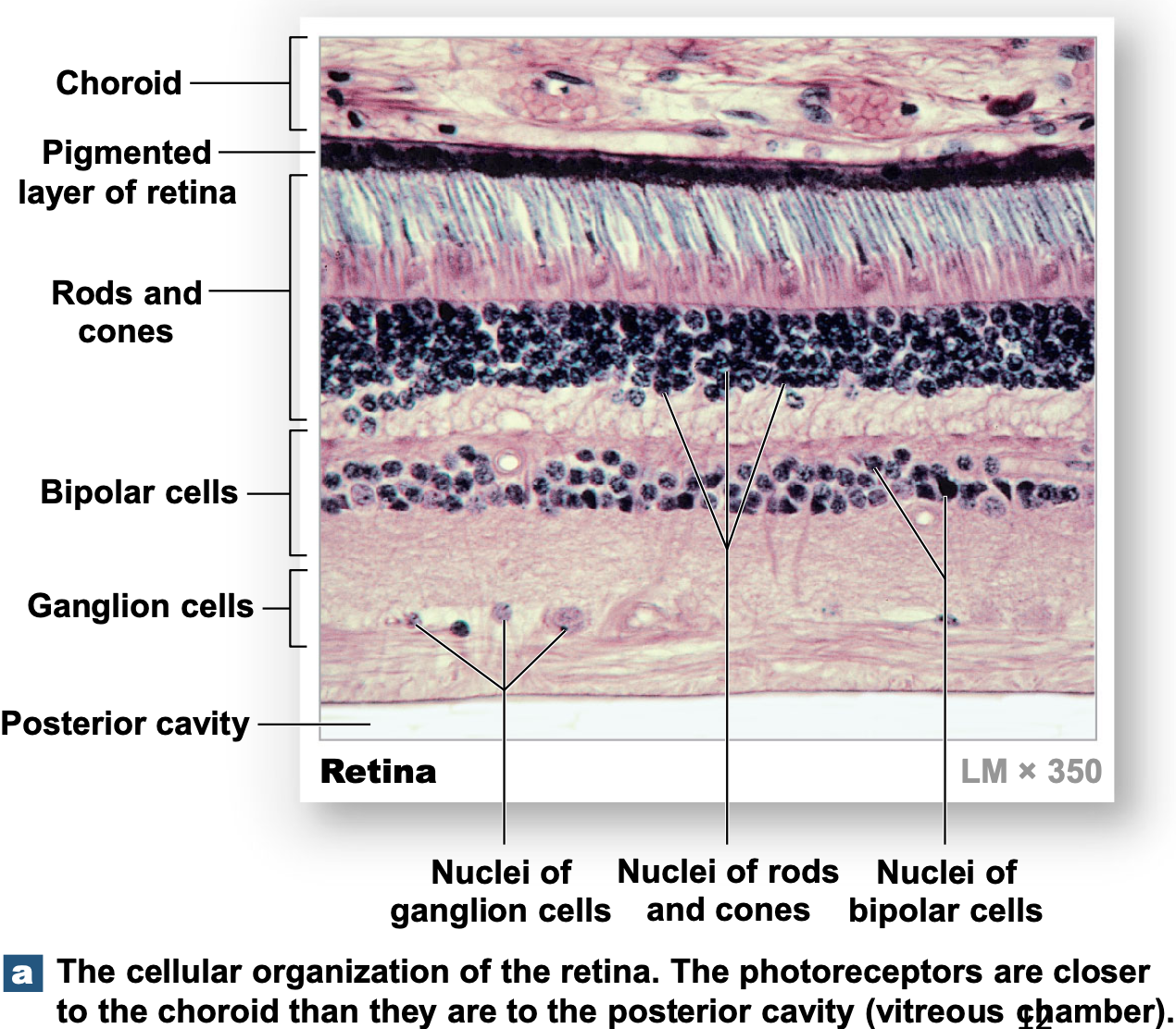
discuss the organisation of the retina and optic nerve
look…. you can try….. but maybe just…. turn the card….
SYSTEM: Nervous System
SUBSECTION: Special Senses - Vision
NOTE: None
CUE: if it helps,
it helps…
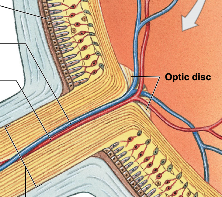
photoreceptive region
have blood vessels, optic nerve also has blood vessels
optic nerve is also covered, and protected by the sclera
where the optic nerve is formed by the axons of retinal ganglion cells.
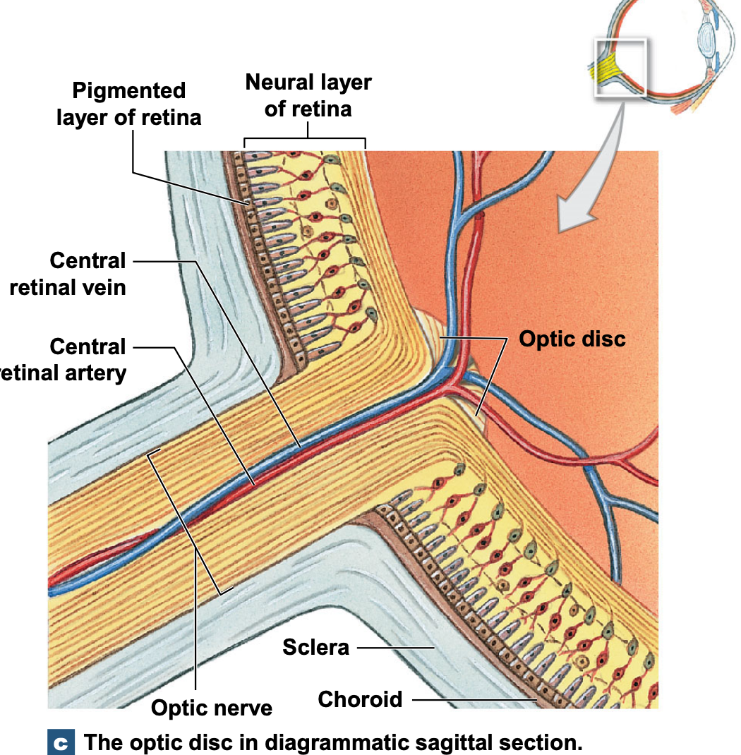

light comes through cornea, through pupil, through lens, through vitreous humour, to hit the neural retina
so light passes through neural layer to hit the layer of photoreceptors
in the diagram are called: rods and cones (since they look like rods and cones).
rods = most present, responsive to low levels of light (not colour)
cones = responsive to particular wavelengths of light
rods and cones are the photoreceptors of the eye
they are connected to bipolar cells of the eye
notice the integration, connections, and networking of the retina
the integration, connections, and networkings of the retina allows the visual field to be mapped.
can integrate the stimulation of individual photoreceptors via synapses, allowing a picture to be produced when interpreting visual information.
retinal ganglion cells are the output cells of the retina
lots of input converges onto them
it is the axons of the retinal ganglion cells that form the optic nerve
amacrine cells either facilitate or inhibit communication between the rods, cones, and ganglion cells
thought to (in this way) alter the sensitivity of the retina.
describe the structure of rods, cones, and the rhodopsin molecule
SYSTEM: Nervous System
SUBSECTION: Special Senses - Vision
NOTE: None
CUE: None
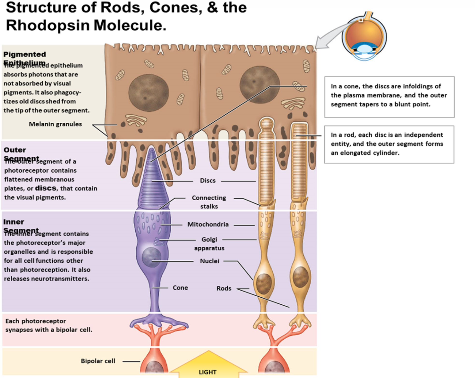
rods and cones are the photoreceptors of the eye
rods = most present, responsive to low levels of light (not colour)
highly sensitive to light
detect the presence/absence of photons
in a rod, each disc is an independent entity, and the outer segment forms the elongated cylinder
in stack of discs, is where photosensitive protein, called opsin, is located.
cones = responsive to particular wavelengths of light (visual pigment is activated by particular frequency) (usually talk about red cones, blue cones, and green cones; meaning they activate particular frequency corresponding to red, blue, or green light).
discs are infoldings of the plasma membrane, forming a tapered segment up to a point
outer segment of photoreceptors: where flattened membranes called discs are; which contain opsin visual pigments
inner segment of photoreceptors: machinery of cells, including mitochondria, Golgi body for protein packaging, nuclei for instructions
photoreceptors synapse with bipolar cells
photoreceptors are most densely populated in the fovea centralis; located at the centre of the macula; site that provides the sharpest colour vision
the ‘visual access’ is a term referring to the line from an object to the fovea. reason we move our eyes, is to attain sharpest image on the fovea, thereby stimulating more photoreceptors, providing sharpest picture
both rods and cones dovetail into the pigmented epithelium
pigmented epithelium is a black, shiny, layer of retina that absorbs photons that are not absorbed by the visual pigments; also phagocytoses old discs that shed from the photoreceptors.
how does colour vision work ? (also mention colour blindness).
SYSTEM: Nervous System
SUBSECTION: Special Senses - Vision
NOTE: None
CUE: None
holy yap: the absorption of photons is the first step of photoreception. this is a transduction mechanism. moving from photons of light, to action potentials. the visual pigments are derivatives of rhodopsin. where opsin is the protein, and retinal is a derivative pigment synthesised from vitamin A. colour vision is produced via activation of red, blue, and green cones. where each cone type has a different kind of opsin that is activated particular wavelengths of light.
colour vision
cones contain opsin (visual pigments) allowing them to be responsive to particular wavelengths of light, activated by particular frequencies.
colour vision is provided by blue cones, green cones, and red cones.
each type has a different form of opsin.
colour blindness
if one or more cones are missing, may culminate in the inability to detect that colour, and/or distinguish it from other colours.
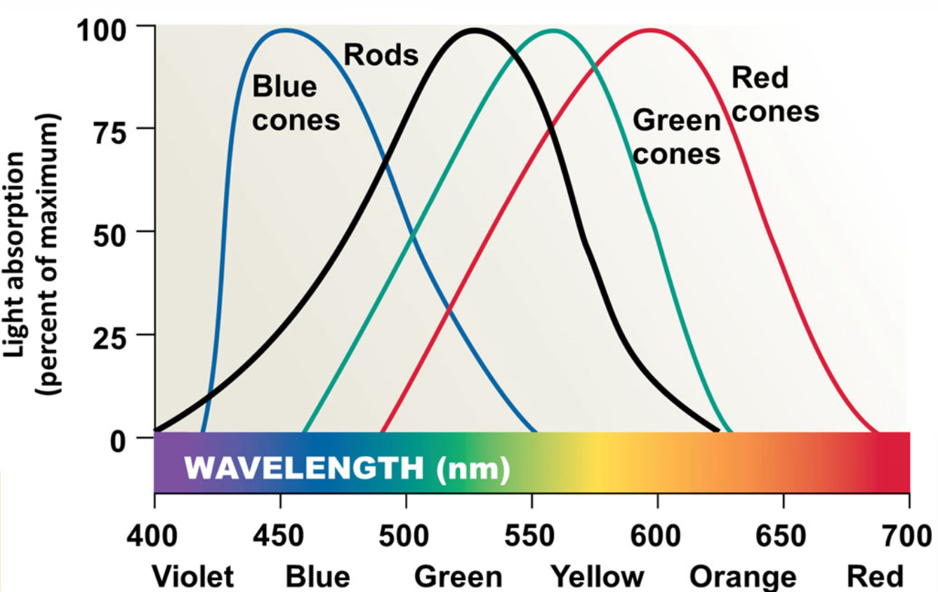
rods are not activated by different colours, but are very sensitive to light; detect the presence of light.
cones respond to particular frequencies:
blue cones: ~450nm
green cones: ~550nm
red cones: ~600nm
describe the steps of photoreception, and discuss what bleaching is.
SYSTEM: Nervous System
SUBSECTION: Special Senses - Vision
NOTE: None
CUE: None
1) Absorption of photo changes retinal from 11-cis to 11-trans form.
which activates opsin.
basically: this allows the photoreceptor to be activated by photon, which changes the shape of retinal molecule, where the change in shape activates the opsin.
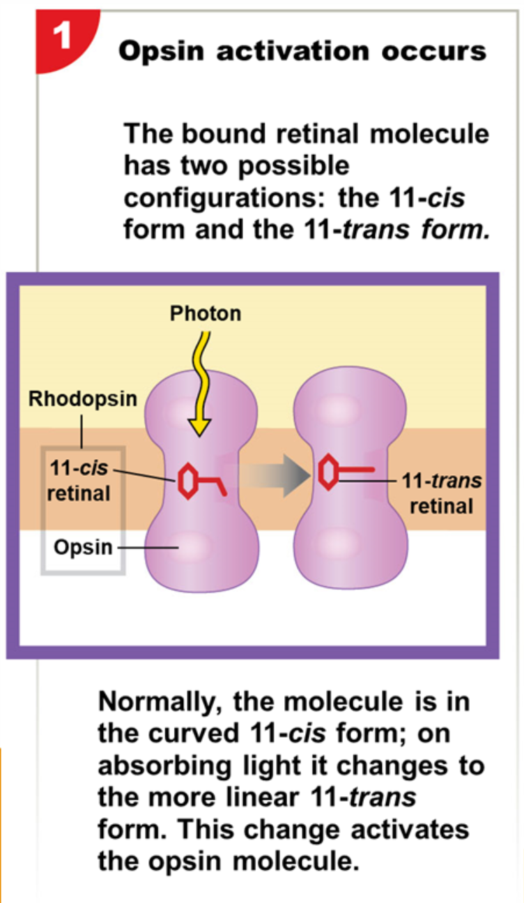
2) opsin activates transducin (a G protein)
which activates phosphodiesterase (PDE)
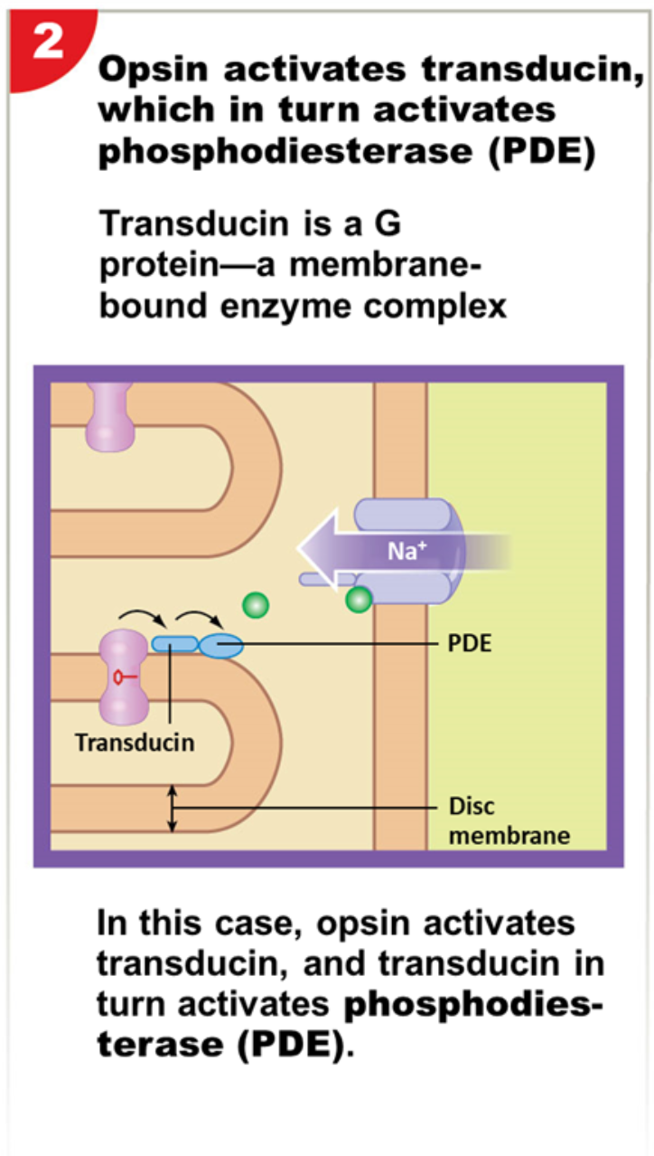
3) PDE reduces levels of cyclic GMP (by breaking down cGMP)
causing chemically gated sodium ion channels close
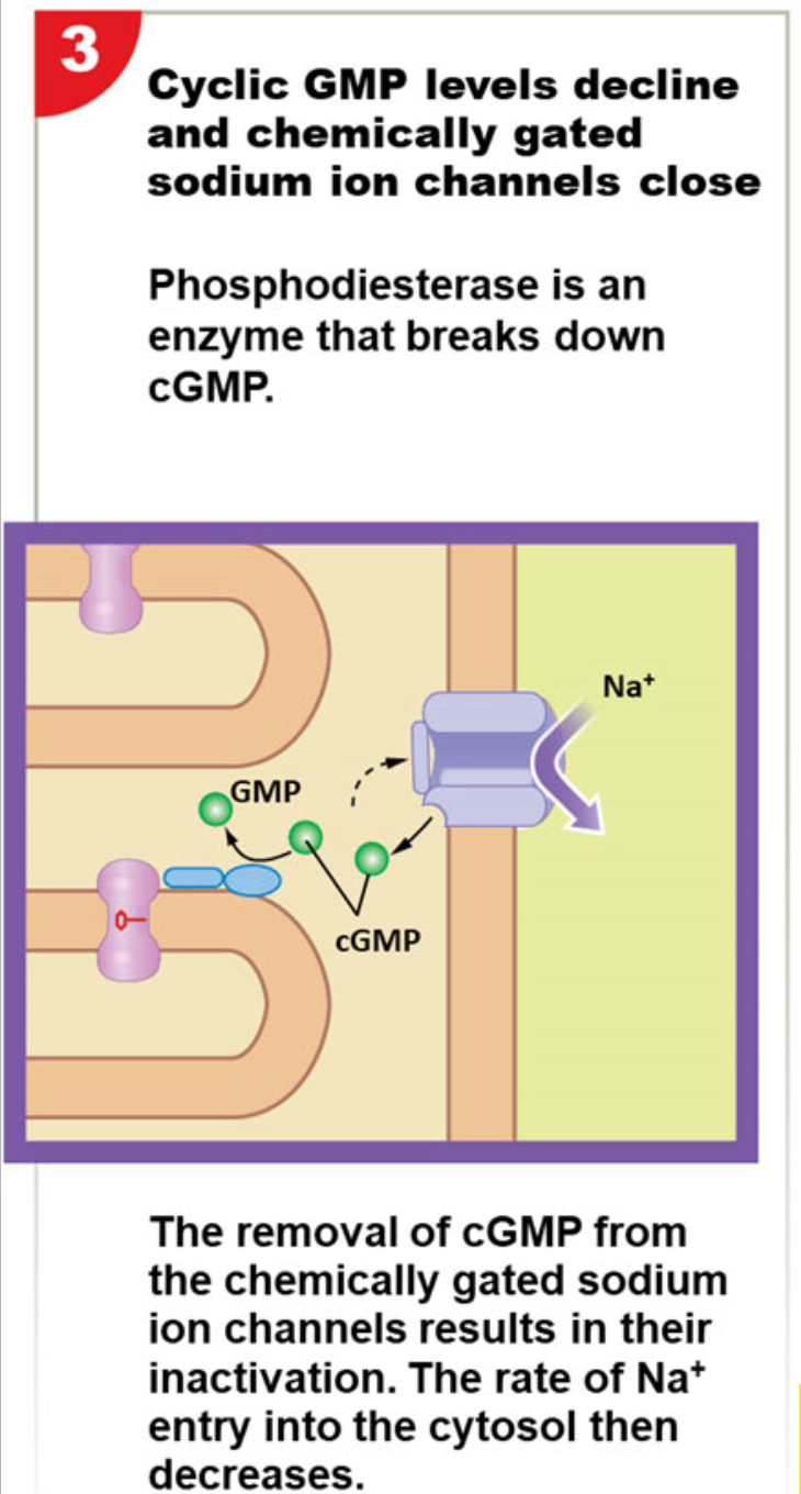
4) dark current is reduced
rate of neurotransmitter release declines
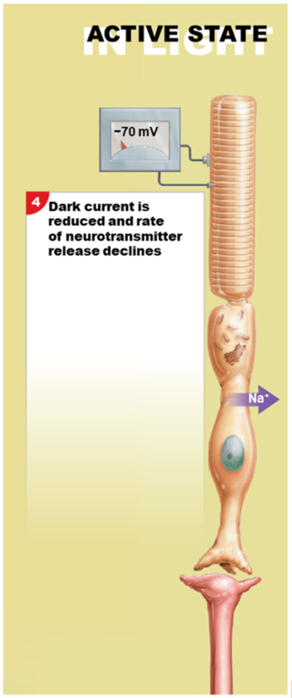
holy yap: when sodium ion channels close, membrane potential drops to ~-70millivolts. as plasma membrane hyperpolarises, rate of neurotransmitter release decreases. this decrease signals to the adjacent bipolar cell that the photoreceptor has been activated, and has absorbed a photon of light. after absorbing the photon, retinal doesn’t spontaneously revert back to its original form; takes a while—entire rhodopsin molecule is broken down into retinal and opsin (bleaching), and then is reassembled—hence, photoreceptor isn’t immediately reactivated; takes time (e.g. being in dark room, then going outside; can be very blinding; takes eyes a moment to readjust; this is bleaching).
bleaching allows the regeneration of visual pigments.
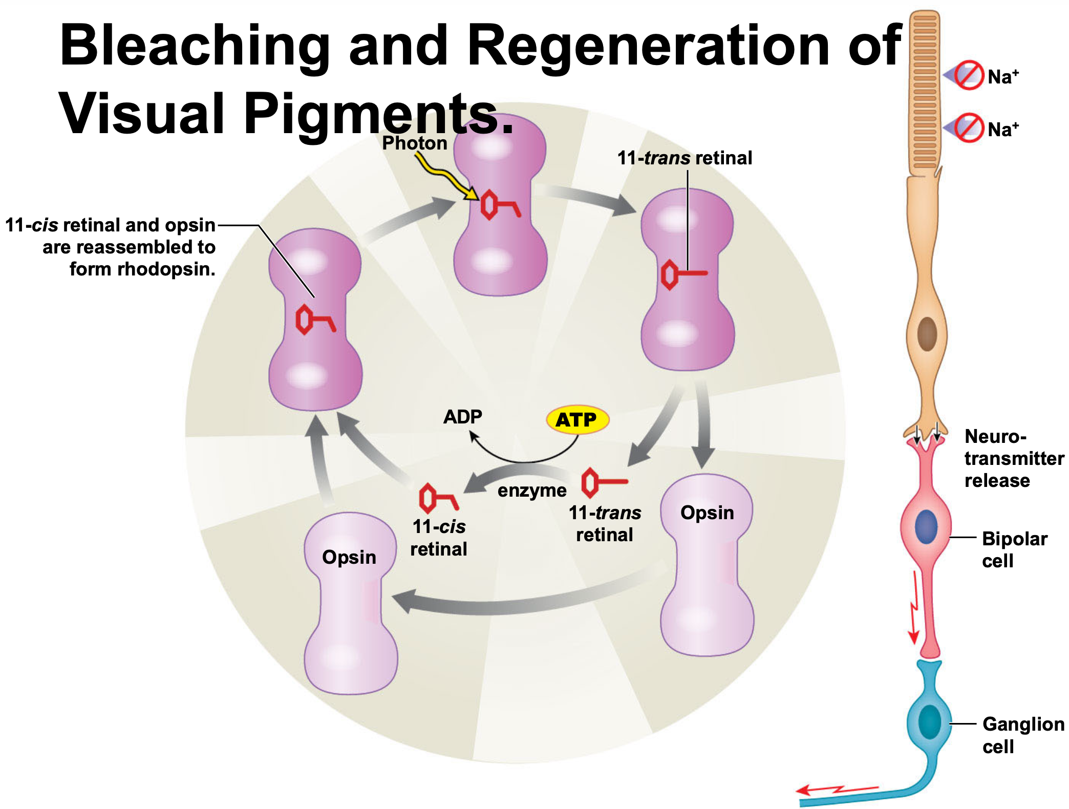
describe visual pathways (generally or detailed)
SYSTEM: Nervous System
SUBSECTION: Special Senses - Vision
NOTE: theres lots of information on this card.
CUE: None
begin at photoreceptors
end at visual cortex of cerebral hemispheres
messages must cross two synapses before moving toward brain
photoreceptor to bipolar cell
bipolar cell to ganglion cell
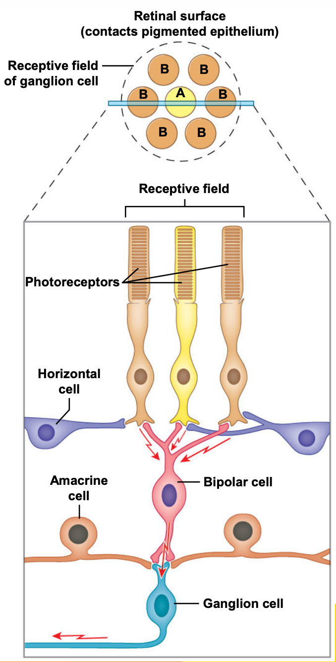
receptive field = field of view
area in which particular retinal ganglion cell is responsive to light
within that, have three cones that can be activated
maps visual world via neurones, coordination of action potentials
central processing of information
holy yap: visual information is processed in many ways and at different levels. firstly from the retinal ganglion cells (converging onto one another causing integration of information). the axons of retinal ganglion cells are the output of the retina; converge at the optic disc. the optic disc is considered the blind spot. there are no photoreceptors there; where the axons converge. axons leave the eye, and proceed as optic cranial nerve (II). have two eyes, hence two optic nerves. nerves cross over partially at optic chiasm. following this, information travels to visual cortex in occipital lobe. this is the primary visual cortex; where visual information gets sorted to go o different areas; projections for memory, particular shapes, colours, moving and stationary images, black and white images, all linked with projections.
axons from ganglion cells converge on optic disc
penetrate wall of eye
axons from ganglion cells converge on the optic disc.
penetrate wall of eye.
proceed towards diencephalon as the optic nerve (II)
two optic nerves reach diencephalon after partial crossover at the optic chiasm
information travels to visual cortex in occipital lobe
optic radiation: bunch of projection fibres linking lateral geniculates with visual cortex.
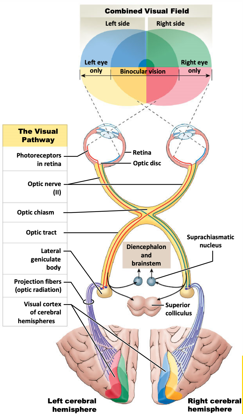
holy yap: combined visual field. at centre of visual field = binocular vision; stuff seen in left eye only, stuff seen in right eye only.
photoreceptors in each eye.
on right eye, red coding is the medial side of retina, but is lateral side of retina on left eye; both end up on left side of body (see the red line in the eye). opposite is true for the blue.
medial left, lateral right, they cross over, come together, then move off to the right visual cortex for processing.
optic nerve leaving each eye, crossing over at optic chiasm, and optic tract carrying information from quadrant of each eye.
relay station: optic tract projects to lateral geniculate body (LGN); where have synapse.
from lateral geniculate nuclei have projection fibres that radiate outwards (optic radiation) to different regions of the occipital cortex.
red and green, blue and yellow correspond to red and green, and yellow and blue in visual field.
other pathways to note: pathway from lateral geniculus to the superior colliculus. and from lateral geniculus to diencephalon and brain stem.
superior colliculus receives information from rods, particularly relating to movement.
also receives auditory and head information.
information for where we orient ourselves, spatially in a moving world.
also involved in driving movement of the extraocular muscles too; eye rotation, and movement tracking.
terminology:
field of vision:
combines visual images from left and right eyes
depth perception:
obtained by comparing relative positions of objects between images received from both eyes.
make assumptions about how far away things are; e.g. if receive information slightly sooner from right eye before left eye can assume that object is closer to the right eye/right side of head than the left.
brainstem and visual processing
photons of light not only provide visual information, but allow humans to perform several autonomic functions, including the circadian rhythm.
circadian rhythm:
daily pattern of activity tied to day-night cycle.
established from visual information.
affects metabolic rate, blood pressure, mental health etc.
all established from visual information (not perceived, directly think about; but light comes in through eyes, activating these processes).
state the three divisions of the ear.
optional: list components of each (not sure if need)
or at least the key components.
SYSTEM: Nervous System
SUBSECTION: Special Senses - Sound
NOTE: None
CUE: None
external ear
auricle (aka. pinna)
funnel-like
funnels auditory information towards meatus to cause vibration of tympanic membrane
elastic cartilages
external acoustic meatus
channel in temporal bone
middle ear
tympanic membrane
three smallest bones of body called the auditory ossicles
internal ear (transition from middle ear to internal ear through oval window).
cochlea
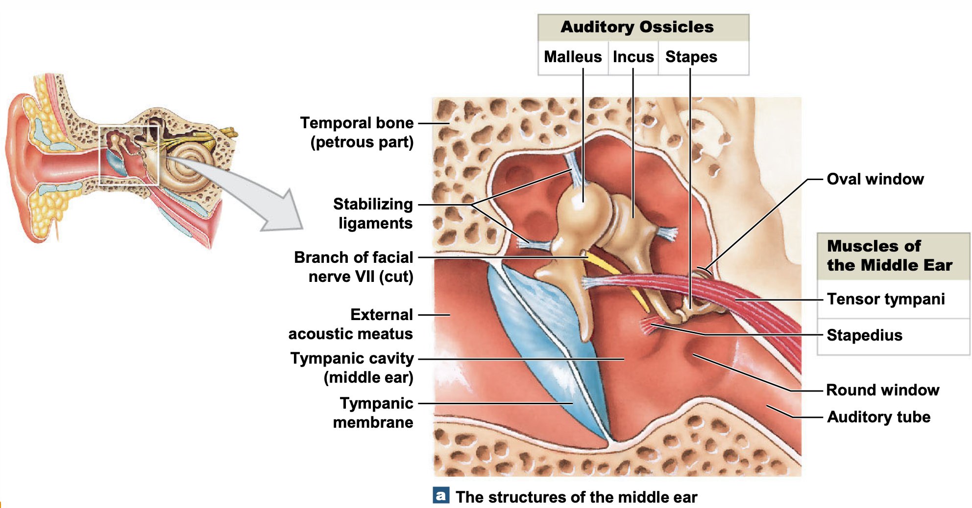
holy yap about diagram: when sound comes in, it is funnelled in through the pinna, into the external acoustic meatus, leading to vibration of the tympanic membrane.
on other side of tympanic membrane is an air-filled space/chamber called the middle ear.
three bones (auditory ossicles): malleus, incus, and stapes transduce vibrational energy from the tympanic membrane to the stapes. role of ossicles is to amplify the vibrational energy of tympanic membrane.
the stapes presses against the oval window (fluid filled space).
muscles of middle ear
stapedius
supports/stabilises position of stapes
tensory tympani
adjusts tautness of the tympanic membrane.
helps to tune in or tune out/increase/decrease receptibility of hearing.
good for dampening sounds of own head (e.g. chewing).
list the auditory ossicles.
SYSTEM: Nervous System
SUBSECTION: Special Senses - Sound
NOTE: None
CUE: aka, the bones of the middle ear.
incus
malleus
stapes
state what happens when sound waves vibrate tympanic muscles, and what protects the ear from loud noises.
SYSTEM: Nervous System
SUBSECTION: Special Senses - Sound
NOTE: None
CUE: None
when sound waves vibrate tympanic membrane
auditory ossicles (incus, malleus, stapes) conduct vibrations to the internal ear.
when tympanic membrane vibrates, ossicles vibrate, amplifying vibration.
two small muscles protect eat from loud noises (muscles of middle ear).
tensor tympani
pulls on malleus and stiffens tympanic membrane.
stapedius
reduces movement of stapes at oval window.
describe structure of internal ear
SYSTEM: Nervous System
SUBSECTION: Special Senses - Sound
NOTE: None
CUE: None
the internal ear
is a winding passageway (called the labyrinth)
aka. bony labyrinth, it surrounds and protects membranous labyrinth (fluid filled space)
two types of fluid
perilymph flows between the two labyrinths
endolymph is within membranous labyrinth
moves within cochlea
bony labyrinth can be subdivided into
vestibule
semicircular canals
cochlea
interest for hearing is the cochlea, others are involved in balance.
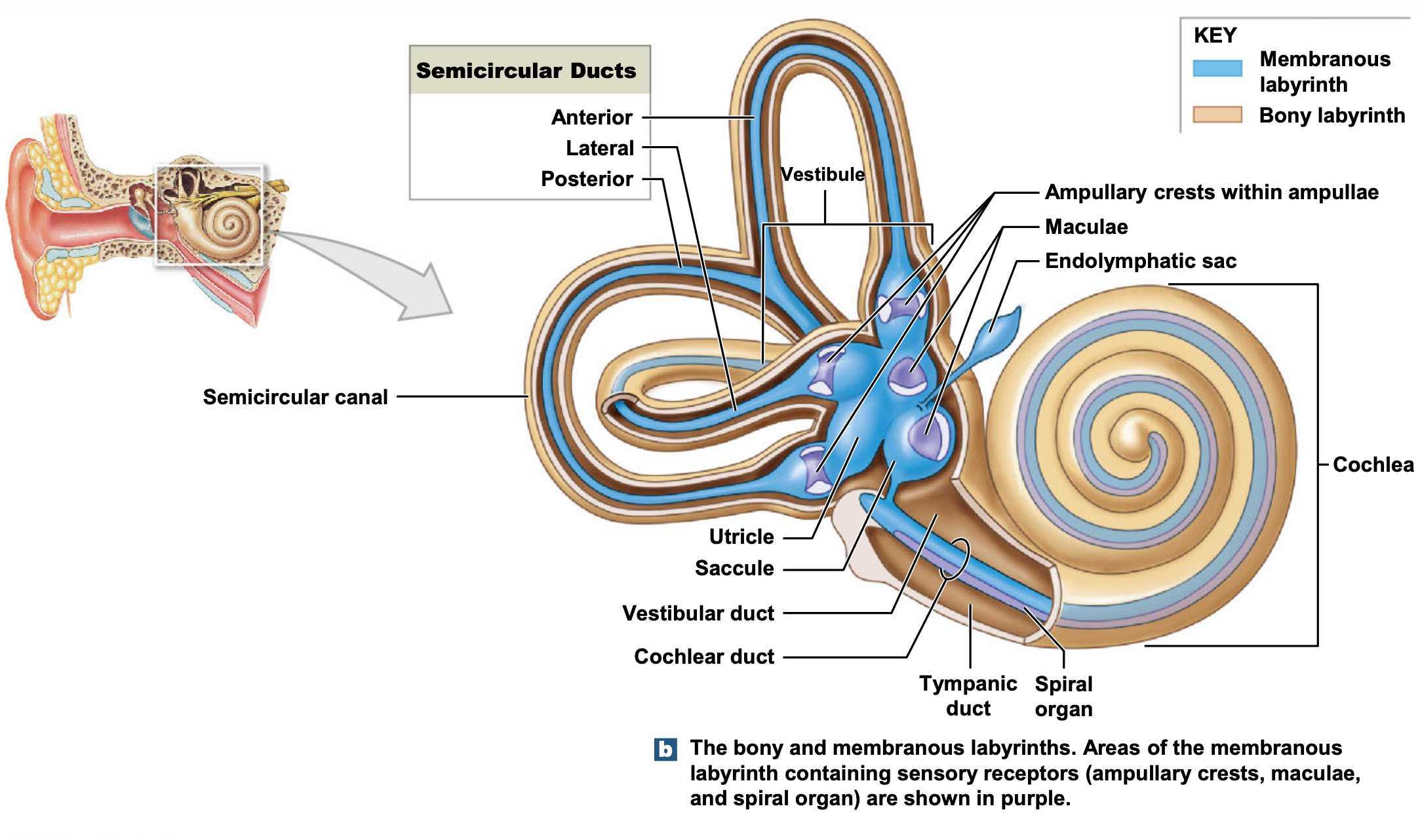
where sound is detected; where transduction occurs
(sound is transduced into action potentials)
holy yap: sound waves have funnelled in through the pinna, caused tympanic membrane to vibrate, amplified by the ossicles, now moving into inner ear where transduction occurs.
this is anchored deep into the temporal bone. semi circle canals are angled, and contain fluid. when moves within, is registered, allowing understanding of where orientation of head is in space. hence why if on a ride, fluid if moving lots, takes a minute for fluid to stop moving once get off ride, hence dizzy feeling despite feet being and telling brain that they are flat on the ground.
vestibular and cochlear duct. curly shaped structure is the cochlear.
describes the process of hearing.
SYSTEM: Nervous System
SUBSECTION: Special Senses - Sound
NOTE: None
CUE: None
sound waves are converted into mechanical movements by vibration and tympanic membrane.
auditory ossicles amplify and conduct vibrations to the internal ear.
vibrations are converted to pressure waves in fluid.
pressure waves are detected by hair cells in cochlear duct. action potentials generation by hair cells are coded, and…
…information is sent to auditory cortex of brain.
describe what a pressure wave is
SYSTEM: Nervous System
SUBSECTION: Special Senses - Sound
NOTE: included state definitions of wavelength and frequency; pitch, amplitude, and intensity (and units where necessary).
CUE: None
pressure waves are s-shaped curves aka. sines waves.
consists of alternating regions where air molecules are crowded together and adjacent to zones where they are farther apart.
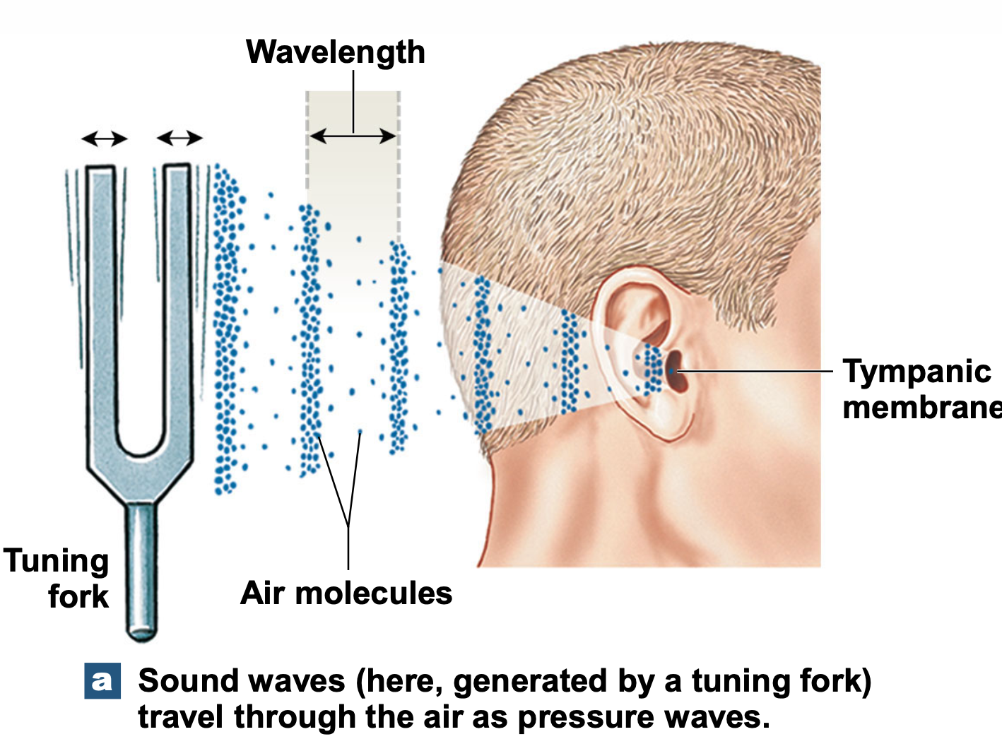
wavelength
distance between two adjacent wave crests (peaks)
frequency
number of waves (cycles) that pass a fixed reference point in a given time. (cycles per unit of time ESSENTIALLY).
measured in hertz (Hz)
denotes: number of cycles per second.
pitch
sensory response to frequency
amplitude
height of sound wave
intensity
magnitude of energy in a sound wave
determines how loud is seems
reported in decibels
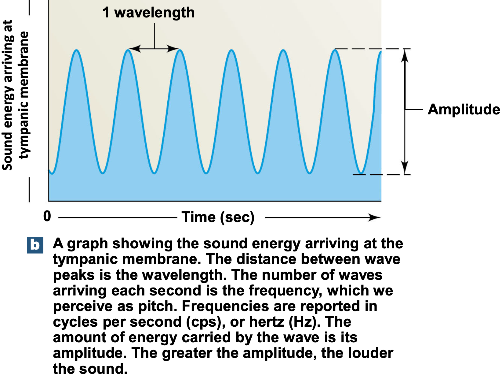
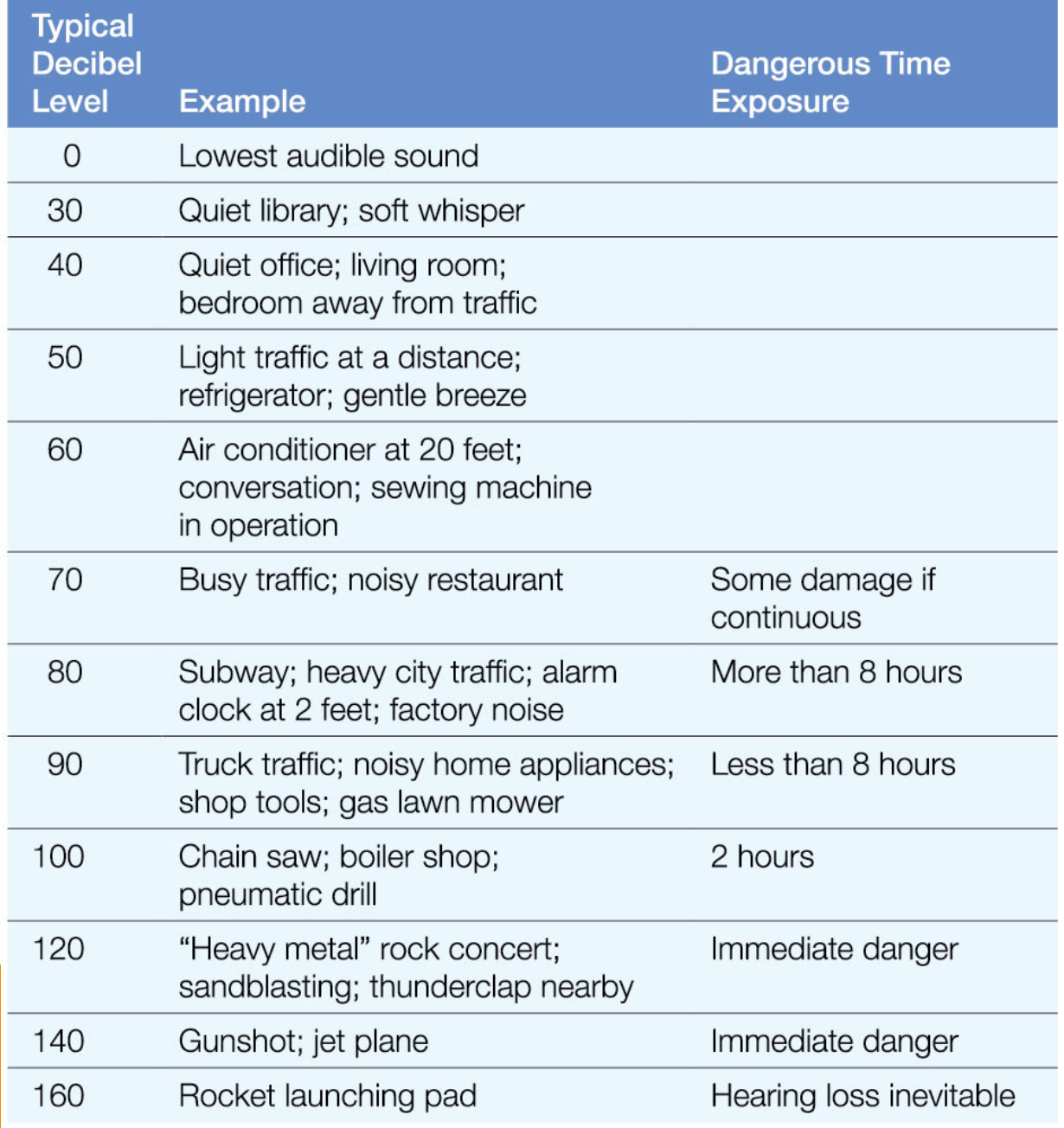
state where cochlear duct and hair cells lie.
SYSTEM: Nervous System
SUBSECTION: Special Senses - Sound
NOTE: None
CUE: None
remember: cochlear is curved, snail-like, hollow tube structure, filled with fluid, with three sections (scala media, scala vestibuli, and scala tympanic).
cochlear duct (aka. scala media)
lies between
scala vestibuli (vestibular duct)
scala tympanic (tympanic duct)
hair cells lie in the spiral organ (aka. organ of Corti)
rests on basilar membrane
separates cochlear duct from scala tympani
it is hair cells that are deformed by pressure changes in the fluid that moves around them.
hair cells lack kinocilia
stereocilia contact overlying tectorial membrane
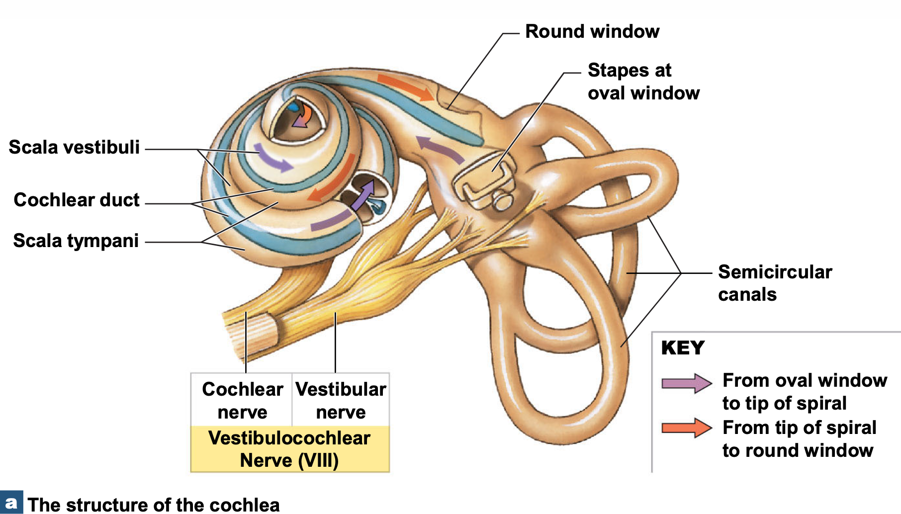
holy yap about diagram:
stapes at oval window: assume sound has sufficient energy and decibels to be coded into action potentials. pressure is forced down and around cochlea. then fluid is moved back from around the cochlea, and pressure is detected at round window. pressure change starts from the pushing of the stapes at the oval window, then fluid is seen moving around chambers of the cochlea, coming around, pressing, and forcing change at the deforming of the round window.
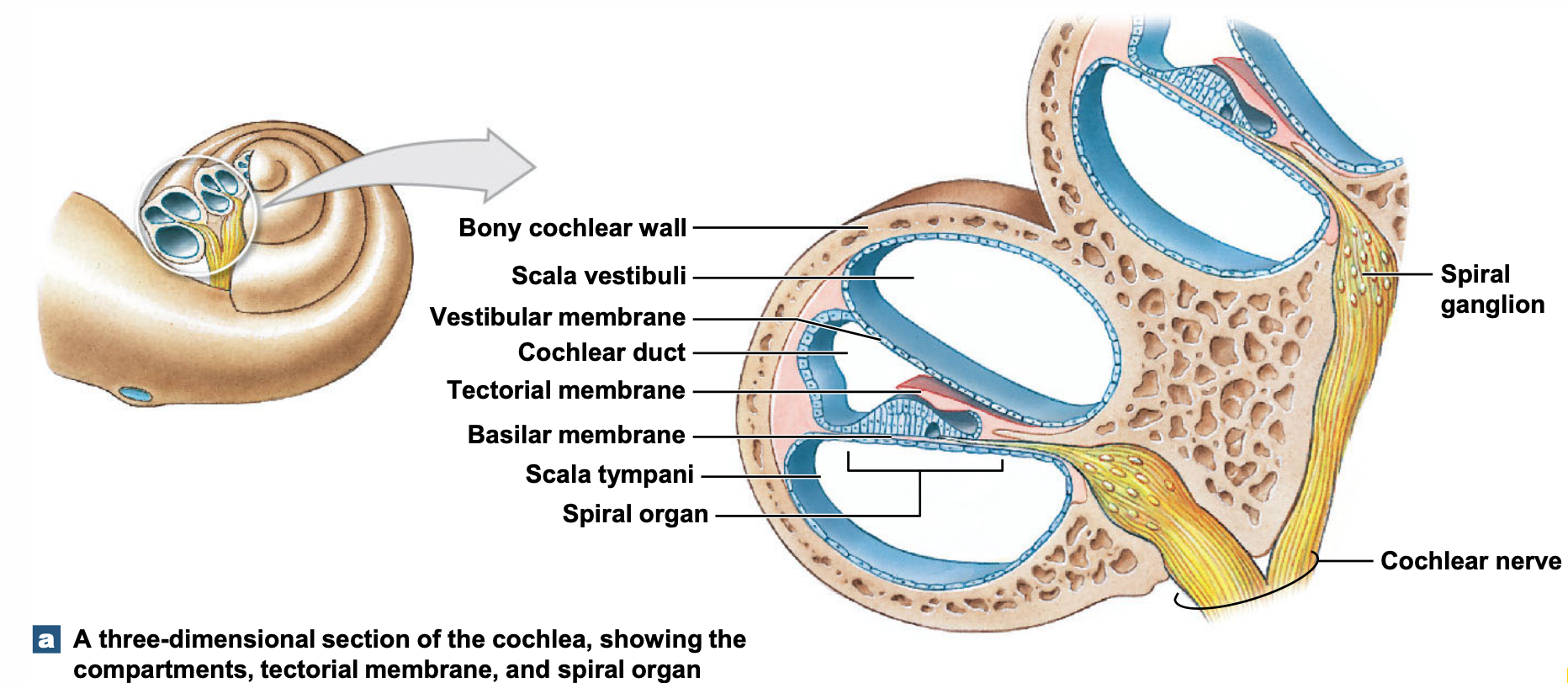
holy yap about diagram:
if cut cochlea in section, can see the three chambers.
on basilar membrane, is where hair cells are situated.
can see from spiral organ; axons coming out to the cochlear nerve (VIII).
cranial nerve VIII has two parts. cochlear nerve and vestibular part. vestibular part is from the semi circular canals of the ear. (vestibular apparatus is associated with balance—another special sense.. not discussed in HLTH1004).
describe auditory discrimination
SYSTEM: Nervous System
SUBSECTION: Special Senses - Sound
NOTE: None
CUE: None
holy yap: term to describe sounds that can be determined; the range from the softest to the loudest tolerable sound. young children have greatest hearing range, with age, damage accumulates.
auditory discrimination
refers to the range from softest to loudest tolerable sound
where the loudest sounds are ~1 trillion times more powerful (in terms of sound energy) than the quietest detectable sounds.
young children have greatest hearing range
with age, damage accumulates, potentially involving:
tympanic membrane gets less flexible
articulations between ossicles stiffen
initial sound waves coming in are less loud; less vibration; less amplification
round window may begin to ossify
hence, response may be lower
loss or stiffening of hair cells
describe the process of hearing
SYSTEM: Nervous System
SUBSECTION: Special Senses - Sound
NOTE: None
CUE: None
sound waves arrive at tympanic membrane
movement of tympanic membrane displaces the auditory ossicles
movement at the stapes at the oval window produces pressure waves in the perilymph of the scala vestibuli.
the pressure waves distort the basillar membrane on their way to the round window of the scale tympani.
vibration of basillar membrane causes vibration of hair cells against the tectorial membrane.
information about the region and intensity of the stimulation is relayed to the CNS via cochlear nerve.

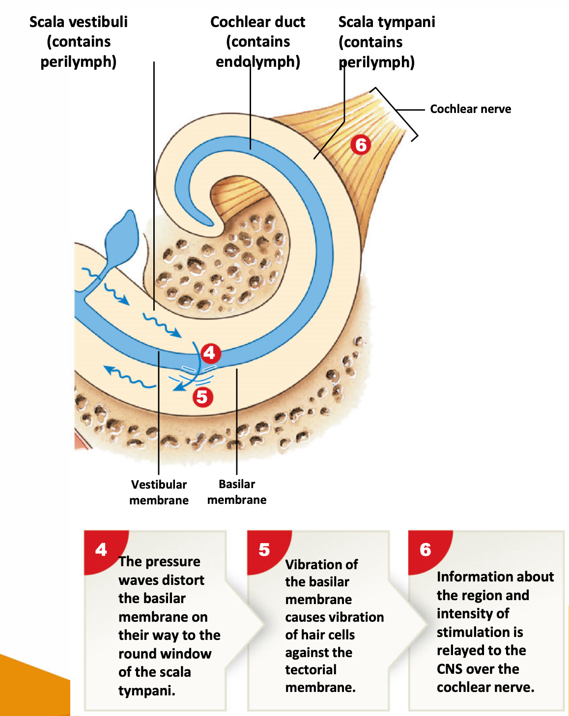
holy yap: middle ear is an air-filled space. can see the pharyngotympanic tube connecting middle ear to pharynx (back of throat). if air filled space, becomes fluid filled space = reduced amplification of sound waves. hence why throat infections and ear infections are often links — reducing hearing and voice range etc.
it is the hair cells that generate the action potentials, and code information about the stimulation to the central nervous system across the cochlear nerve (VIII). spiral ganglia contains hair cells of bipolar sensory that monitor cochlear hair cells.
or this one (or any other relevant explanations).
sound waves are converted into mechanical movements by vibration and tympanic membrane.
auditory ossicles amplify and conduct vibrations to the internal ear.
vibrations are converted to pressure waves in fluid.
pressure waves are detected by hair cells in cochlear duct. action potentials generation by hair cells are coded, and…
…information is sent to auditory cortex of brain.
describe the auditory pathway
SYSTEM: Nervous System
SUBSECTION: Special Senses - Sound
NOTE: None
CUE: cochlear nerve, cochlear nucleus
sound energy information is captured by the cochlear, then auditory pathways from cochlear leave the cochlear from the cochlear nerve (VIII).
afferent fibres of sensory neurons in spiral ganglion form the cochlear nerve
axons enter medulla oblongata and synapse with the cochlear nucleus
information ascends to:
superior olivary nuclei within the pons, and the
inferior colliculi of midbrain
(superior colliculus is associated with vision, inferior colliculi are paired nuclei that sit just inferior to the superior colliculi).
where the midbrain coordinates unconscious motor responses to sound
e.g. jumping at loud noises.
ascending auditory sensations synapse in the medial geniculate body of the thalamus.
projection fibres deliver information to the auditory cortex of the temporal lobe.
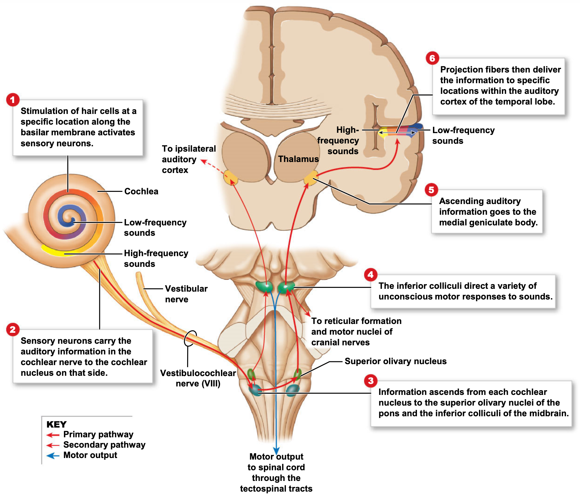
holy yap about diagram:
can see cochlear.
colour coded by frequency of sound
hair cells respond in those regions to those particular frequencies of sound
sensory neurons carry auditory information in cochlear nerve to cochlear nucleus on that side.
some cross-over occurs ipsilaterally or contralaterally.
information ascends from each cochlear nucleus to the superior olivary nuclei within the pons, and the inferior colliculi of midbrain.
inferior colliculi directs various unconscious motor responses to sound.
including jumping to loud noises (serves to be protective)
projections occur to medial geniculate body of thalamus, and then to the auditory cortex.
similar mapping of auditory information (similar to visual) can be seen as well (colour coding of cochlear with colour coding of auditory cortex based on particular frequencies).
WEEK 8: RESPIRATORY SYSTEM
learning objectives
• Describe the anatomical structure and function of the respiratory system components
• Understand the difference between breathing and respiration
• Differentiate between the different lung volumes and capacities
• Describe exchange of gases at tissues
• Describe exchange of gases at lungs
• Understand the factors that can regulate respiration in both short and long term
• Understand the relationship between the respiratory system and the cardiovascular system
• Explain how the nervous system controls breathing
Describe the structural and functional classifications of the respiratory system.
SYSTEM: Respiratory System
SUBSECTION: Respiratory System
NOTE: None
CUE: None
Structural classification.
Upper respiratory system
nose, nasal cavity, pharynx, and associated structures
Lower respiratory system
larynx, trachea, bronchi, and lungs
Functional classification.
Conducting zone
consists of: interconnected cavities and tubes both inside and outside the lungs, including the nose, nasal cavity, pharynx, larynx, trachea, bronchi, bronchioles, and terminal bronchioles.
function to: filter, warm, and humidify incoming air while directing it into the lungs.
Respiratory zone
consists of: the structures within the lungs where gas exchange occurs, including respiratory bronchioles, alveolar ducts, alveolar sacs, and alveoli.
function to: serve as the primary sites for oxygen and carbon dioxide exchange between the air and blood stream.
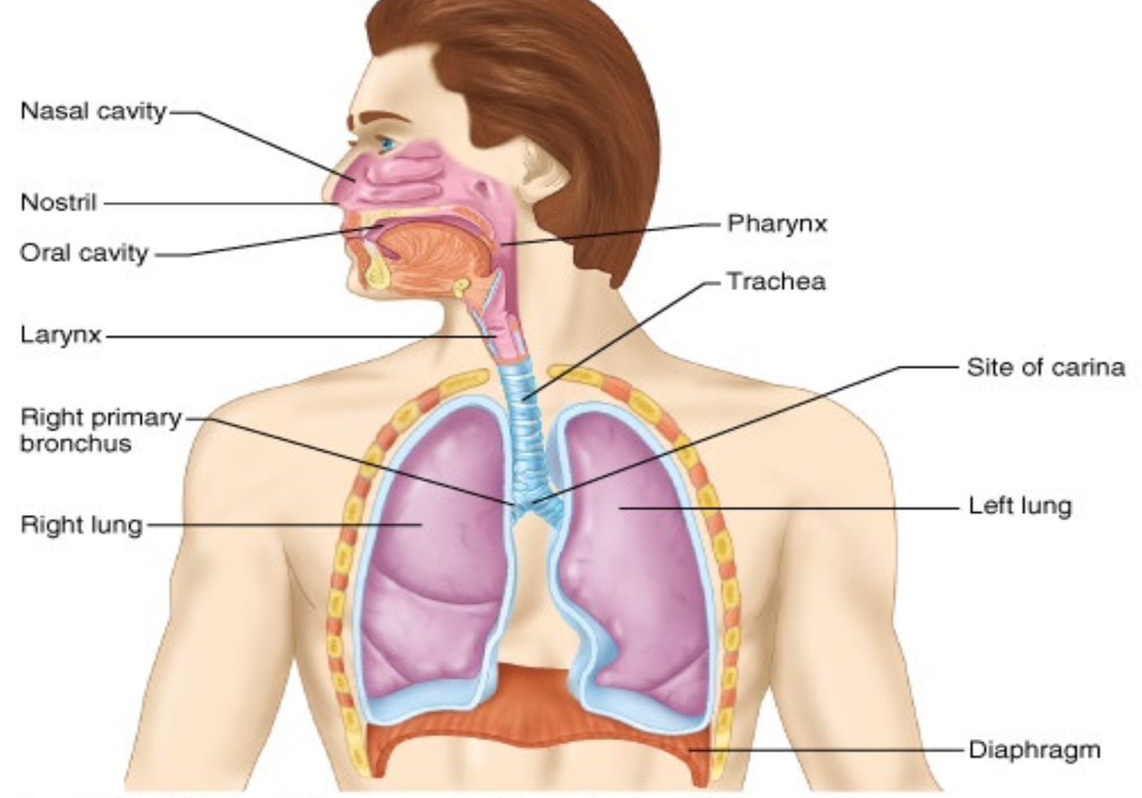
describe the functions of the respiratory system.
SYSTEM: Respiratory System
SUBSECTION: Respiratory System
NOTE: None
CUE: None
provides for gas exchange: intake of O2 for delivery to body cells, and removal of CO2 produced by body cells.
helps regular blood pH.
contains receptors for sense of smell, filters inspired air, produces vocal sounds (as phonation), and excretes small amounts of water and heat.
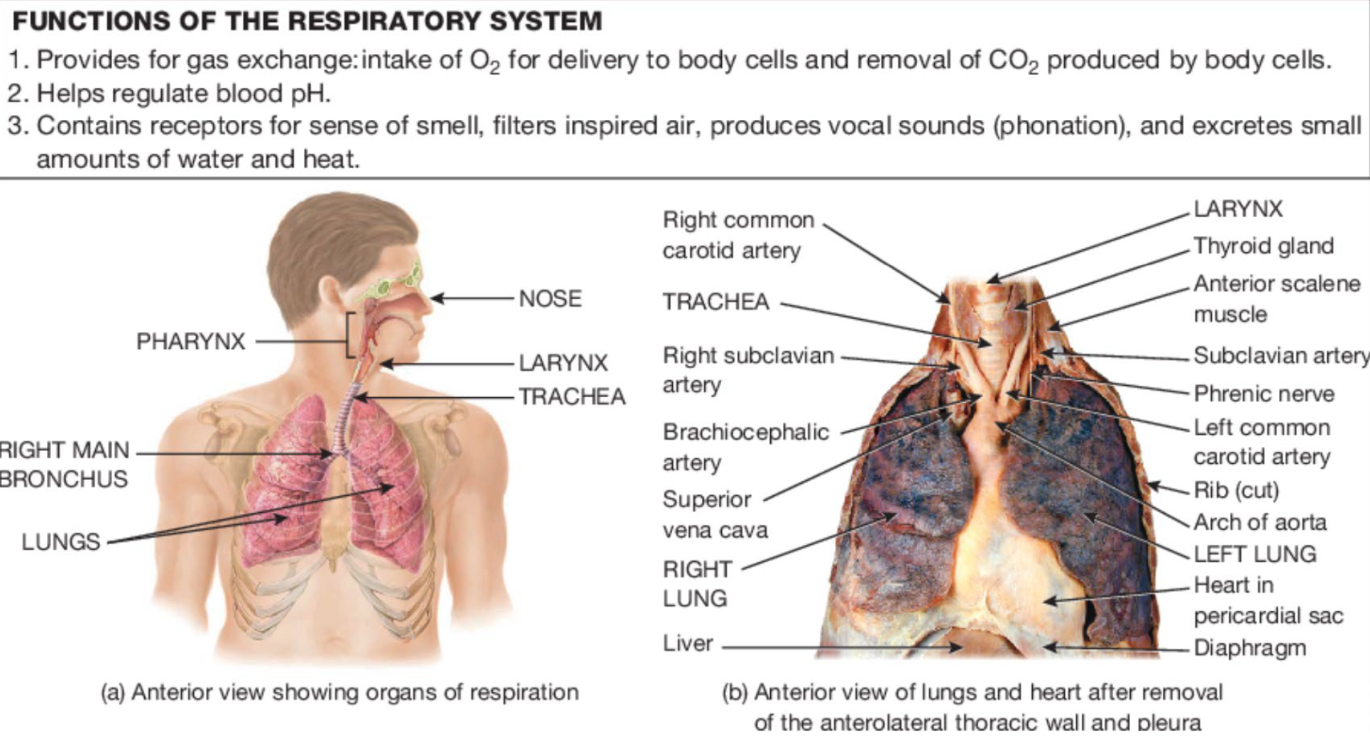
State the functions of the nose (particularly the internal structures of the nose)
SYSTEM: Respiratory System
SUBSECTION: Nose
NOTE: None
CUE: None
nose functions as an essential gateway to the respiratory system, ensuring that air is properly conditioned before reaching the lungs, this involves:
conditioning incoming air:
filtering, warming, and humidifying inhaled air before it reaches lungs
olfaction:
detecting olfactory stimuli as smell
speech modification:
modifying speech vibrations
resonating sound by prolonging, amplifying, or altering vibrations as they pass through the nasal cavity.
describe the structural components of the
external nose
internal nasal cavity.
SYSTEM: Respiratory System
SUBSECTION: Nose
NOTE: None
CUE: bony framework, cartilaginous framework, nostrils, nasal conchae, nasal meatuses, olfactory region, paranasal sinuses & nasolacrimal ducts, internal nares.
introduction:
The nose is a specialised organ at the entrance of the respiratory system, consisting of a visible external portion and an internal nasal cavity within the skull.
nose functions as an essential gateway to the respiratory system, ensuring that air is properly conditioned before reaching the lungs, this involves:
filtering, warming, and humidifying inhaled air (conditioning incoming air)
detecting olfactory stimuli as smell (olfaction)
modifying speech vibrations (speech modification)
External Nose
External nose refers to the visible part of the nose, and consists of a supporting framework of bone and hyaline cartilage, covered by muscle, skin, and a mucous membrane.
Structural components:
Bony Framework:
formed by the frontal bone, nasal bones, and maxillae.
Cartilaginous Framework:
composed of multiple hyaline cartilage pieces connected to each other, and certain skull bones by fibrous connective tissue.
functions to: provide flexibility and shape
Nostrils (aka. External Nares):
openings that allow air to enter the nasal cavity.
Internal Nasal Cavity
The nasal cavity is a large, air-filled space inside the skull. It is located beneath the nasal bones, and above the oral cavity. To trap debris and humidify air, the internal nasal cavity is lined with muscle and a mucous membrane.
The cavity is divided into right and left sections by the nasal septum, which is consists of the vomer, the perpendicular plate of ethmoid bone, the maxillae, palatine bones, and hyaline cartilage.
Structural components:
Nasal Conchae (aka. Turbinates):
three bony projections (superior, middle, and inferior).
function to: increase surface area, create airflow turbulence, and enhance air filtration, warming, and humidification.
Nasal Meatuses:
three bony projections (super, middle, and inferior).
are narrow passageways between each concha.
function to: direct airflow through the nasal cavity.
Olfactory Region:
located in the superior portion of the nasal cavity, contains sensory receptors for smell.
Paranasal Sinuses & Nasolacrimal Ducts:
open into the nasal cavity
function to: facilitate drainage of mucus from the sinuses and tears from the eyes.
Internal Nares (aka. Choanae; aka Internal Naris):
openings at the back of the nasal cavity that connect to the pharynx.
nose ends at internal nares.
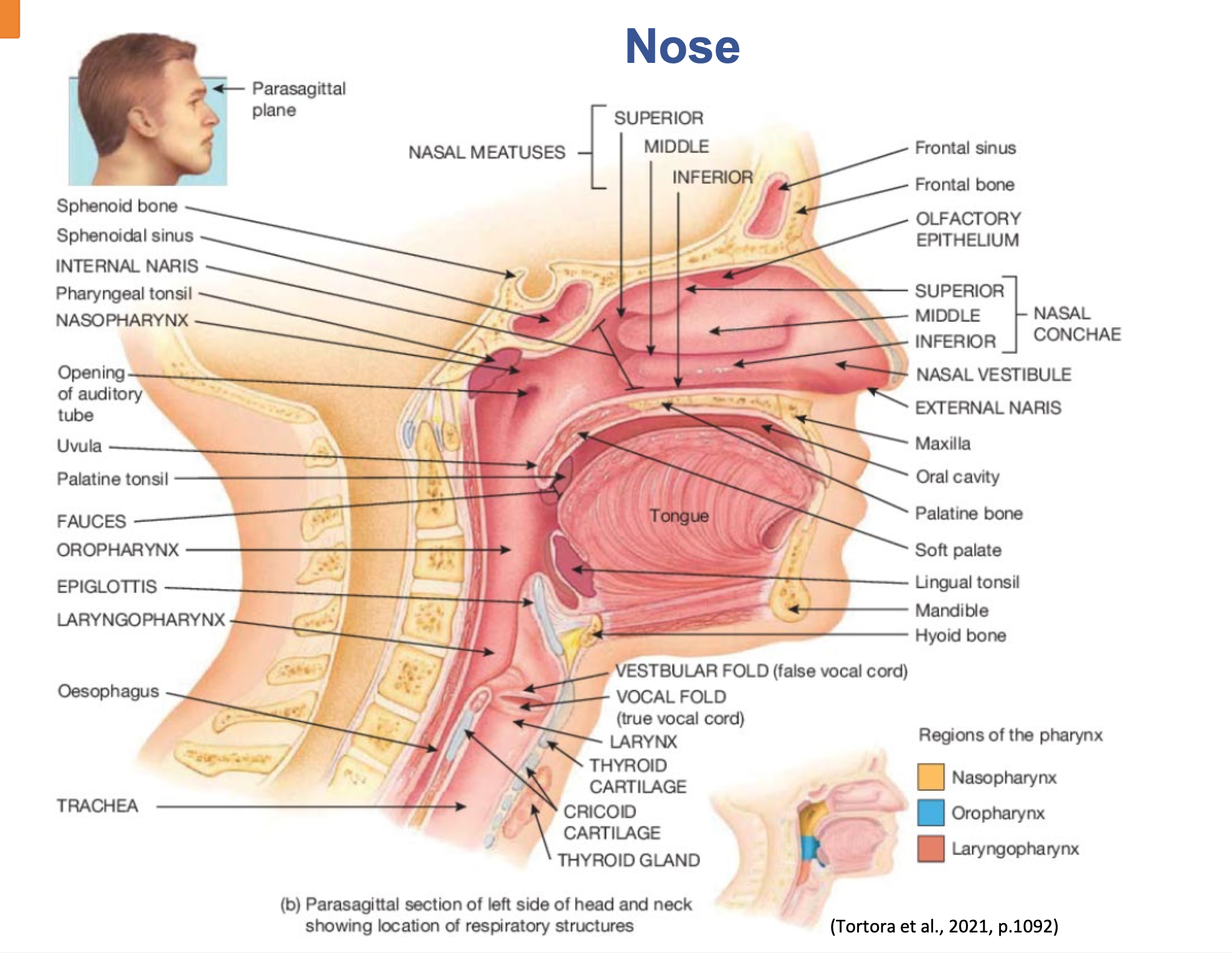
describe the pharynx, include:
shape and length
location
what its wall is made of
what assists in deglutition (swallowing)
pharynx function
the three anatomical divisions of the pharynx
SYSTEM: Respiratory System
SUBSECTION: Pharynx
NOTE: None
CUE: None
after the nose is the pharynx. nose ends at the internal nares (or naris).
pharynx, aka throat.
funnel-shaped tube
~13cm (5in) long
starts at the internal nares, and extends to the cricoid cartilage (which is the most inferior cartilage of the larynx—voice box).
pharynx lies just posterior to the nasal and oral cavities, superior to the larynx, and just anterior to the cervical vertebrae.
Its wall is made of skeletal muscles and is lined with a mucous membrane.
remember: skeletal muscles=can be controlled voluntarily
contraction of skeletal muscles assists in deglutition (swallowing) .
pharynx functions as:
a passage way for air and food
provides a resonating chamber for speed sounds
houses the tonsils, which participate in immunological reactions against foreign invaders.
pharynx can be divided into three anatomical regions
nasopharynx
oropharynx
laryngopharynx
describe the larynx, include:
what it connects
location
what the wall is made of
what is the most important cartilage
what the epiglottis is, including shape and what it is covered with.
what is parts of the epiglottis are attached to [something] and not attached to.
do the pharynx and larynx rise of fall during swallowing
SYSTEM: Respiratory System
SUBSECTION: Larynx
NOTE: None
CUE: None
larynx, aka voice box
short passageway that connects the laryngopharynx with the trachea.
located in the midline of the neck, anterior to the oesophagus, and the 4th-6th cervical vertebrae (C4-C6).
the wall of the larynx is composed of nine pieces of cartilage.
three occur singly
thyroid cartilage aka. adams apple
epiglottis
cricoid cartilage
three occur in pairs (arytenoid, cuneiform, and corniculate cartilages).
of the paired cartilages, the arytenoid cartilages are the most important, because they influence changes in position and tension of the vocal folds (plays a role in true vocal cords for speech)
the epiglottis is a large, leaf-shaped piece of elastic cartilage, that is covered with epithelium.
The ‘stem’ of the epiglottis is the tapered inferior portion that is attached to the anterior rim of the thyroid cartilage.
The broad superior ‘leaf-shaped’ portion of the epiglottis is unattached, and is free to move up and down—like a trap door.
the larynx and pharynx rise during swallowing.
elevation of the pharynx widens it to receive food or drink
elevation of the larynx causes the epiglottis to move down and form a lid over the glottis, closing it off.
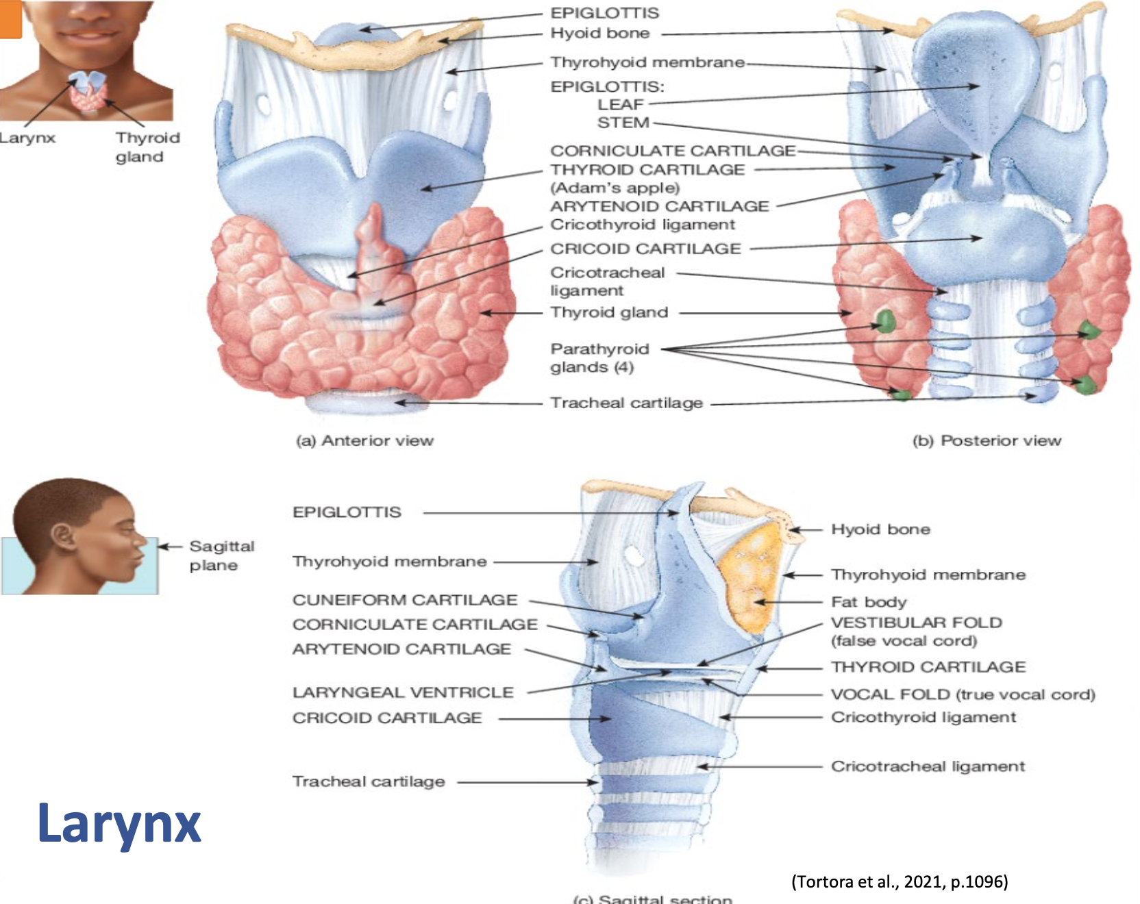
describe the larynx, include:
what are the two folds that the mucous membrane of the larynx forms.
hence, what is the space called between the superior pair
what are the functions of the superior pair
function of inferior pair
what does contracting and relaxing muscles in the inferior pair do (also mention how the muscles attach)
how is sound produced (phonation)
what causes the variation of pitch, and loudness of sound.
SYSTEM: Respiratory System
SUBSECTION: Larynx
NOTE: None
CUE: None
larynx, aka voice box
the mucous membrane of the larynx forms two pairs of folds:
a superior pair called the vestibular folds
false vocal chords
an inferior pair called the vocal folds
true vocal chords.
the space between the vestibular folds is known as the rima vestibuli
vestibular folds do not function in voice production, but when the folds are brought together, they function in
holding the breath against pressure in the thoracic cavity when brought together.
e.g. when a person strains to lift a heavy object
vocal folds are the principle structures of voice production.
Deep to the mucous membrane of the vocal folds, are bands of elastic ligaments stretched between the rigid cartilages of the larynx—like the strings on a guitar.
Intrinsic laryngeal muscles attach to both the rigid cartilages and the vocal folds.
When the muscles contract, they move the cartilage, which pulls the elastic ligaments tight, which in turn, stretches the vocal folds out into the airways so that the rima glottidis is narrowed.
Contracting and relaxing the muscles varies the tension in the vocal folds—like loosening or tightening a guitar string.
related to the variation in pitch of sound.
Air passing through the larynx vibrates the vocal folds, and produces sound (phonation) by setting up sound waves in the column of air in the pharynx, nose, and mouse.
The variation in the pitch of sound is related to the tension in the vocal folds.
The greater the pressure of air, the louder the sound produced by the vibrating vocal folds.
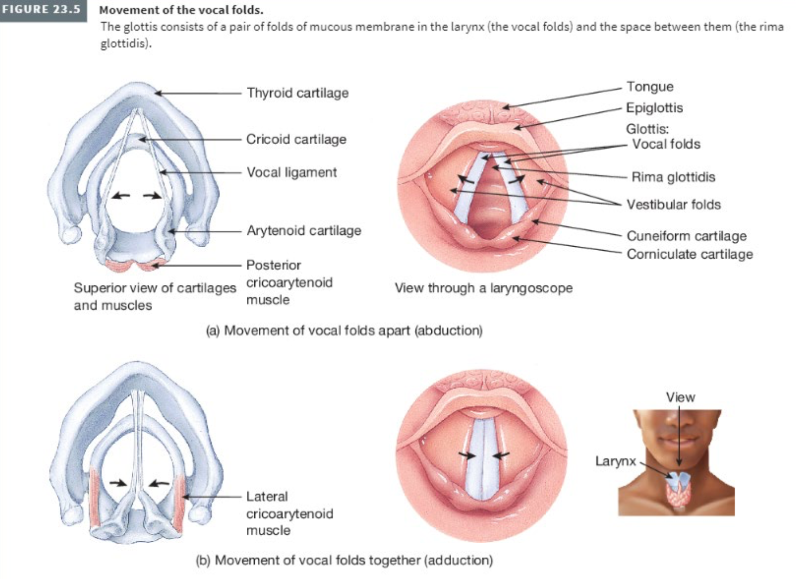
describe the trachea, include:
state shape, structure
length, diameter
location
is it apart of conducting zone or respiratory zone
what provides structural support to the trachea, preventing collapse during breathing.
what does the trachea divide into
SYSTEM: Respiratory System
SUBSECTION: Trachea
NOTE: None
CUE: None
trachea, aka. windpipe.
sturdy, tube-like structure that serves as a passageway for air.
~12cm length, 2.5cm diameter
located anterior to the oesophagus, and extends from the larynx to the superior boarder of the fifth thoracic vertebrae (T5), where it branches into the right and left primary bronchi.
apart of conducting zone of respiratory system.
Trachea is reinforced by 16-20 C-shaped rings of hyaline cartilage (stacked one above the other and connected by dense connective tissue). This provides structural support and prevents collapse during breathing.
At the superior border of the fifth thoracic vertebra, the trachea divides into a right main (primary) bronchus which goes into the right lung, and a left main (primary) bronchus which goes into the left lung.
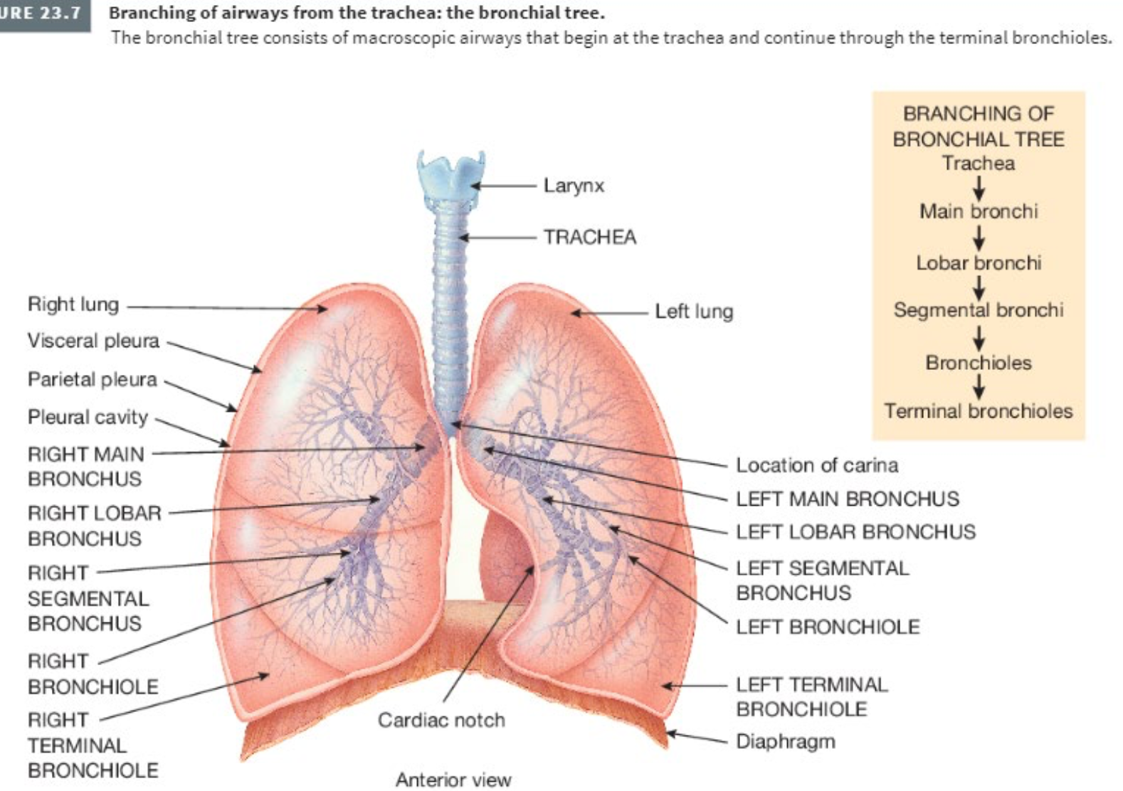
describe the bronchi, including:
how the primary bronchi divide from the trachea
shape of right vs left main bronchi
what similarity do the trachea and bronchi share
what do the main bronchi branch into (include all)
state what secondary bronchi supply
state what cells bronchioles contain, and their function.
are the bronchi apart of conducting zone or respiratory zone.
what does the branching of bronchi represent, what is it commonly known as (optional question).
SYSTEM: Respiratory System
SUBSECTION: Bronchi
NOTE: None
CUE: None
At the superior boarder of the fifth thoracic vertebra, the trachea divides into a right main (primary) bronchus which goes into the right lung, and a left main (primary) bronchus which goes into the left lung.
the right main bronchus is more vertical, shorter, and wider than the left.
like the trachea, the main bronchi contain incomplete rings of cartilage (C-shaped rings).
so that air can go in and stuff like that.
branching of bronchi
the main bronchi branch into smaller airways called the lobar (secondary) bronchi, with one for each lung lobe.
three in the right lung, and two in the left lung.
the secondary bronchi further branch/divide into segmental (tertiary) bronchi.
these supply specific bronchopulmonary segments within each lobe.
segmental bronchi continue branching into bronchiole, which repeatedly divide into smaller tubes, ending in the terminal bronchioles.
These bronchioles contain club (Clara) cells, which help protect against inhaled toxins, produce surfactants, and function as stem cells for epithelial repair.
The terminal bronchioles mark the end of the conducting zone of the respiratory system.
(hence, bronchi are apart of the conducting zone).
this extensive branching, from the trachea to the terminal bronchioles, represents and inverted tree, and is commonly known as the bronchial tree.
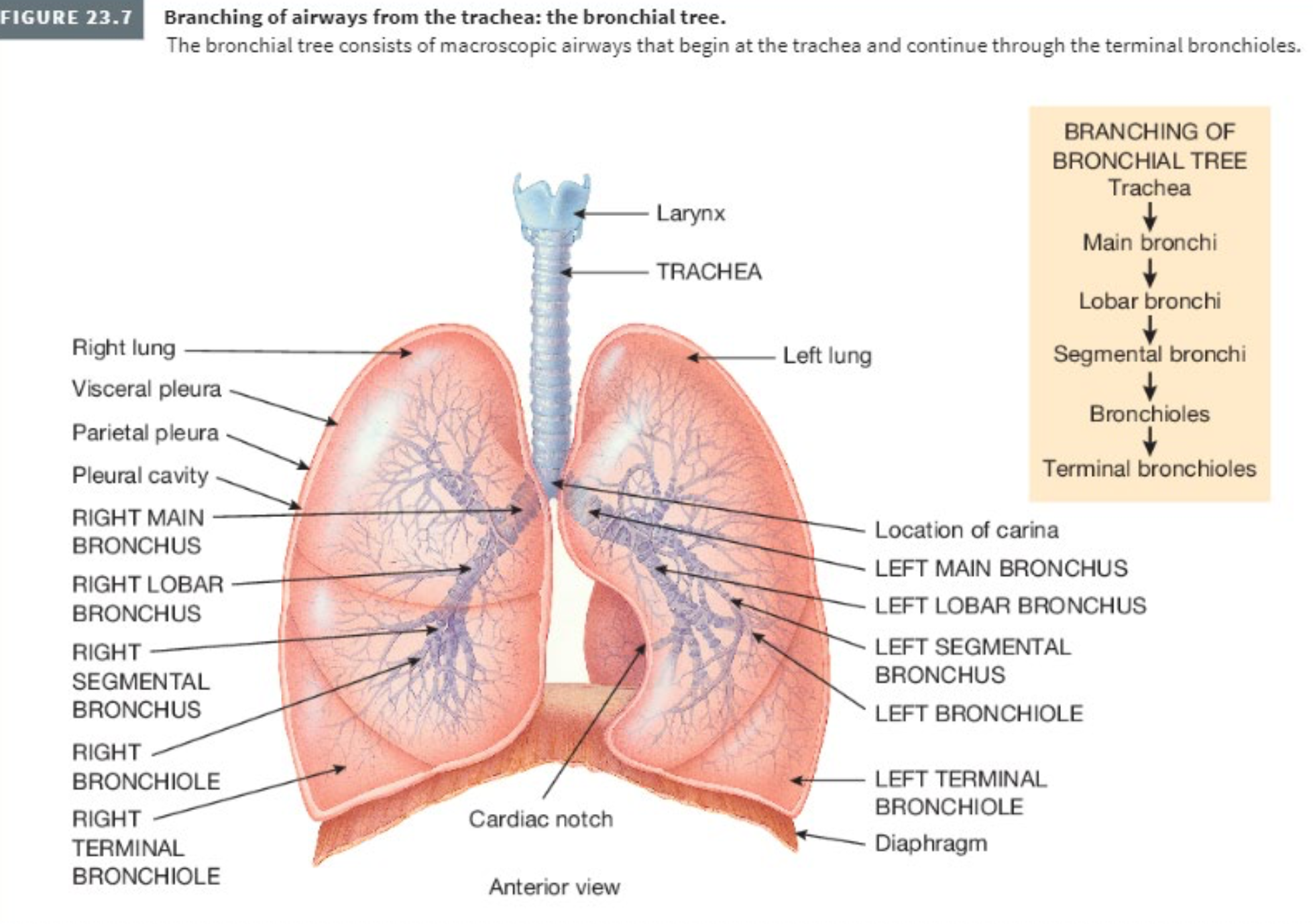
The terminal bronchioles subdivide into microscopic branches called respiratory bronchioles. This begins the respiratory zone of the respiratory system.
state description of lungs, include:
shape
location
function
lobes of lungs (include purpose)
SYSTEM: Respiratory System
SUBSECTION: Lungs
NOTE: None
CUE: None
two spongy, cone-shaped organs
located in the thoracic cavity
function to exchange gas, supplying oxygen to bloodstream, and removing carbon dioxide.
each lung is divided into lobes, allowing efficient airflow and ventilation
three lobes in right lung
two lobes in left lung
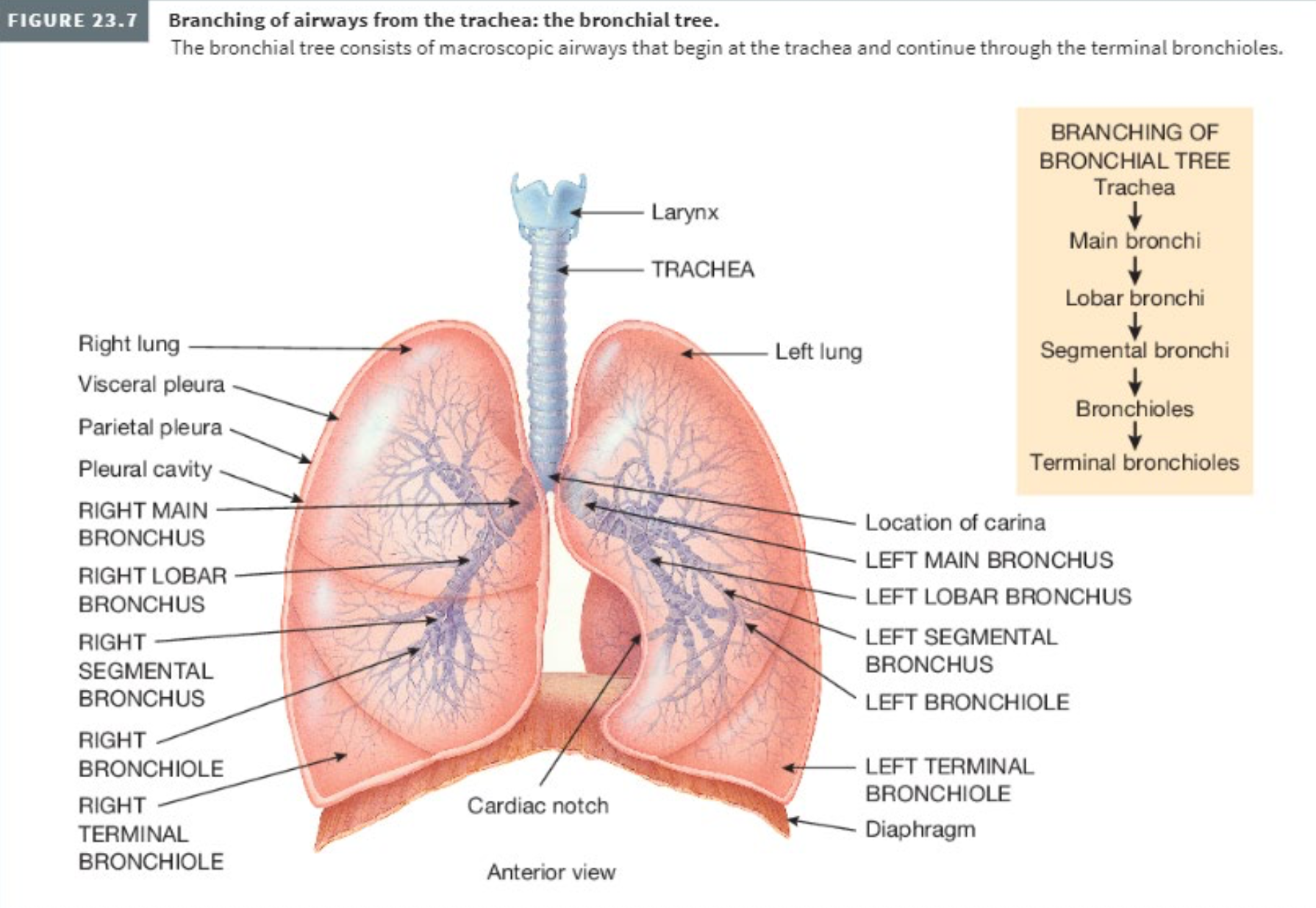
what is the cardiac notch, and what is its location and its purpose?
SYSTEM: Respiratory System
SUBSECTION: Lungs
NOTE: None
CUE: None
The cardiac notch is a concave indentation (as a shallow groove) located on the anterior surface of the left lung.
The cardiac notch accommodates the space occupied by the heart; is allows the left lung to fit around the heart, and contributes to the left lung having two lobes, compared to the three lobes of the right lung.
Due to the space occupied by the heart, the left lung is ~10% smaller than the right lung.

how are the left and right lung separated ?
why is it good that the lungs are separated ?
SYSTEM: Respiratory System
SUBSECTION: Lungs
NOTE: None
CUE: None
the lungs are separated from each other by the heart, and other structures of the mediastinum, which divides the thoracic cavity into two anatomically distinct chambers.
It is good that the lungs are separated in the case of trauma.
If trauma causes one lung to collapse, the other map remain expanded.
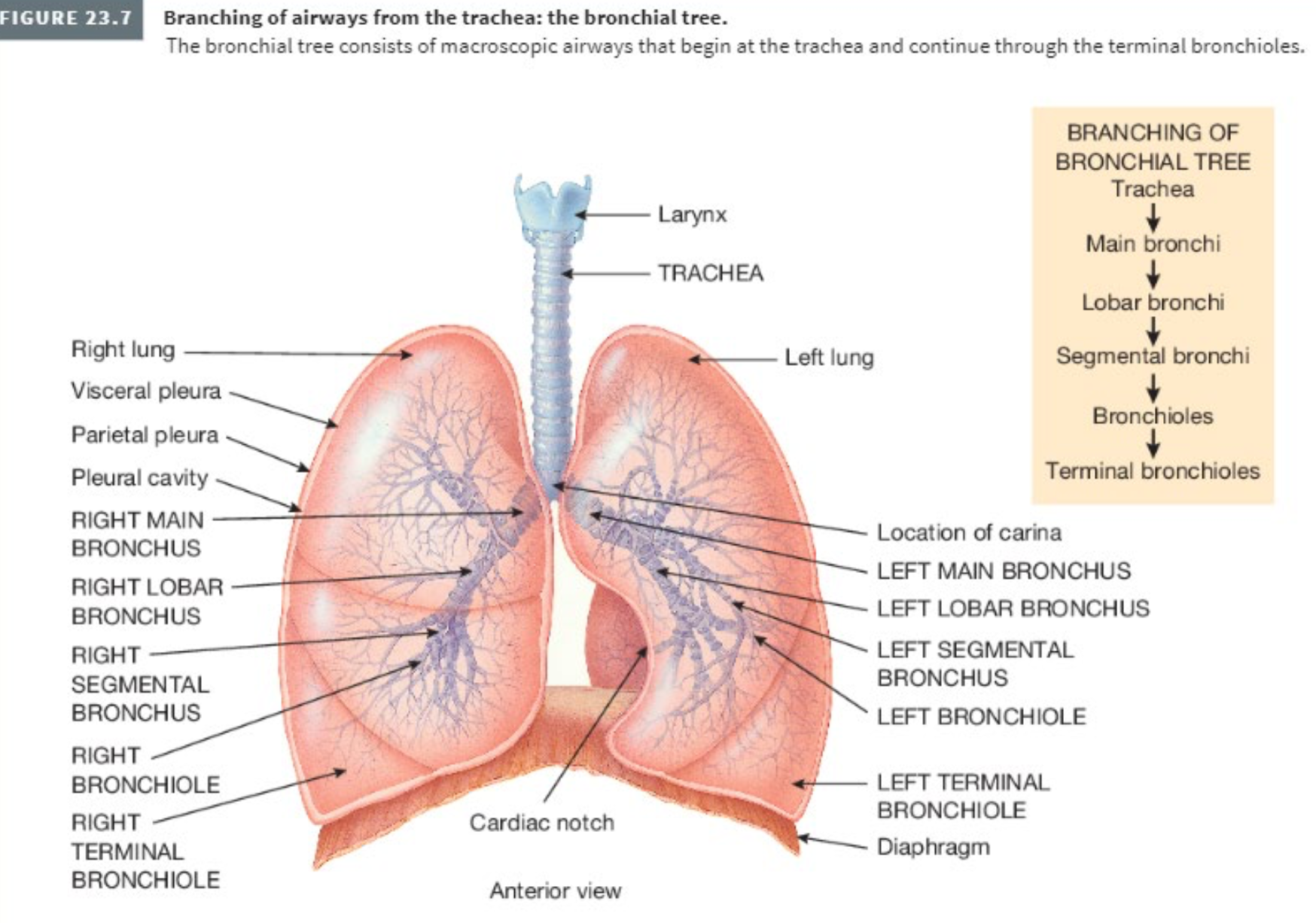
describe the pleural membrane that encloses the lungs.
talk about where pleural fluid comes from, and its function.
hence, briefly discuss pleuritis (inflammation of the pleural membrane), and pleural effusion.
SYSTEM: Respiratory System
SUBSECTION: Lungs
NOTE: None
CUE: pleural membrane
Each lung is enclosed and protected by a double-layered serous membrane called the pleural membrane.
the superficial layer is called the parietal pleura. this lines the wall of the thoracic cavity.
the deep layer is called the visceral pleura. this covers the lungs themselves.
Between the visceral and parietal pleurae is a small space called the pleural cavity.
separate pleural cavities surround the left and right lung.
the pleural cavity contains a small amount of lubricating fluid secreted by the membranes.
pleural fluid reduces friction between the membranes, allowing them to slide easily over each other during breathing.
pleural fluid also produces surface tension, causing the two membranes to adhere to one another (just as a film of water causes two glass microscope slides to sick together)
pleuritis refers to the inflammation of the pleural membrane.
where, in its early stages, pleuritis may be associated with pain due to friction between the parietal and visceral layers of the pleura.
if the inflammation persists, excess fluid accumulates in the pleural space, a condition known as pleural effusion.
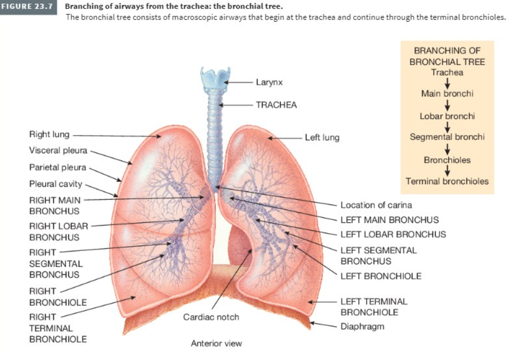
describe the lungs; the parts that are and/or make up the… areas of the lung.
convex
apex
costal surface
medial surface
cardiac notch
the extension of the pleura
lung lobes
SYSTEM: Respiratory System
SUBSECTION: Lungs
NOTE: There is a lot of random information in this card. So just, say whatever…
CUE: None
location:
The lungs extend from the diaphragm to just-slightly-superior to the clavicles, and lie against the ribs anteriorly and posteriorly.
shape:
convex
The broad inferior portion of the lung, the base, is concave and fits over the convex area of the diaphragm.
The base of the lungs extends from the 6th costal cartilage anteriorly to the spinous process of the tenth thoracic vertebra posteriorly.
apex:
The narrow superior portion of the lung is the apex.
The apex of the lungs lies super to the medial third of the clavicles. This is the only area that can be palpated.
costal surface:
The surface of the lung that lies against the ribs is called the costal surface.
It matches the rounded curvature of the ribs.
The anterior, lateral, and posterior surfaces of the lungs lie against the ribs.
collectively are referred to as the costal surface.
mediastinal (medial) surface:
the medial surface of each lung contains a region called the hilum (an area/depression/pit where blood vessels and nerves enter of leave an organ).
bronchi, pulmonary vessels, lymphatic vessels, and nerves enter and exit the lung through the hilum.
the hilum is held together by the pleura and connective tissue, and constitutes the root of the lung.
cardiac notch (did not add everything, can refer to cardiac notch card for more).
medially, the left lung also contains a concavity, called the cardiac notch. this is where the apex of the heart lies.
Due to the space occupied by the heart, the left lung is ~10% smaller than the right lung.
While the right lung is thicker and broader, is it also somewhat shorter than the left lung since the diaphragm is higher on the right side, accommodating the liver that lies inferior to it.
The pleura extends ~5cm below the base from the sixth costal cartilage anteriorly to the twelfth rib posteriorly. Hence, the lungs do not completely fill the pleural cavity in this area.
provides more space during inhalation and exhalation.
The lungs almost fill the thorax.
Each lung is divided into lobes, allowing efficient airflow and ventilation
three lobes in right lung
two lobes in left lung
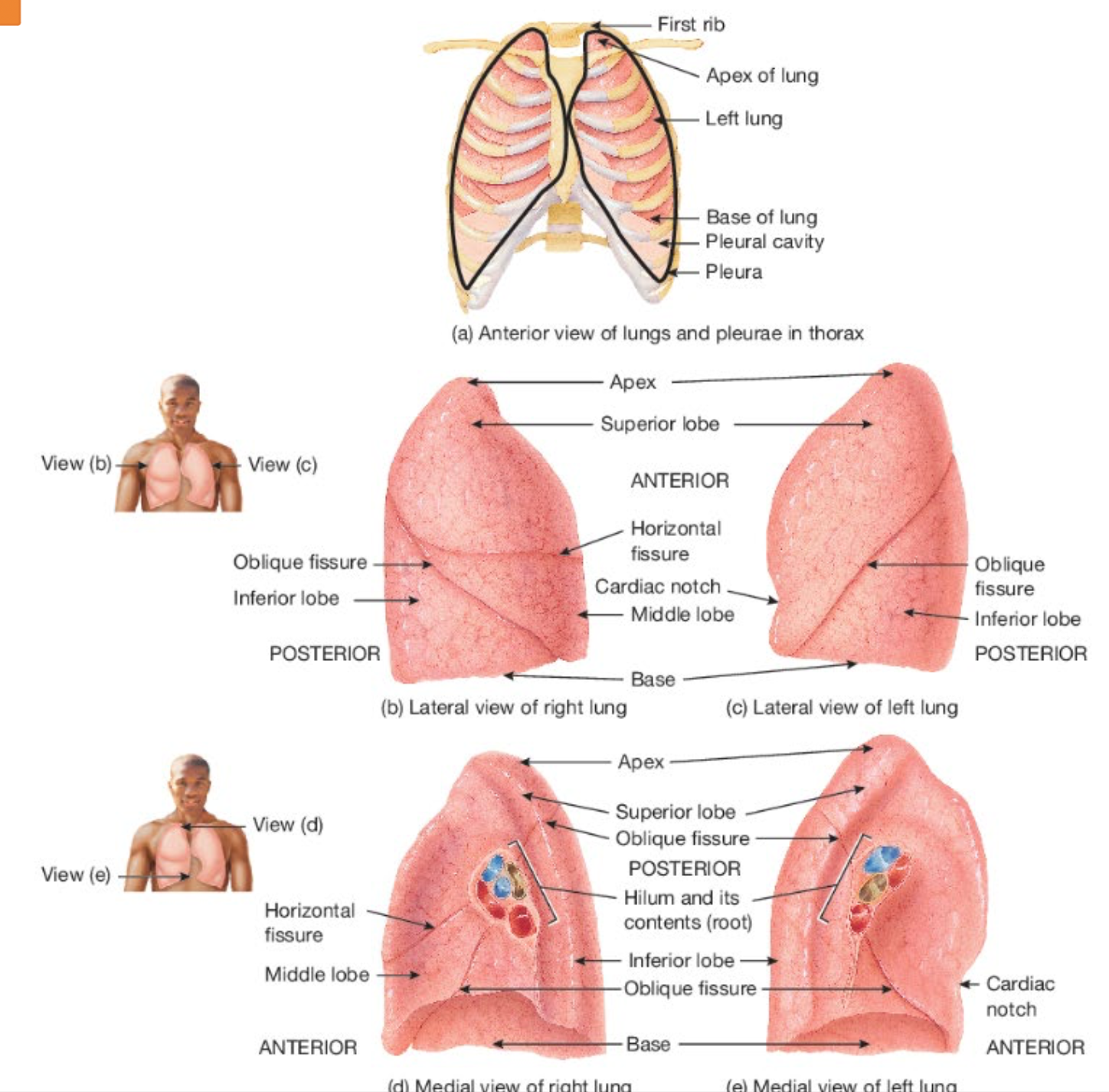
how can excessive fluid in the pleural cavity be removed ?
SYSTEM: Respiratory System
SUBSECTION: Lungs
NOTE: Not sure to what extend need to know this.
CUE: None
excessive fluid in the pleural cavity can be removed by inserting a needle anteriorly through the seventh intercostal space.
this does not injure lung tissue.
the needle is passed along the superior border of the lower rib to avoid damage to the intercostal nerves and blood vessels.
where inferior to the seventh intercostal space, is the danger of penetrating the diaphragm.
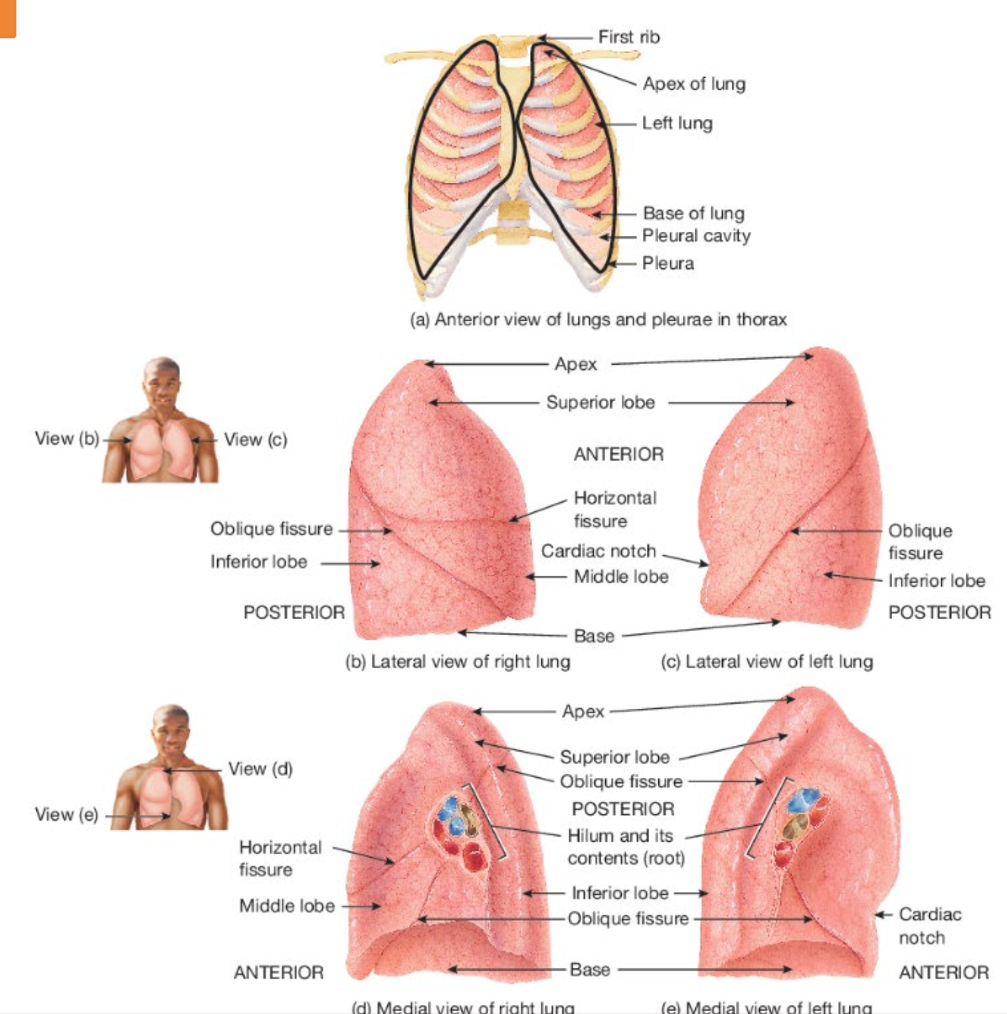
describe respiratory bronchioles, include:
whether they are apart of the conducting zone or respiratory zone, and why
what they divide into
what is an alveolus
what are alveolar macrophages
SYSTEM: Respiratory System
SUBSECTION: Lungs—Microscopic anatomy of lung lobules
NOTE: None
CUE: alveoli
Terminal bronchioles subdivide into microscopic branches called respiratory bronchioles. These begin the respiratory zone zone of the respiratory system.
respiratory bronchioles have alveoli budding from their walls.
alveoli participate in gas exchange (hence, why respiratory bronchioles begin the respiratory zone).
respiratory bronchioles subdivide into several (2-11) alveolar ducts.
where, around the circumference of the alveolar ducts are numerous alveoli and alveolar sacs.
alveolus
an alveolus is a cup-shaped outpouching supported by a thin elastic basement membrane called an alveolar sac.
outpouchings=sac-like protrusion or pocket
an alveolar sac consists of two or more alveoli that share a common opening.
alveolar macrophages
associated with the alveolar wall are alveolar macrophages (dust cells). these are phagocytes that remove dust particles and other debris from the alveolar spaces (via phagocytosis).
also present are fibroblasts, which produce reticular and elastic fibres.
describe the four layers of the respiratory membrane, and their role in gas exchange.
also discuss the thickness of the respiratory membrane.
SYSTEM: Respiratory System
SUBSECTION: Lungs—Microscopic anatomy of lung lobules
NOTE: None
CUE: None
int. the exchange of O2 and CO2 between the air in the lungs and the bloodstream occurs through diffusion (high to low) across the respiratory membrane.
this process enables O2 to enter the bloodstream, while CO2 is removed (from the bloodstream) to be exhaled.
the respiratory membrane is formed by the thin walls of the alveoli and the capillaries, and consists of four distinct layers.
Alveolar wall:
first, outermost layer
comprised of type I and type II alveolar cells (crucial for gas exchange and surfactant production, respectively), and alveolar macrophages (help remove particles and pathogens from alveolar spaces).
Epithelial basement membrane:
second layer
located just beneath the alveolar cells.
provides structural support and anchors the alveolar cells to underlying tissue.
Capillary basement membrane:
third layer
located beneath the capillary endothelial cells, and is often fused with the epithelial basement membrane.
helps to reduce the distance for gas diffusion between the alveolar air space and the capillary blood.
Capillary endothelium:
the innermost layer of the respiratory membrane
comprised of endothelial cells
allows for rapid exchange of gases between the alveoli and the blood; as O2 moves into the blood, and CO2 moves out.
Thinness of respiratory membrane:
The layers of the respiratory membrane collectively form an extremely thin layer, allowing for efficient and rapid diffusion of gases between the alveolar air and the bloodstream; essential for maintaining proper oxygenation of the body.
despite having several layers, respiratory membrane is very thin.
~0.5µm thick, ~1/16th the diameter of a red blood cell
this facilitates rapid diffusion of gases.
It has been estimated that lungs contain 300 million alveoli, providing immense surface area of ~72m2 for gas exchange (~the size of a racquetball court).
define the following terms
breathing (aka. pulmonary ventilation)
inspiration
expiration
quiet breathing
forced breathing
ventilation
respiration
internal respiration
external respiration
SYSTEM: Respiratory System
SUBSECTION: Mechanisms of Breathing
NOTE: None
CUE: None
Breathing (or pulmonary ventilation):
the physical act of inhaling (taking air into the lungs) and exhaling (expelling air from the lungs).
it is the mechanical process that moves air in and out of the lungs, allowing for gas exchange.
inspiration (breathing in):
air enters the lungs to supply O2expiration (breathing out):
air is expelled, removing CO2
Quiet breathing:
passive process, relies on diaphragm and elastic lung recoil.Forced breathing:
active process, involves additional muscle recruitment to increase airflow, like during exercise.
Ventilation:
the overall process of air moving in and out of the lungs (breathing), but also describes the efficiency of this airflow in relation to gas exchange.
ventilation is concerned with how well air reaches the lungs, ensuring O2 is delivered and CO2 is removed.
Respiration:
encompasses the entire process of gas exchange in the body, including:
External respiration:
the exchange of O2 and CO2 between the air in the lungs and the blood in the capillaries.(air crosses between the capillaries; O2 enters capillaries, CO2 leave capillaries into alveoli, and later on, exhaled).
Internal respiration:
the exchange of gases between the blood and the tissues of the body, where O2 is delivered to cells, and CO2 is removed for transport back to the lungsO2 reaches capillaries, is carried by red blood cells, reaches tissues and cell—usually for cellular respiration; CO2 from cells is carried by red blood cells to the capillaries, to the lungs, and then exhaled).
explain the process of gas exchange in the body (respiration)
SYSTEM: Respiratory System
SUBSECTION: Mechanisms of Breathing
NOTE: None
CUE: three steps
pulmonary ventilation (breathing):
the inhalation (inflow) and exhalation (outflow) of air, and involves the exchange of air between the atmosphere and the alveoli of the lungs.air flows between the atmosphere and the alveoli of the lungs because of the alternating pressure differences created by the contraction and relaxation of respiratory muscles.
the rate of airflow, and the amount of effort needed for breathing are also influenced by alveolar surface tension, compliance of the lungs, and airway resistance.
external (pulmonary) respiration:
gas exchange between the alveoli of the lungs and the blood in pulmonary capillaries across the respiratory membrane. this is where pulmonary capillary blood gains O2 and loses CO2.internal (tissue) respiration):
gas exchange between blood in systemic capillaries and tissue cells. this is where blood loses O2 and gains CO2.
within cells, the metabolic reactions that consume O2 and form CO2 during ATP production are termed cellular respiration.
what does Boyle’s law state
SYSTEM: Respiratory System
SUBSECTION: Mechanisms of Breathing
NOTE: None
CUE: None
Boyle’s Law explains pulmonary ventilation (breathing), and describes the relationship between pressure and volume.
Boyle’s law states that at constant temperature for a fixed mass, the absolute pressure and the volume of a gas are inversely proportional.
summary:
pressure is inversely proportional to volume.
gases flow from high to low pressure.
inspiration:
↑ volume, ↓ pressure = air flows in
expiration:
↓ volume, ↑ pressure = air flows out
Boyle’s law details how air moves in and out of the lungs based on pressure differences.
during inhalation, lung volume increases, decreasing the pressure inside, drawing air in.
air moves into the lungs when the air pressure inside the lungs is less than the air pressure in the atmosphere.
breathing in=inhalation/inspiration.
just before each inhalation, the air pressure inside the lungs is equal to the air pressure in the atmosphere (which at sea level is ~760mmHg or 1atm).mmHg = millimetres of mercury
atm = atmosphere
for air to flow into the lungs, the pressure inside the alveoli must become lower than the atmospheric pressure; this condition is achieved by increasing the size of the lungs.
(if size/volume increases, pressure decreases)
during exhalation, lung volume decreases, increasing the pressure inside, forcing air out.
air moves out the lungs when the air pressure inside the atmosphere is less than the air pressure inside the lungs.