M6 Cardiac_Ventriculography *
1/44
There's no tags or description
Looks like no tags are added yet.
Name | Mastery | Learn | Test | Matching | Spaced | Call with Kai |
|---|
No analytics yet
Send a link to your students to track their progress
45 Terms
What is the goal of catheter selection in ventriculography? often what french size?
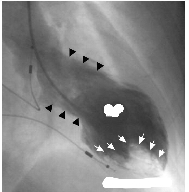
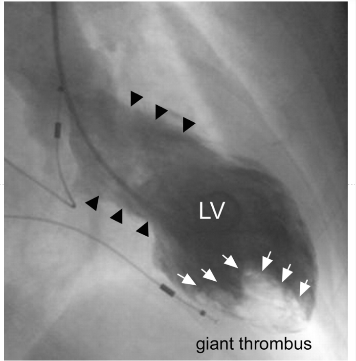
what do you see in this vgram
thrombus
What are the 3 types of catheters used in ventriculography?
Pigtail Catheters,
Straight Tip Catheters,
Balloon Tip Catheters.
function of the sideholes on the pigtail catheter
They have a pigtail curl and multiple side holes to prevent the contrast jet from damaging the endocardium.
Imaging ventricles, pulmonary arteries, and pulmonary veins anatomy.
dependent on the placement of side holes whether distal or prox depends on if its intended for wedge or ventricle
What is the optimal catheter position in ventriculography?
50% - 70%.
What is the EF of mild hypokinesis?
What is the typical cause of endocardial staining?
Lack of wall motion; the wall doesn't contract.
Define akinesia as it relates to ventricular analysis.
Abnormal wall motion, where the wall moves in the opposite direction during contraction.
What is dyskinetic wall motion?
Catheter position and rate of contrast injection to avoid endocardial staining. due to straight tip
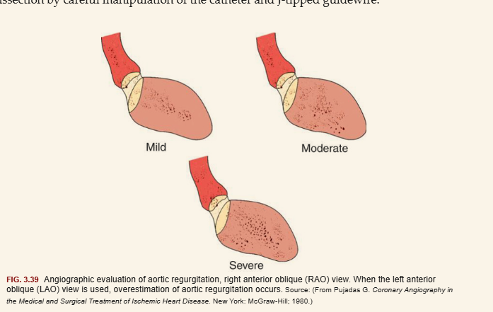
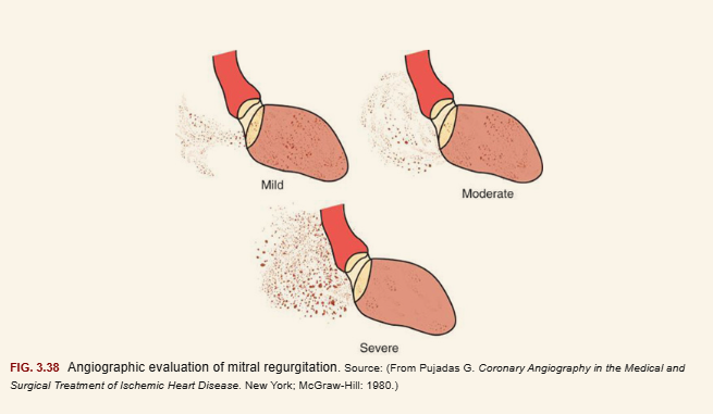
Mitral Valve Regurgitation (MVR) assessment graded from what to what
graded from 1 - 4
1 being best
4 being severe
What is the technique (type of catheters) for imaging in the right heart system?
______________ ______ occurs when there is improper catheter positioning during ventriculography, leading to the contrast media causing discoloration of the heart
endocardial staining
Catheters used for coronary angiography from the radial approach are generically called __________ catheters because they can engage both the _____ and the _____ coroanry ostia
universal catheters
engage both left and right
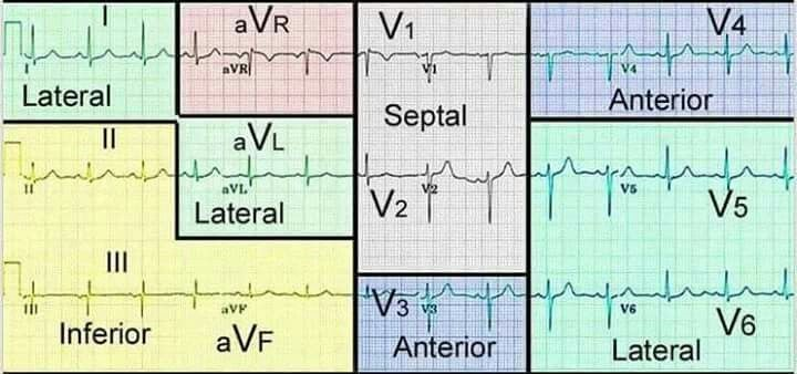
left ventriculography best view for
anterior
apical
inferior
sides of the heart is
30 RAO
in L ventriculography
best view for
septal
lateral
posterior
sides of the heart is
60 LAO
in LVG 60 LAO is best view for what sides of the heart
3x
septal
lateral
posterior
in LVG 30 RAO
is best view for what sides of the heart
3x
anterior
apical
inferior
for RVG
what is the best view to see the RVOT & central pulmonary arteries
AP CRA
For RVG in AP CRA
you can see what sides of the hearts
RVOT (infundibulum)
CENTRAL PA
what is the orientation on the left and right
L LAO
R RAO

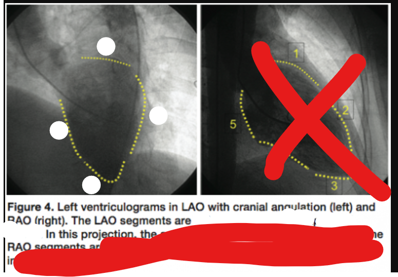
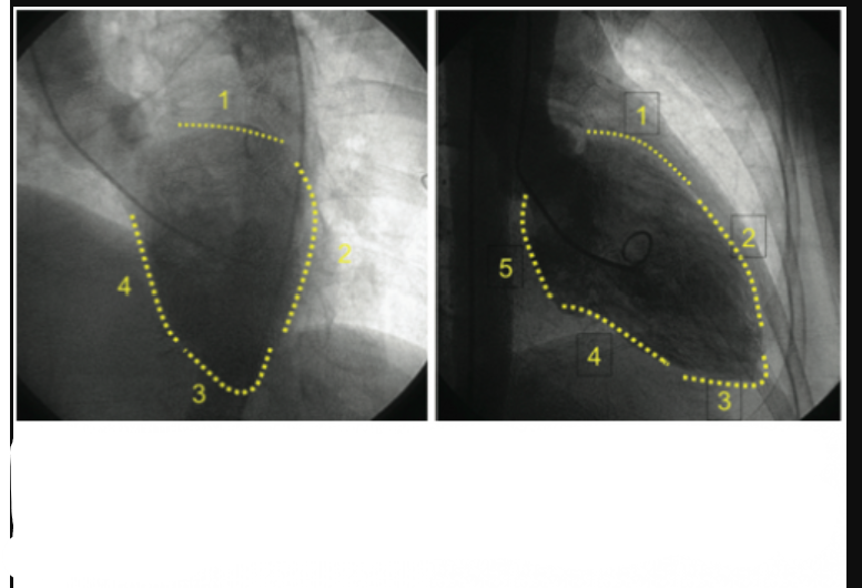
label the heart surface in this LAO angiographic image
basal/ posterior
lateral
apical
septal
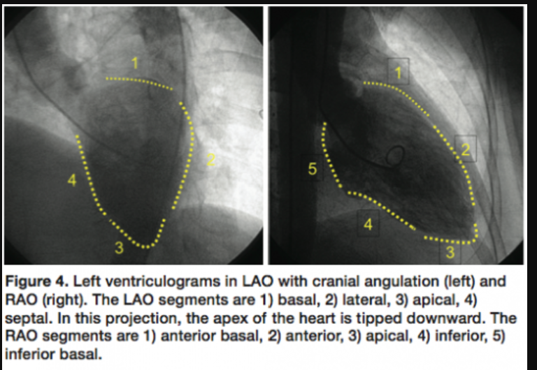
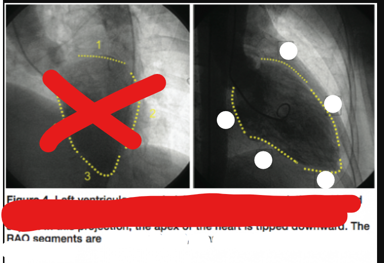
label the heart surface in this RAO angiographic image
anterior basal
anterior
apical
inferior
inferior basal


review this image
review this
image to assess cardiac function and identify any abnormalities in the ventricles.

review this


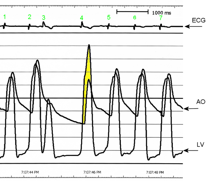
found in HOCM. is a sign of left ventricular outflow tract obstruction. It's characterized by a drop in arterial pressure after a premature heartbeat.
Brockenbrough phenomenon
Paradoxical Drop in Arterial Pulse Pressure:
Instead of generating a strong post-PVC pulse (which happens in normal hearts), the LVOT obstruction blocks blood flow, leading to:
Higher LV pressure (because blood is retained due to the obstruction).
Lower aortic pressure (because less blood is ejected).
This results in a decreased pulse pressure (lower SBP) after the PVC.
more volume leads to greater obstruction due to the hypertrophied septum and systolic anterior motion (SAM) of the mitral valve
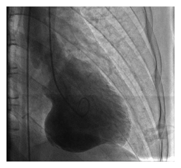
what cardiomyopathy
apical HCM

projection?
RAO
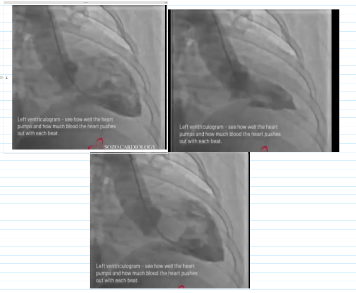
WALL MOTION ASSESSMENT
ESTIMATED EF
NORMAL
55-60% EF
projection
Wall motion assessment
Estimated ejection fraction
Any valvular disease visualized
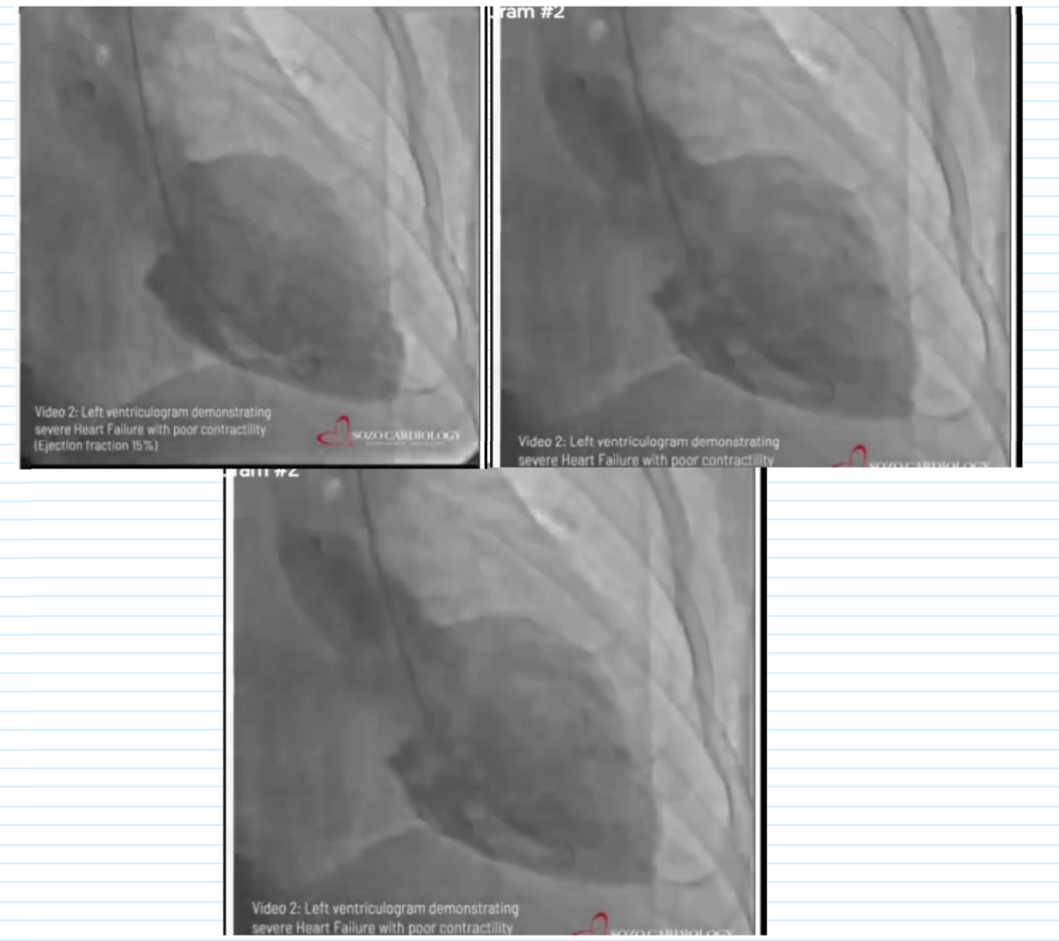
Wall motion assessment: Severe global hypokinesis
Estimated ejection fraction: ~20%
Any valvular disease visualized: none
projection
Wall motion assessment
Estimated ejection fraction
Any valvular disease visualized
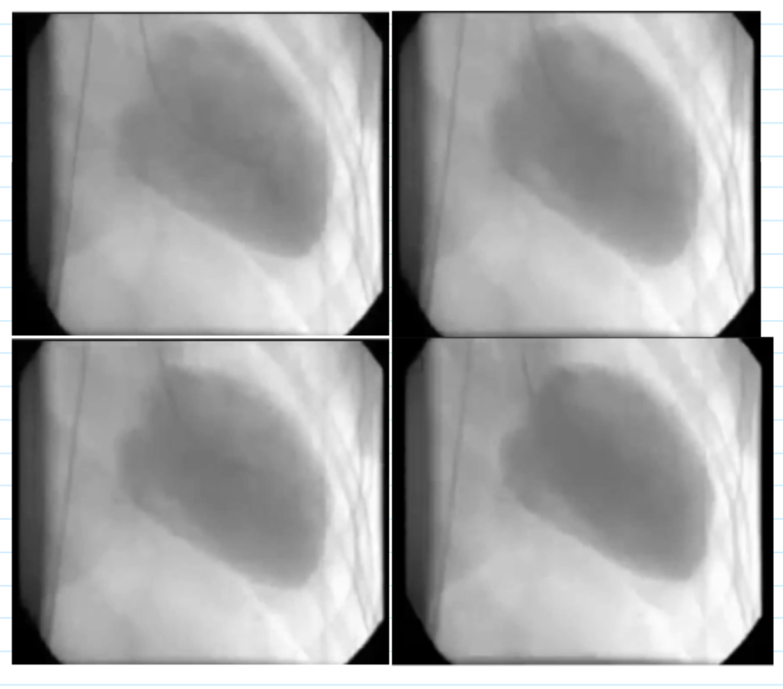
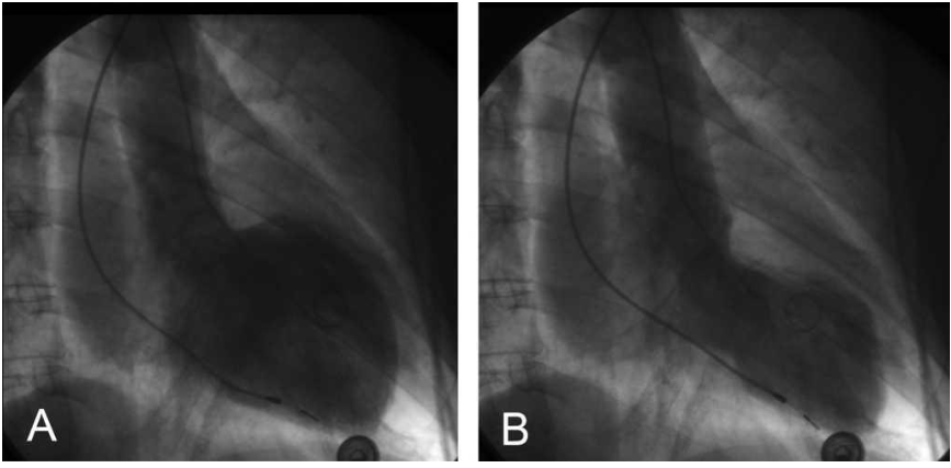
Projection (RAO/LAO): RAO
Wall motion assessment: Severe global hypokinesis
Estimated ejection fraction: ~20%
Any valvular disease visualized: none
Projection (RAO/LAO)
Wall motion assessment
Estimated ejection fraction
Any valvular disease visualized
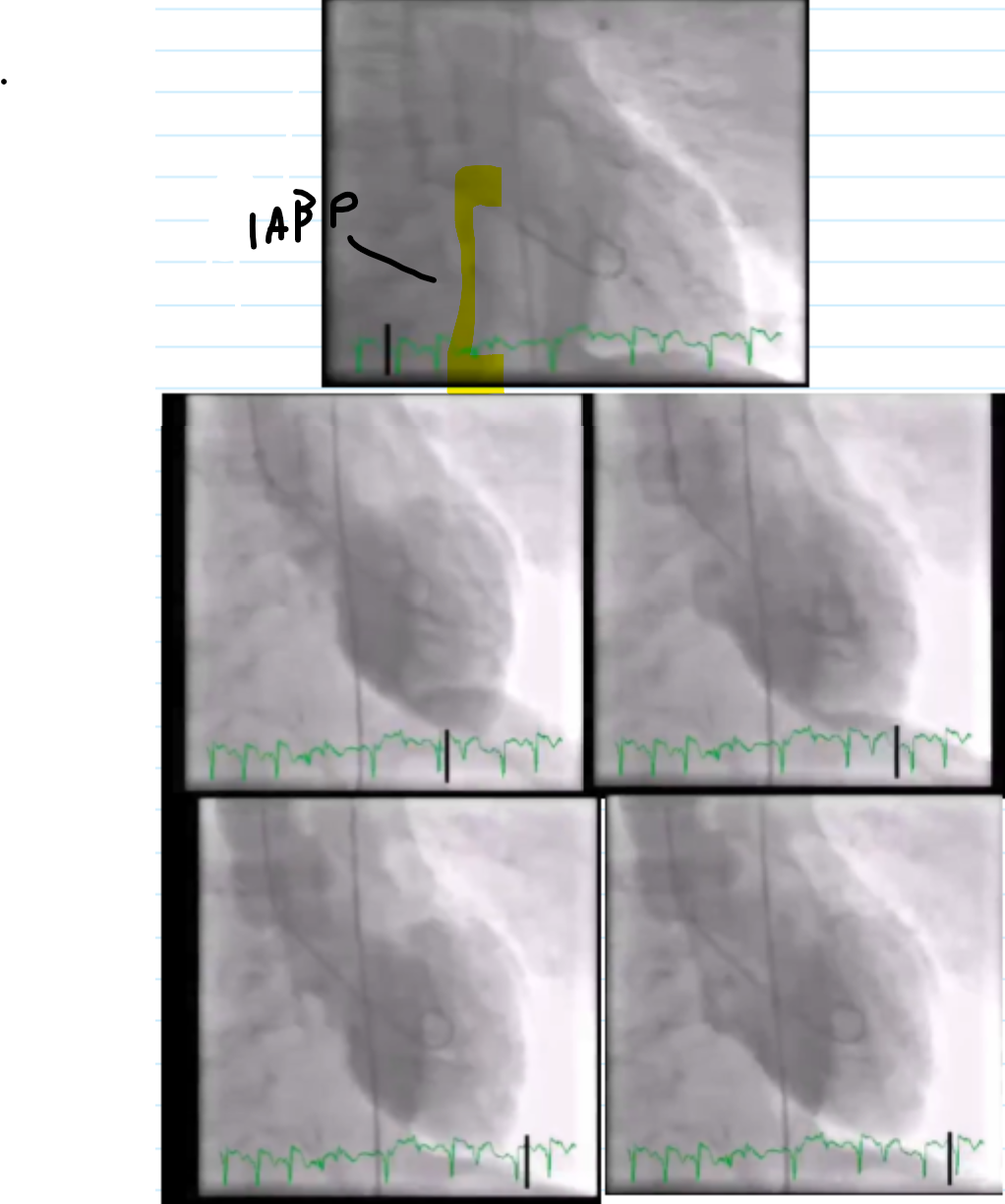
Projection (RAO/LAO): RAO
Wall motion assessment: Takotsubo (anterior, apical and inferior akinesis)
Estimated ejection fraction: ~30%
Any valvular disease visualized: none
IABP present
Ventriculogram: Poor EF Secondary to Massive M.I. 42 second loop.
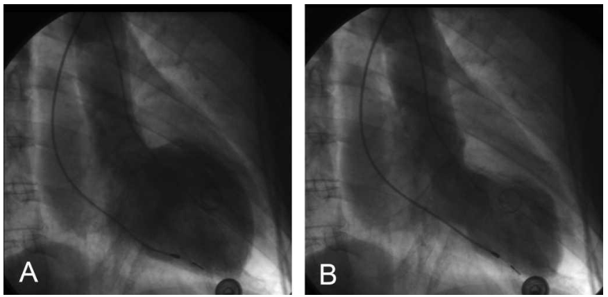
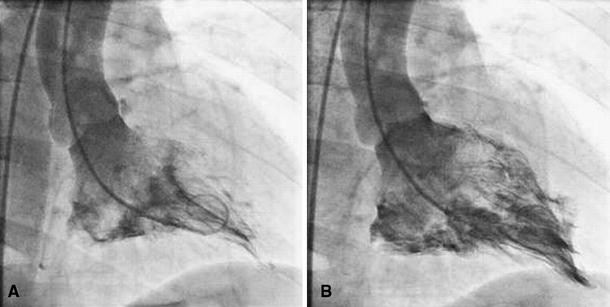
Review the image carefully and give the abnormality seen in the image.
4+ MR
what is the rating of mitral regurg
4+ M.R.
Review the image carefully and give the abnormality seen in the image.
LV ANEURYSM