Diencephalon, Basal Ganglia and Cerebral Cortex
 Call Kai
 Learn
Learn Practice Test
 Spaced Repetition
 Match
Match Flashcards
 Knowt Play
1/92

There's no tags or description
Looks like no tags are added yet.
Name | Mastery | Learn | Test | Matching | Spaced | Call with Kai | Chat |
|---|
No analytics yet
Send a link to your students to track their progress
93 Terms
Locate and identify the key structures of the diencephalon
Recall the components of the basal ganglia and explain their general functional roles in motor control
Locate and name the major gyri and sulci (landmarks) of the cerebral hemispheres
Describe the key functional cortical areas and integrate this knowledge with previously learned concepts (i.e., descending motor pathways and ascending sensory pathways).
Describe the Brodmann system for cortical mapping and the sensory and motor homunculi
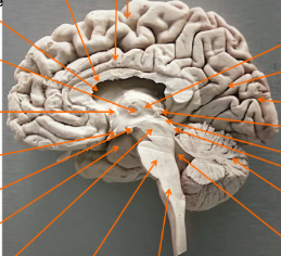
Label this image and identify the uncus
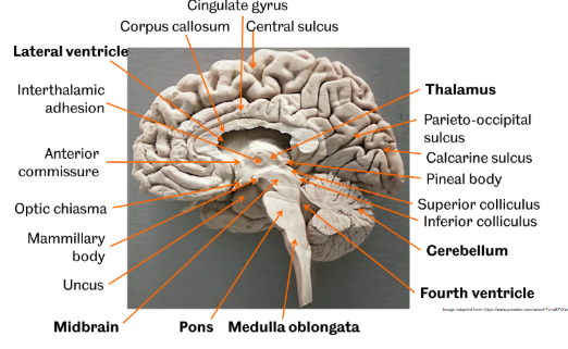
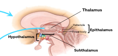
Define the diencephalon
key division of the forebrain situated between the telencephalon and midbrain, surrounding the third ventricle. Comprising the thalamus, hypothalamus, epithalamus, and subthalamus, it acts as a central relay/processing hub for sensory information, endocrine control, and emotional regulation
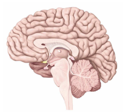
Locate the diencephalon in this right brain midsagittal section
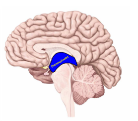
What are subdivisions of the diencephalon?
hypothalamus, thalamus, subthalamus, epithalamus (made up of habenula and pineal body)
Define the telencephalon
comprising 85% of total human brain weight. It includes the cerebral cortex, subcortical structures (hippocampus, basal ganglia), and olfactory bulbs
What are the 3 nuclear groups of the thalamus?
anterior, medial, lateral
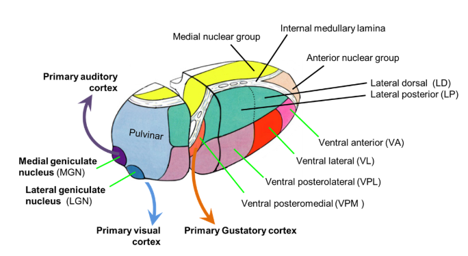
Role of the anterior nuclear group of the right thalamus
Involved in memory and emotion (part of the limbic system)
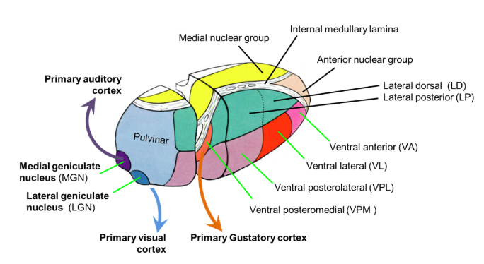
Role of the medial nuclear group of the right thalamus
Associated with individual awareness and emotional processing
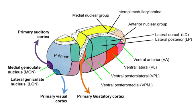
Role of the lateral nuclear group of the right thalamus
The largest group, which handles most sensory and motor relays
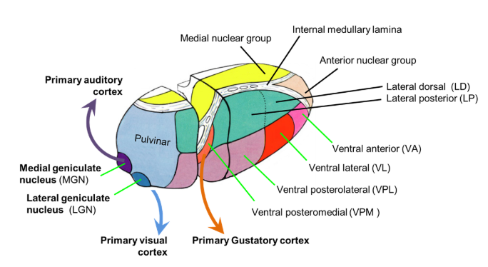
What are the subdivisions of the lateral nuclear group of the right thalamus? What does each section connect with?
Lateral Dorsal (LD) and Lateral Posterior (LP) nuclei are part of the lateral nuclear group of the thalamus, primarily functioning in cognitive and sensory association
LD nucleus connects with the limbic system (cingulate gyrus) and plays roles in emotional processing
LP nucleus (related to the pulvinar) acts as a higher-order relay for visual, parietal, and sensory inputs
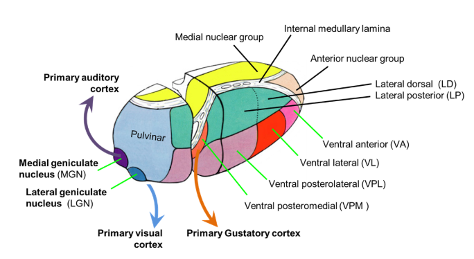
Functions of the thalamus
It’s a bit like directory enquiries!
Sensory relay
Support for Motor systems
Mental operation regulation
Which sensory pathways pass through the thalamus?
reactive and discriminative ie spinothalamic and dorsal column-medial lemniscus pathways
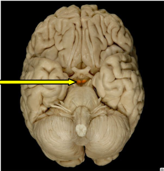
What is this arrow pointing to?
mammillary body, pair of small, round nuclei located on the inferior surface of the posterior hypothalamus, part of the diencephalon and the limbic system. They are crucial for memory, spatial navigation, and emotion, receiving inputs from the hippocampus
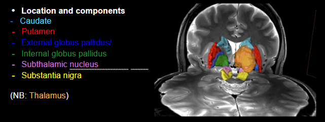
What makes up the basal ganglia?
Caudate
Putamen
External globus pallidus
Internal globus pallidus
Subthalamic nucleus
Nucleus accumbens
Substantia nigra
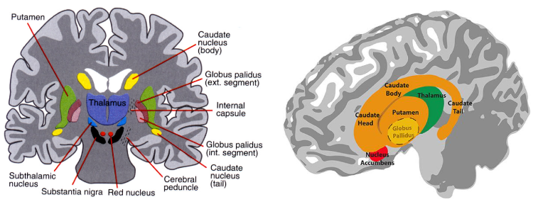
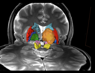
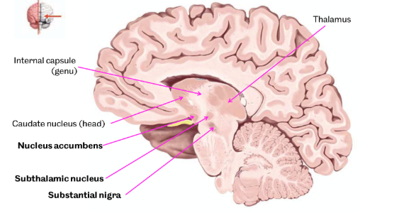
Locate the structures that make up the basal ganglia
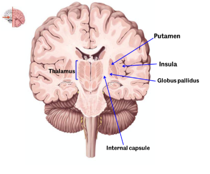
What is the external capsule sandwiched between?
between putamen and claustrum
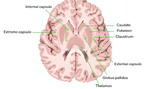
What type of matter are the internal and external capsules?
white matter
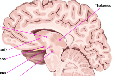
label this right brain parasagittal view of the brain with the structures making up the basal ganglia
thalamus, nucleus accumbens, subthalamic nucleus and substantia nigra evident
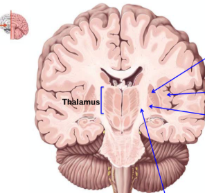
Label this coronal anterior section of the brain
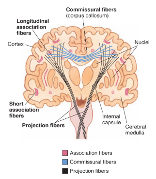
What are the 3 types of cerebral white matter/fibres?
association, commissural and projection fibres
Which pathways pass through the basal ganglia?
motor pathways - corticospinal and corticonuclear
What is the function of basal ganglia?
Direct: facilitates movements by Disinhibition.
Indirect: Inhibition of unwanted movements.
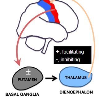
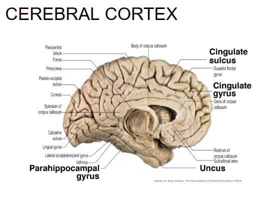
What do these structures make up?
cerebral cortex
What does the limbic system include?
parts of the cerebral cortex, basal nuclei, the thalamus, the hypothalamus, and the olfactory cortex
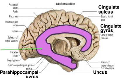
What does the limbic system control?
visceral functions through the autonomic nervous system and the endocrine system and is involved in emotions and memory, reproduction and nutrition.
What are the different areas of cortical function ?
motor control (frontal), sensory processing (parietal), auditory processing (temporal), and vision (occipital): eg
Primary Motor Cortex (Frontal Lobe): Initiates and controls voluntary movements.
Prefrontal Cortex (Frontal Lobe): Involved in high-level cognitive processes such as planning, decision-making, and personality.
Broca’s Area (Frontal Lobe): Responsible for speech production and language processing.
Primary Somatosensory Cortex (Parietal Lobe): Processes sensory information regarding touch, temperature, pressure, pain, and body position.
Explain the Brodmann method of numbering
a system of numbering regions of the cerebral cortex based on their cytoarchitecture
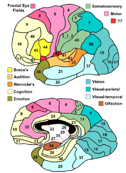
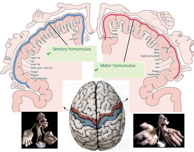
What are the homunculi? Which 2 homunculi are visible on this image?
Sensory and Motor Homunculi (Representations of the body mapped onto the primary somatosensory and motor cortices, illustrating the proportion of cortex dedicated to different body parts)
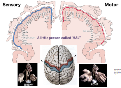
What are sulci and gyri and their function?
The human brain's surface is characterised by ridges known as “gyri” (singular: gyrus) and grooves called “sulci” (singular: sulcus). These structures increase the brain's surface area, allowing for a greater number of neurons and enhanced cognitive abilities.
Describe key neural connection pathways in the brain?
The key neural connections (ascending and descending pathways) between the cortex, basal ganglia, brainstem, spinal cord, and cerebellum, including Spinothalamic tract, Dorsal column-medial lemniscus pathway, Corticospinal tract and Corticobulbar tract.
Summarise the basal ganglia
group of subcortical nuclei involved in motor control and other functions. The main components include Caudate Nucleus, Putamen and Globus Pallidus, Nucleus accumbens, Subthalamic Nucleus and Substantia Nigra. Their modulatory roles in motor control are primarily achieved through two opposing internal circuits, the Direct Pathway (which promotes movement) and the Indirect Pathway (which inhibits movement)
Other names for diencephalon
interbrain and tweenbrain
Where is the diencephalon located?
nestled deep between the cerebral hemispheres, positioned superior to the brain stem.
Function of the diencephalon
serves as a vital connection point, relaying virtually all information traveling between the cerebrum (the largest part of the brain) and the rest of the nervous system, with the sole exception of the sense of smell (olfaction).
What forms the wall of the third ventricle? What is this latter structure?
diencephalon
fluid-filled cavity in the brain, underscoring its central and protected location within the skull
What are the 4 subdivisions of the diencephalon?
Thalamus, the Hypothalamus (inferior and anterior to the thalamus), the Epithalamus (posterior to the thalamus, containing the pineal gland), and the Subthalamus (ventral to the thalamus).
What is the largest part of the diencephalon? What does it consist of?
thalamus is the largest part, making up about 80% of the diencephalon's volume, and consists of paired ovoid structures on either side of the third ventricle
Role of the diencephalon
plays an indispensable role in maintaining essential bodily functions and consciousness
Role of thalamus
acts as the major relay station and processing center for almost all sensory and motor signals destined for the cerebral cortex
Role of hypothalamus
primary centre for regulating homeostasis, controlling the autonomic nervous system, body temperature, hunger, thirst, sleep, and emotional responses, and it directly controls the endocrine system through the pituitary gland.
Role of epithalamus
via the pineal gland, secretes melatonin to regulate the sleep-wake cycle (circadian rhythm)
Role of subthalamus
involved in modulating motor functions and is highly interconnected with the basal ganglia
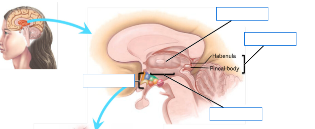
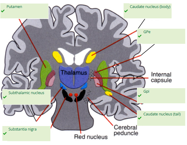
Label this image
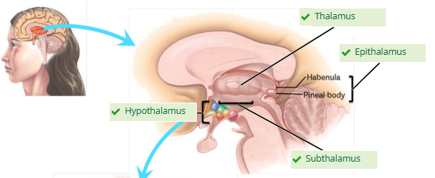
What are the primary components of the basal ganglia?
striatum (composed of the caudate nucleus and the putamen), the globus pallidus (divided into the external segment, GPe, and the internal segment, GPi), the subthalamic nucleus (STN), and the substantia nigra (SN)
Where are components of the basal ganglia found?
cluster of subcortical nuclei located deep within the white matter of the forebrain, diencephalon, and midbrain
What is the main input structure of the basal ganglia and what does it receive?
striatum, receiving excitatory signals from the entire cerebral cortex
What forms the output nuclei?
GPi and a part of the substantia nigra, the pars reticulata (SNr)
What do the output nuclei send projections to?
send inhibitory projections to the thalamus
Which intrinsic nuclei are essential for modulating flow of information?
STN, GPe, and the substantia nigra pars compacta (SNc)
What is the primary functional role of basal ganglia?
primarily as a gate-keeping mechanism for the initiation and execution of voluntary motor movements. It acts as a tonic "brake," constantly inhibiting the motor circuits in the thalamus and cortex, thereby preventing unwanted movements.
Define disinhibition/action selection
process of initiating a desired movement involves lifting this inhibitory brake on the specific motor program required
Which basal ganglia nucleus is central to its role of disinhibition?
striatum is central to this, integrating cortical signals and determining which motor program to "select" and allow through the basal ganglia-thalamo-cortical loop
Which opposing neural circuits achieve disinhibition?
direct pathway: directly inhibiting the output nuclei (GPi/SNr), which in turn disinhibits the thalamus, allowing it to excite the motor cortex
indirect pathway suppresses movement by ultimately increasing the inhibitory output of the GPi/SNr via a circuit involving the GPe and STN, which results in more inhibition of the thalamus
What is the consequence of dysfunction of basal ganglia disinhibition dysfunction?
Dysfunction in this system, such as dopamine loss in Parkinson's disease, leads to characteristic movement disorders.
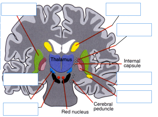
Label this image
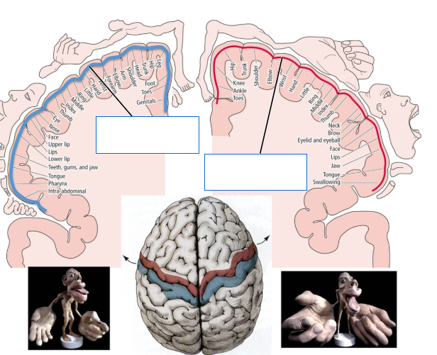
Label this image
What increases the surface area of the cerebral hemispheres?
defined by an intricate network of ridges, the gyri, and grooves, the sulci, which significantly increase the cortical surface area
What are major ridge landmarks?
Central Sulcus (fissure of Rolando), which separates the frontal and parietal lobes
Lateral Sulcus (fissure of Sylvius), which separates the temporal lobe from the frontal and parietal lobes.
How are key functional areas of the cerebral hemispheres mapped?
using the Brodmann system based on cytoarchitecture
What are other names for the primary motor cortex?
precentral gyrus and brodmann area 4
Which pathway originates at the primary motor cortex ? What does it control?
the origin of the descending Corticospinal Tract (Pyramidal Pathway) for voluntary movement, controlling the contralateral body
Which pathways terminate at the primary somatosensory cortex ? What does it control?
termination site for the ascending Dorsal Column-Medial Lemniscal Pathway (fine touch, vibration, proprioception) and the The Spinothalamic Tract (also known as Anterolateral System; pain, temperature, crude touch), conveying sensory information from the contralateral side of the body.
What are other names for the primary somatosensory cortex?
Postcentral Gyrus and Brodmann Areas 1, 2, 3
Where are the motor and sensory homunculi located? Define these
cortical areas located in the primary motor and somatosensory cortexes
distorted topographic maps where the size of the body part is proportional to the density of motor or sensory innervation, resulting in disproportionately large representations for the hands, lips, and face
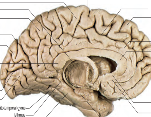
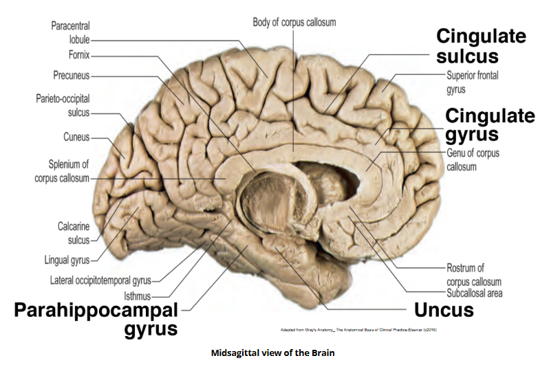
Locate the cingulate sulcus, gyrus, parahippocampal gyrus and uncus on this image
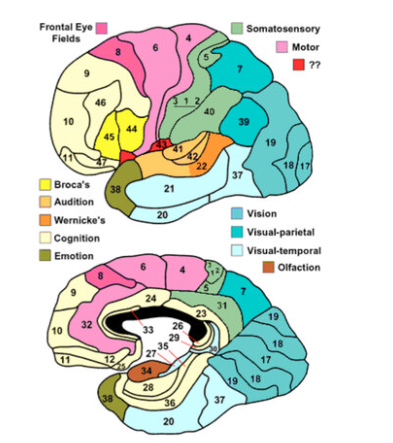
What does the Brodmann map refer to?
system of dividing the cerebral cortex into 52 numbered regions based on differences in cellular structure under a microscope rather than just gross anatomy or function, with each area having distinct cell layers and densities that correlate to specific functions, making it a foundational though sometimes refined map in neuroscience for localising brain activity
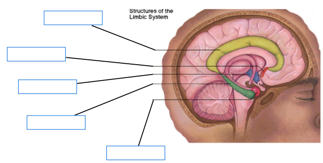
Label this image
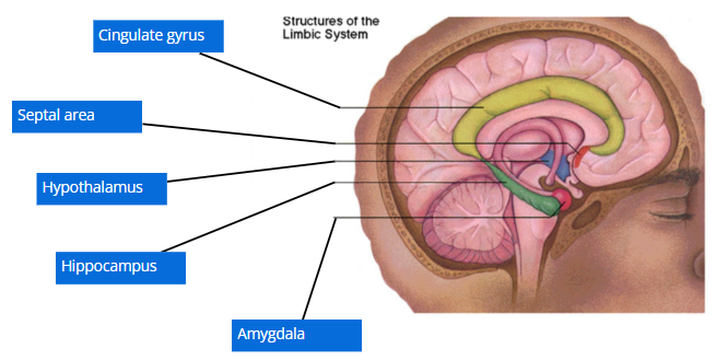
What are the main components of the limbic lobe?
Cingulate Gyrus (Brodmann Areas 23, 24, 33), located above the corpus callosum
Parahippocampal Gyrus (Brodmann Area 28, the Entorhinal Cortex, is key for memory), which curls into the temporal lobe and contains the Hippocampus (crucial for memory formation) and the Amygdala (central for emotion and fear memory)
What is the limbic lobe and what role does it play?
C-shaped ring of cortex on the medial surface of the cerebrum, playing a central role in emotion, memory, and motivation
What is Brodmann area 6 and its function?
Premotor and Supplementary Motor Areas involved in motor planning
Brodmann area 44 and 45 and function
Broca’s area for speech production
Broca area 22 and function
Wernicke's Area for language comprehension
Broca area 17 and function
primary visual cortex - occipital lobe
Broca area 41 and 42 and function
primary auditory cortex, temporal lobe
difference between wernicke's area and broca's area
two key regions in the brain's left hemisphere responsible for language. Broca’s area controls speech production and articulation, while Wernicke’s area governs language comprehension.
A patient understands speech but can no longer coordinate and articulate it, what area is damaged?
broca’s area
A patient no longer comprehends speech, what area is damaged?
wernicke’s area
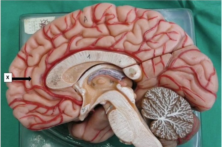
Which one statement is true concerning structure X?
This is the corpus callosum
This is the cingulate sulcus
This is the calcarine sulcus
This is the cingulate gyrus
This is the parieto-occipital sulcus
This is the cingulate sulcus
What is the gyrus immediately above the corpus callosum?
cingulate gyrus
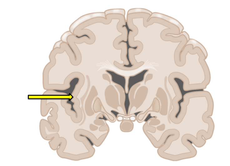
It is the claustrum and little is known about its function
It is the temporal lobe, which plays a role in processing auditory information
It is the insula, which plays a role in interoception (internal sense) and social emotion
It is the parietal lobe, which plays a role in integrating sensory information
It is part of the basal ganglia, which plays a role in the coordination of movement
It is the insula, which plays a role in interoception (internal sense) and social emotion
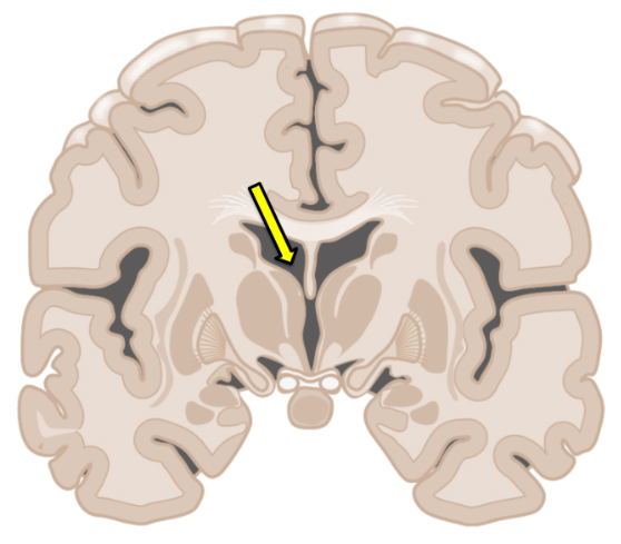
The structure indicated by the arrow is involved in cerebrospinal fluid (CSF) circulation. What is the next immediate step in the process?
The CSF flows down the cerebral aqueduct
Additional CSF is produced in the third ventricle
CSF is resorbed into the dural venous sinuses
CSF flows through the interventricular foramina
The CSF flows down to the fourth ventricle
CSF flows through the interventricular foramina
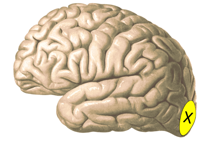
The Brodmann area X is concerned with:
reading words - this is the primary visual cortex
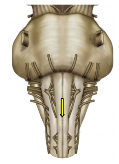
The arrow indicates the decussation of tracts which originate in the:
motor cortex
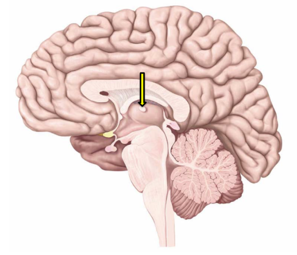
Which statement(s) is/are true regarding the structure indicated by the arrow?
i. Is part of the thalamus.
ii. Is part of the hypothalamus.
iii. Is the interthalamic adhesion (massa intermedia).
iv. Is the infundibulum.
i and iii
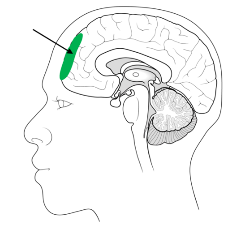
The area of the brain indicated by the arrow (highlighted in green) is associated with:
making decisions and personality
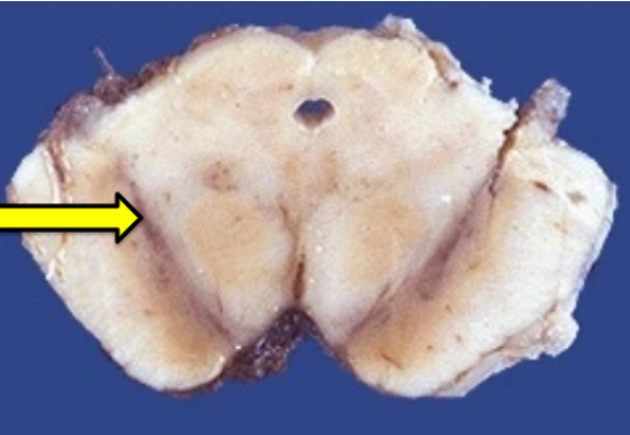
Which of the following is the most complete and accurate answer concerning the structure indicated by X?
It is the substantia nigra
It is the substantia nigra and it is located in the tectum of the midbrain
It is the substantia nigra and is considered to be part of the basal ganglia
It is the substantia nigra and is considered to be part of the subthalamus
It is the substantia nigra and contains mostly serotonergic neurons
It is the substantia nigra and is considered to be part of the basal ganglia
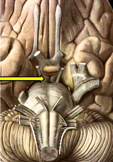
What structure is indicated by the arrow?
A mammillary body
The optic tract
The head of the caudate nucleus
The body of the caudate nucleus
A part of the thalamus
A mammillary body
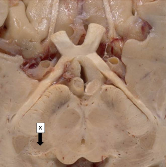
Which one statement best describes structure X?
This is the lateral geniculate nucleus and is a thalamic relay between the inferior colliculus and the auditory cortex
This is the lateral geniculate nucleus and is a thalamic relay between the optic tracts and the visual cortex
This is the medial geniculate nucleus and is a thalamic relay between the inferior colliculus and the auditory cortex
This is the medial geniculate nucleus and is a thalamic relay between the optic tracts and the visual cortex
This is the medial geniculate nucleus and is a thalamic relay between the superior colliculus and the auditory cortex
This is the medial geniculate nucleus and is a thalamic relay between the inferior colliculus and the auditory cortex