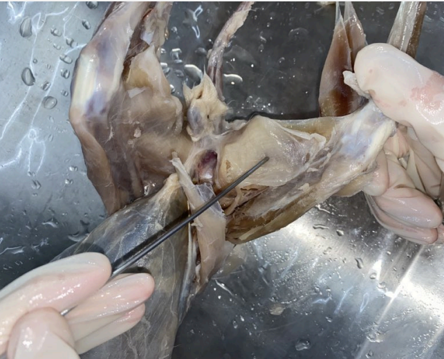
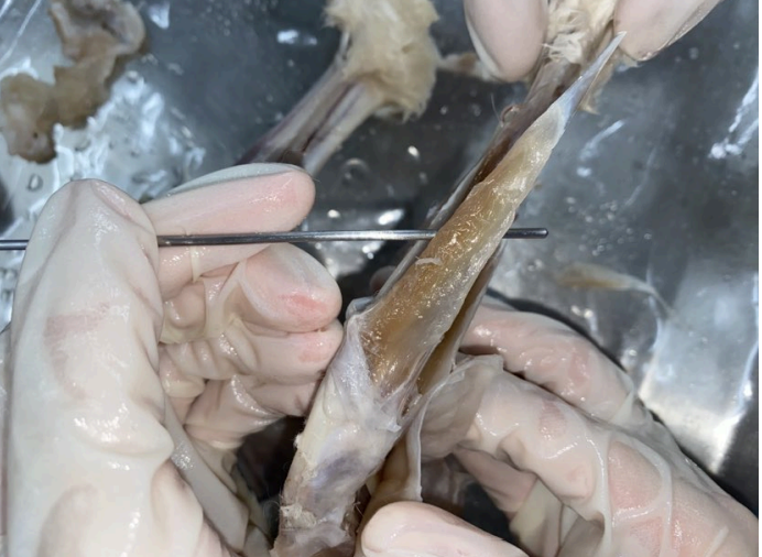
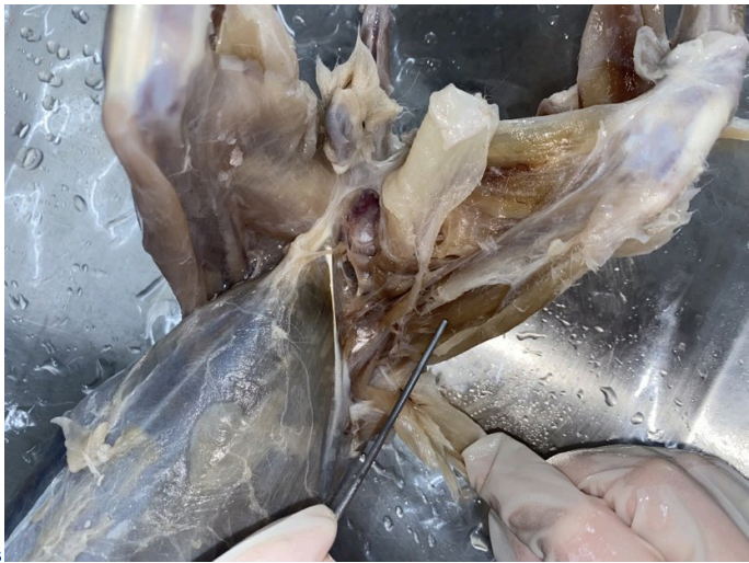
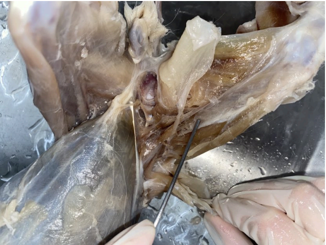
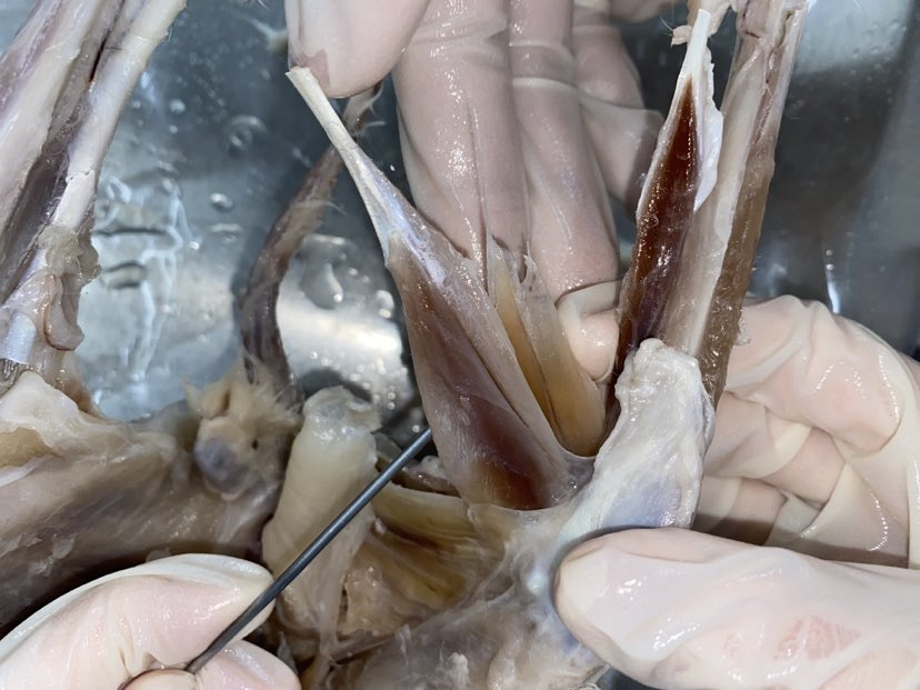
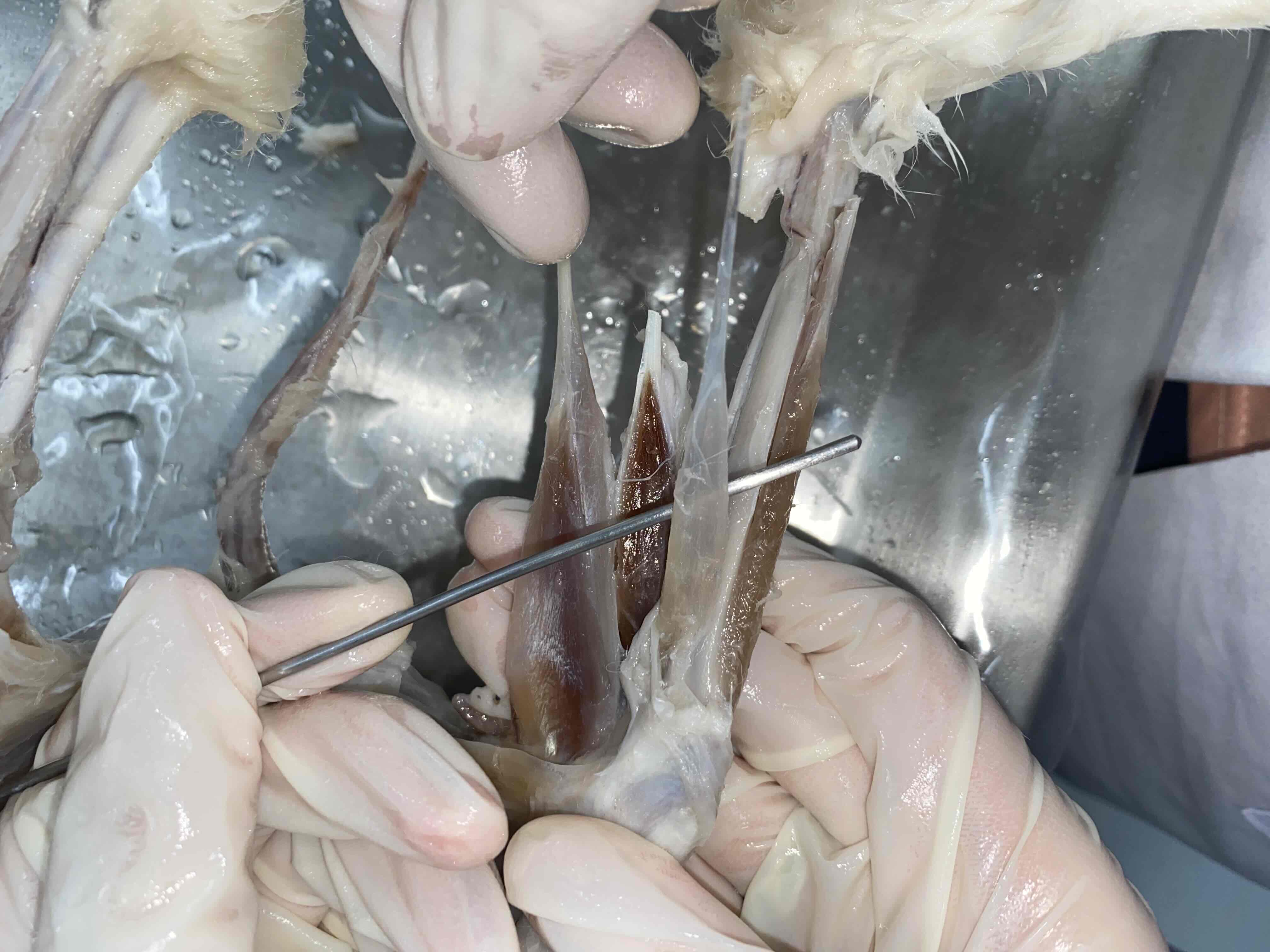
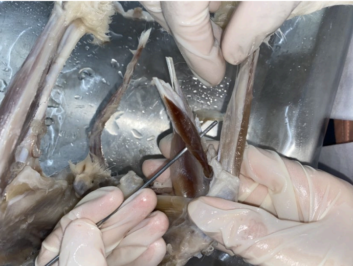
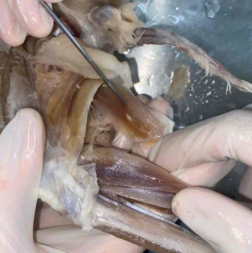
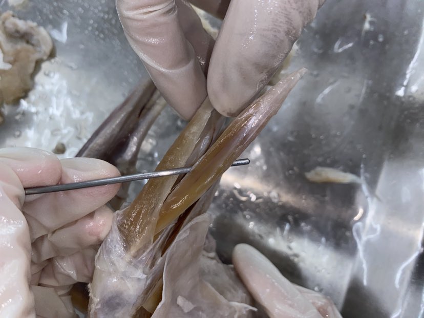
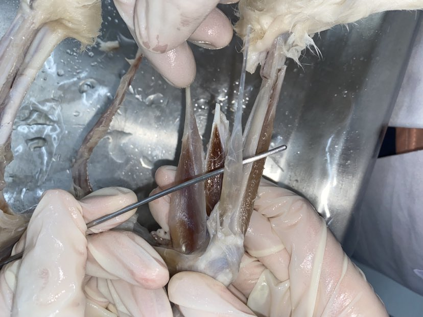
FRONT THIGH AND LEG
1/12
There's no tags or description
Looks like no tags are added yet.
Name | Mastery | Learn | Test | Matching | Spaced | Call with Kai | Chat |
|---|
No analytics yet
Send a link to your students to track their progress
13 Terms
Vastus lateralis
O: Greater trochanter and surface of the femur
I: Patella and adjacent ligaments
A: Extend the shank
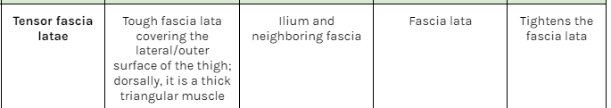
Tensor fascia latae
O: iliium and neighboring fascia
I: fascia lata
A: tightens the fascia lata
Sartorius
O: Crest and ventral border of the ilium
I: Proximal end of the tibia; patella; fascia and
ligaments in between
A: Adduct and rotate thigh; extend shank
Gracilis
O: Ischial and pubic symphyses
I: Tibia (via an aponeurosis) long bone
A: adduct leg
Tibialis anterior
O: Proximal parts of tibia fibula
I: First metatarsal via a strong tendon
A: Extend (dorsiflex) foot
Rectus femoris
O: Ilium, near the acetabulum
I: Patella and adjacent ligaments
A: Extend the shank
Vastus medialis
O: Femur
I: Patella and adjacent ligaments
A: Extend the shank
gastrocnemius
O: Surface fascia; femur; tendon and fascia of the plantaris muscle
I: Calcaneus (via the Achilles' tendon)
A: Ventroflex the foot
plantaris
O: Patella and femur
I: Digits (via a tendon divided into four)
A: Flex digits; extend ankle
Soleus
O: fibula
I: Calcaneus (joins tendon of the gastrocnemius)
A: Ventroflex the foot
(with the gastrocnemius)
semitendinosus
Extensor digitorum longus
Lateral epicondyle of the femur | Stout tendon which diverges into four tendons inserted on each digit | Extends digits and dorsiflexes the foot |
Flexor Digitorum Longus
Tibia, fibula, adjacent fascia | Slender tendons that unite to form one, which then divides into 4 tendons inserted on the digits | Flexes the digits |